Abstract
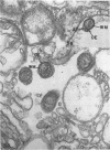
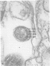
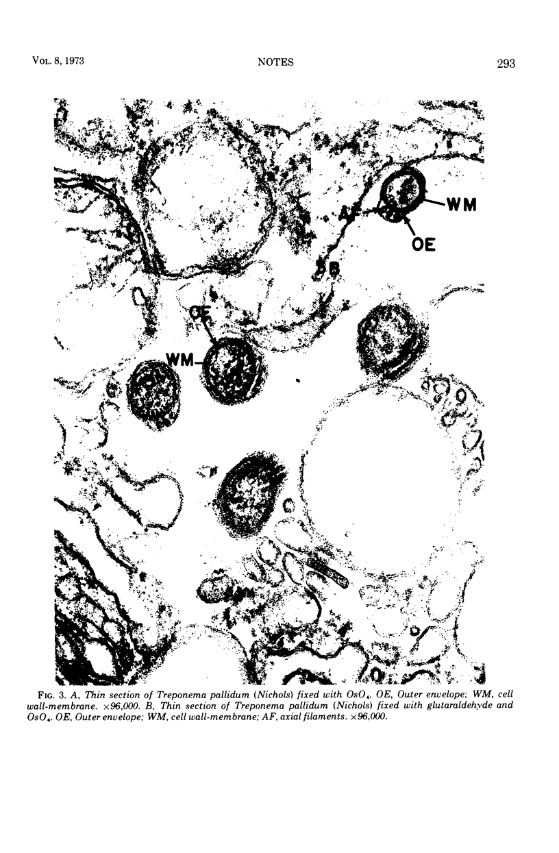
Ultrathin sections of virulent Treponema pallidum (Nichols strain) were examined with the electron microscope, and the presence of an outer cell envelope was documented.
Full text
PDF
Images in this article
Selected References
These references are in PubMed. This may not be the complete list of references from this article.
- Azar H. A., Pham T. D., Kurban A. K. An electron microscopic study of a syphilitic chancre. Engulfment of Treponema pallidum by plasma cells. Arch Pathol. 1970 Aug;90(2):143–150. [PubMed] [Google Scholar]
- Drusin L. M., Rouiller G. C., Chapman G. B. Electron microscopy of Treponema pallidum occurring in a human primary lesion. J Bacteriol. 1969 Feb;97(2):951–955. doi: 10.1128/jb.97.2.951-955.1969. [DOI] [PMC free article] [PubMed] [Google Scholar]
- Hasegawa T. Electron microscopic observations on the lesions of condyloma latum. Br J Dermatol. 1969 May;81(5):367–374. doi: 10.1111/j.1365-2133.1969.tb14000.x. [DOI] [PubMed] [Google Scholar]
- Johnson R. C., Wachter M. S., Ritzi D. M. Treponeme outer cell envelope: solubilization and reaggregation. Infect Immun. 1973 Feb;7(2):249–258. doi: 10.1128/iai.7.2.249-258.1973. [DOI] [PMC free article] [PubMed] [Google Scholar]
- KELLENBERGER E., RYTER A., SECHAUD J. Electron microscope study of DNA-containing plasms. II. Vegetative and mature phage DNA as compared with normal bacterial nucleoids in different physiological states. J Biophys Biochem Cytol. 1958 Nov 25;4(6):671–678. doi: 10.1083/jcb.4.6.671. [DOI] [PMC free article] [PubMed] [Google Scholar]
- LUFT J. H. Improvements in epoxy resin embedding methods. J Biophys Biochem Cytol. 1961 Feb;9:409–414. doi: 10.1083/jcb.9.2.409. [DOI] [PMC free article] [PubMed] [Google Scholar]
- Lauderdale V., Goldman J. N. Serial ultrathin sectioning demonstrating the intracellularity of T. Pallidum. An electron microscopic study. Br J Vener Dis. 1972 Apr;48(2):87–96. doi: 10.1136/sti.48.2.87. [DOI] [PMC free article] [PubMed] [Google Scholar]
- Ovcinnikov N. M., Delektorskij V. V. Current concepts of the morphology and biology of Treponema pallidum based on electron microscopy. Br J Vener Dis. 1971 Oct;47(5):315–328. doi: 10.1136/sti.47.5.315. [DOI] [PMC free article] [PubMed] [Google Scholar]
- Sykes J. A., Miller J. N. Ultrastructural studies of treponemes: location of axial filaments and some dimensions of Treponema pallidum (Nichols strain), Treponema denticola, and Treponema reiteri. Infect Immun. 1973 Jan;7(1):100–110. doi: 10.1128/iai.7.1.100-110.1973. [DOI] [PMC free article] [PubMed] [Google Scholar]
- Wiegand S. E., Strobel P. L., Glassman L. H. Electron microscopic anatomy of pathogenic Treponema pallidum. J Invest Dermatol. 1972 Apr;58(4):186–204. doi: 10.1111/1523-1747.ep12539907. [DOI] [PubMed] [Google Scholar]


