

Abstract
Follicular occlusion tetrad is a symptom complex consisting of four conditions having a similar pathophysiology. It includes Hidradenitis suppurativa, acne conglobata, dissecting cellulitis of the scalp and pilonidal sinus. The exact pathogenesis of this group of disease is unknown but evidence suggests that they share the same pathological process initiated by follicular occlusion in apocrine gland bearing areas. Though each of these conditions is commonly encountered singly, follicular occlusion tetrad as a symptom complex has been rarely reported in the literature.
Keywords: Acne conglobata, dissecting cellulitis of the scalp, follicular occlusion tetrad, Hidradenitis suppurativa, pilonidal sinus
INTRODUCTION
Follicular occlusion tetrad is a complex consisting of four conditions having a similar pathophysiology. It includes Hidradenitis suppurativa, acne conglobata, dissecting cellulitis of the scalp and pilonidal sinus. Though each of these conditions are commonly encountered on their own, as a symptom complex follicular occlusion tetrad has rarely been reported in the literature. Here we present a case of Hidradenitis suppurativa in a 36-year-old male patient who also had the above mentioned associations.
CASE REPORT
A 36-yeasr-old male patient presented to us with a history of recurrent boils since18 years. They initially started in the groin and buttocks with boils and gradually broke down to ooze pus. The lesions were associated with pain. The boils healed with scarring after weeks to months. The process progressively started to involve the axillae and then the chest. The patient was treated with multiple antibiotics for over a decade that gave only symptomatic relief. The patient was diagnosed with pilonidal sinus at the age of 28 and he underwent surgery for the same. There was no history of recurrent fever or weight loss. There was no history of abnormal bowel movements or any other systemic complaints. There was no family history of similar complaints, though his father suffered from psoriasis.
On examination, vitals were stable. The patient was obese at 96 kg. There was no pallor, edema or koilonychia. The cardiovascular and respiratory systems were normal. On general examination, there were multiple areas of cicatricial alopecia on the scalp [Figure 1] and nodular scars on lower half of the face [Figure 2]. On query, the patient gave history of severe acne with nodules and pustules healing with scars on the face. He also had patchy swellings over the scalp region associated with pain in the past, which was diagnosed as folliculitis by a physician. They recurred for about 4-5 years and gradually healed with scarring alopecia.
Figure 1.
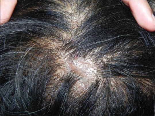
Patch of cicatricial alopecia on the occiput region of the scalp
Figure 2.
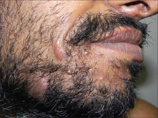
Pustules, cysts and nodule - reminiscent of acne conglobata - on the lower half of the face
On local examination, there were skin colored-to-erythematous nodules, grouped comedones and band like scars in the groin, buttocks, axillae and chest [Figure 3 and 4]. A few nodules were tender with oozing of pus. The patient had difficulty in lifting arms due to pain and scars in the axillae. There was no associated lymphadenopathy. On the basis of the above findings a diagnosis of Hidradenitis suppurativa was made.
Figure 3.
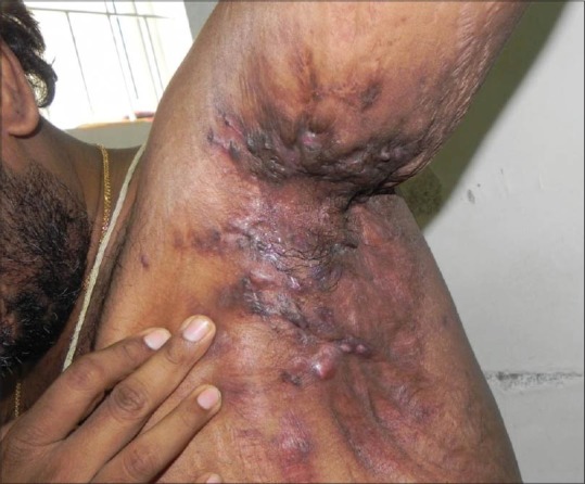
Multiple nodules, cysts and band like scars in the axillary region
Figure 4.
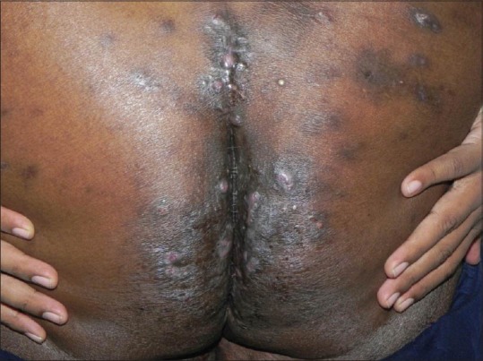
Multiple nodules and pustules over the buttocks with surgical scar (done for pilonidal sinus) seen in the upper part of gluteal cleft
A thorough general examination was followed by lab work-up. Hemogram revealed normocytic, normochromic anemia, lymphocytopenia and leukocytosis. Urine microscopy was insignificant. Liver function tests and renal function tests were within normal limits and so was FBS, PPBS and fasting lipid profile.
As the patient was obese, he was advised to follow a strict diet chart to lose weight. The patient was started on oral Cephalosporins and topical mupirocin to combat infection. We also started him on oral isotretinoin 0.5 mg/kg body weight once daily. The patient came back after six weeks for prescription refill but was thereafter lost for follow-up.
DISCUSSION
Follicular occlusion tetrad is a condition that includes Hidradenitis suppurativa (HS), acne conglobata, dissecting cellulitis of the scalp and pilonidal sinus. Hidradenitis suppurativa was first described in 1839 by Velpeau;[1] Verneuil[2] gave it its name in 1854 and associated it with the sweat glands. Later, HS was classified as a member of the follicular occlusion triad, along with acne conglobata and dissecting cellulitis of the scalp. In 1975, pilonidal cyst was added as a member to this triad, forming the follicular occlusion tetrad.[3] In 1989, Plewig and Steger introduced the term acne inversa to substitute the term HS.
Hidradenitis suppurativa or acne inversa is a chronic, inflammatory, recurrent, debilitating skin disease that usually presents after puberty with painful, deep-seated, inflamed lesions in the apocrine gland-bearing areas of the body, most commonly the axillae, inguinal and anogenital regions. The exact pathogenesis of this group of diseases is unknown but evidence suggests that they share the same pathological process initiated by follicular occlusion. Hidradenitis suppurativa (HS) usually starts at or soon after puberty. Women are three times more commonly affected than men. The pathogenesis and aetiology are unknown, but is thought to originate from poral occlusion of the pilosebaceous units.
Diagnostic criteria of Hidradenitis suppurativa (adopted by the 2nd International Conference on Hidradenitis suppurativa, March 5,2009, San Francisco, CA US).
Typical lesions, i.e. deep-seated painful nodules: ‘blind boils’ in early lesions; abscesses, draining sinus, bridged scars and ‘tombstone’double-ended pseudo-comedones in secondary lesions
Typical topography, i.e. axillae, groins, perineal and perianal region, buttocks, infra and inter mammary folds
Chronicity and recurrences.
All three criteria must be met for establishing the diagnosis.
Dissecting cellulitis(also called Perifolliculitis capitis abscedens et suffodiens) manifests with perifollicular pustules, nodules, abscesses and sinuses that evolve into scarring alopecia. It predominantly occurs in African American men between 20-40 years of age, but can occasionally affect other races and women too. Associated musculoskeletal findings are sometimes reported. Its course is chronic and relapsing, and treatment is often difficult. Dissecting cellulitis must be distinguished from several other scalp conditions. The tendency of dissecting cellulitis to cause severe alopecia, fluctuant nodules, and sinus tracts helps to distinguish it from acne keloidalis nuchae.[4] It differs from pseudopelade of Brocq by its lack of atrophy and “foot prints in the snow” alopecia morphology. Unlike tinea capitis, culture of dissecting cellulitis does not produce a positive fungal culture and nuchal palpation does not reveal palpable lymph nodes,[5] though reports have noted an inflammatory tinea capitis (kerion) that mimicked dissecting cellulitis in adolescents.[6]
Folliculitis decalvans starts with areas of perifollicular erythema. Its follicular papules and pustules spread peripherally, leaving central scarred patches of alopecia without nodules or sinuses. Tufted folliculitis resolves with patches of scarring alopecia within which multiple hair tufts emerge from dilated follicular orifices.
Medical therapies include isotretinoin, antibiotics, and prednisone. Destructive therapies include X-ray therapy, surgical excision, and skin grafting. Laser epilation of hair follicles is a promising new therapy for dissecting cellulitis.
Acne conglobata is an uncommon nodulocystictype of acne vulgaris that is often resistant to therapy. This disorder typically begins in adulthood and presents as numerous comedones, papules, pustules, nodules, abscesses, and draining sinus tracts involving the chest, back and buttocks. These lesions frequently become secondarily infected with gram-positive bacteria and often heal with scarring. Pathology usually reveals inflammatory infiltrate around follicles, which can often disrupt the normal dermal architecture. Acne conglobata is particularly disfiguring and socially detrimental to patients because of its chronicity, severity, and treatment challenge.
Acne conglobata (AC) resembles acne fulminans because both cause numerous inflammatory nodules on the trunk. Acne conglobata produces polyporus comedones and noninflammatory cysts, while acne fulminans does not. Unlike acne conglobata, large nodules of acne fulminans tend to become painful ulcers with overhanging borders surrounding exudative necrotic plaques that become confluent. The therapy of Choice for AC includes isotretinoin 0.5-1 mg/kg for 4-6 months. Simultaneous use of systemic steroids such as prednisone 1 mg/kg/d for 2-4 weeks may also prove beneficial, particularly if systemic symptoms are evident. Alternatives include oral tetracycline, minocycline, or doxycycline. Oral tetracycline antibiotics should not be combined with oral isotretinoin because of an increased risk of pseudotumor cerebri. For treatment-resistant cases, dapsone 50-150 mg/d is recommended; this treatment should be carefully monitored. Treatment of acne conglobata with infliximab has been reported.[7]
Pilonidal sinus was described as far back as 1833 when Mayo described a hair-containing cyst located just below the coccyx.[8] Incidence of pilonidal disease is about 26 per 100,000 population. Pilonidal disease occurs predominantly in males at a ratio of about 3-4:1.[9] No specific laboratory studies or tests are needed to diagnose pilonidal disease and its sequelae or differentiate it from other disease entities; it is a clinical diagnosis best elicited by history and physical examination findings. Treatment of infection or abscess with oral antibiotics followed by surgical excision of the sinus is practiced.
Footnotes
Source of Support: Nil
Conflict of Interest: None declared.
REFERENCES
- 1.Aissele VA. In: Dictionnairede medicine, a general directory of Sciences Medicals under the theoretical report and practice. BechetJeune Z, editor. Vol. 2. Paris: 1839. pp. 1839–91. [Google Scholar]
- 2.Verneuil A. Studies on tumors of the skin; after few diseases of the perspiratory glands. Arch GenMed. 1854;4:447–68. [Google Scholar]
- 3.Plewig G, Kligman AM. Berlin: Springer-Verlag; 1975. Acne: Morphogenesis and Treatment; pp. 192–3. [Google Scholar]
- 4.Luz Ramos M, Muñoz-Pérez MA, Pons A, Ortega M, Camacho F. Acne keloidalisnuchae and tufted hair folliculitis. Dermatology. 1997;194:71–3. doi: 10.1159/000246063. [DOI] [PubMed] [Google Scholar]
- 5.Padilha-Gonçalves A. Inflammatory tineacapitis (kerion) mimicking dissecting cellulitis. Int J Dermatol. 1992;31:66. doi: 10.1111/j.1365-4362.1992.tb03526.x. [DOI] [PubMed] [Google Scholar]
- 6.Sperling LC. Inflammatory tineacapitis (kerion) mimicking dissecting cellulitis. Occurrence in two adolescents. Int J Dermatol. 1991;30:190–2. doi: 10.1111/j.1365-4362.1991.tb03849.x. [DOI] [PubMed] [Google Scholar]
- 7.Shirakawa M, Uramoto K, Harada FA. Treatment of acne conglobata with infliximab. J AmAcadDermatol. 2006;55:344–6. doi: 10.1016/j.jaad.2005.06.008. [DOI] [PubMed] [Google Scholar]
- 8.Hull TL, Wu J. Pilonidal disease. SurgClin North Am. 2002;82:1169–85. doi: 10.1016/s0039-6109(02)00062-2. [DOI] [PubMed] [Google Scholar]
- 9.da Silva JH. Pilonidal cyst: Cause and treatment. Dis Colon Rectum. 2000;43:1146–56. doi: 10.1007/BF02236564. [DOI] [PubMed] [Google Scholar]