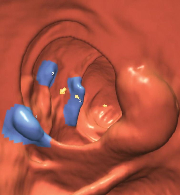
Figure 2b:

(a–d) Images show cecal carpet lesion detected at screening CT colonography in 50-year-old man (patient 3). (a) Three-dimensional colon map and (b) endoluminal CT colonography view show three CAD marks in the cecum (yellow dots in a; blue regions with arrows in b), which identify focal areas of a 3.5-cm carpet lesion. The lesion is located across from the normal-appearing ileocecal valve. (c, d) Transverse 2D images in (c) polyp and (d) soft-tissue windows confirm a flat soft-tissue lesion (arrows). Note the etching of positive oral contrast material on the surface of the lesion, which is better seen in d. (e) The lesion was confirmed at same-day endoscopy and proved to be a tubulovillous adenoma without high-grade dysplasia after laparoscopic right hemicolectomy.
