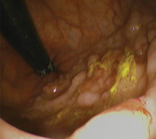
Figure 3c:

(a–c) Images show rectal carpet lesion detected at CT colonography in 65-year-old woman (patient 4). (a) Three-dimensional endoluminal CT colonography image (obtained with the patient in the prone position) that simulates a retroflexed rectal view at endoscopy shows a large 8-cm multinodular lesion carpeting the lower rectum, with multiple individual CAD marks (blue regions with arrows). Red line = automated centerline for navigation. (b) Sagittal 2D image confirms a flat soft-tissue lesion (arrow), with an area of contrast material coating (arrowheads). (c) This large yet relatively subtle lesion was confirmed at same-day endoscopy and proved to be a villous adenoma without high-grade dysplasia after transanal surgical excision.
