
Figure 2.
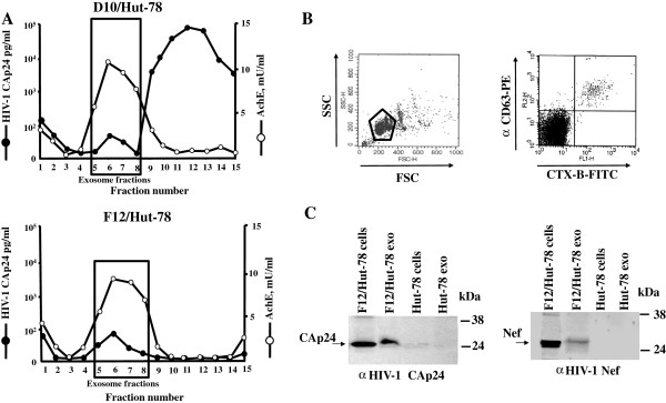
Characterization of exosomes from supernatants of D10/Hut-78 and F12/Hut-78 cells. A. HIV-1 Gag contents and AchE activity measured in fractions from 6-18% iodixanol gradients loaded with vesicles obtained by differential centrifugations of supernatants from D10/Hut-78 and F12/Hut-78 cells. The results are the mean values of duplicate conditions, and are representative of two independent experiments. B. Detection by FACS of GM1 and CD63 on pools of AchE positive fractions from iodixanol gradients loaded with vesicles from F12/Hut-78 cells. Vesicles were bound to aldehyde/sulfate latex beads and then labeled with both FITC-CTX-B and PE-conjugated anti-CD63 monoclonal antibody. Both FSC/SCC (on the left) and fluorescence (on the right) dot plots are reported. The gate on the left panel includes both single and doublet beads considered for the fluorescence analysis. The results are representative of three independent experiments carried out on vesicles from three iodixanol gradient preparations. C. Detection of HIV-1 CAp24 (on the left) and Nef (on the right) by western blot analysis of both cells and exosomes from F12/Hut-78 and uninfected, parental Hut-78 cells. On the left of each panel, arrows indicate the specific signals, while on the right molecular markers are given in kDa. Results are representative of two (for CAp24 detection) and six (for Nef detection) independent experiments.