

Abstract
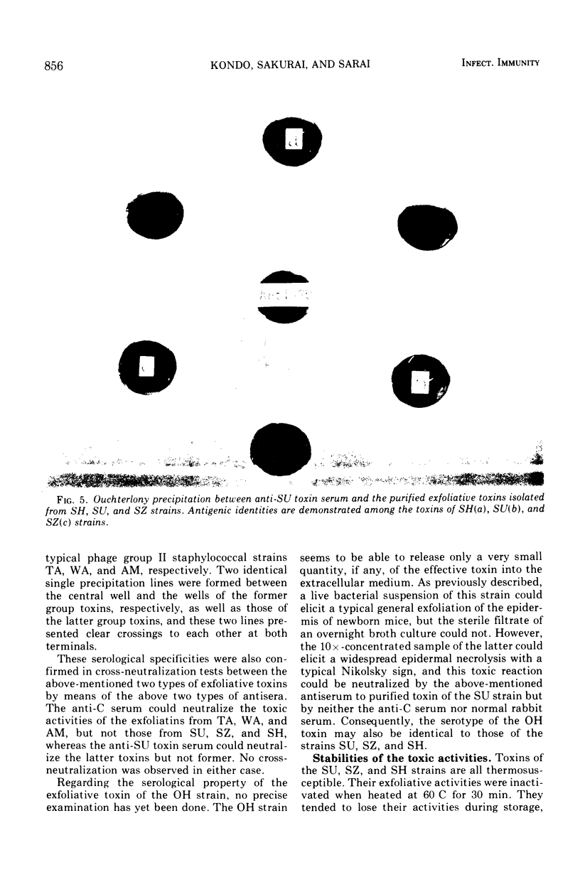
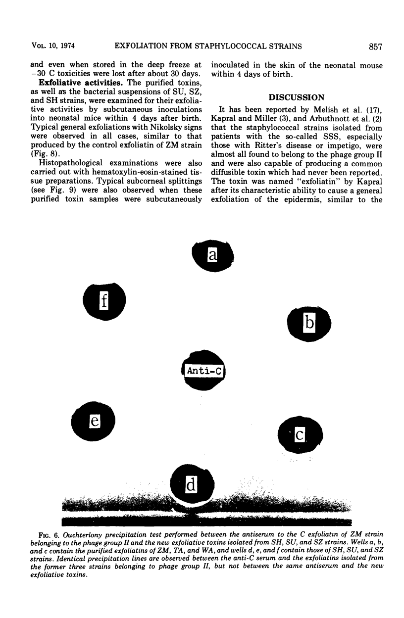
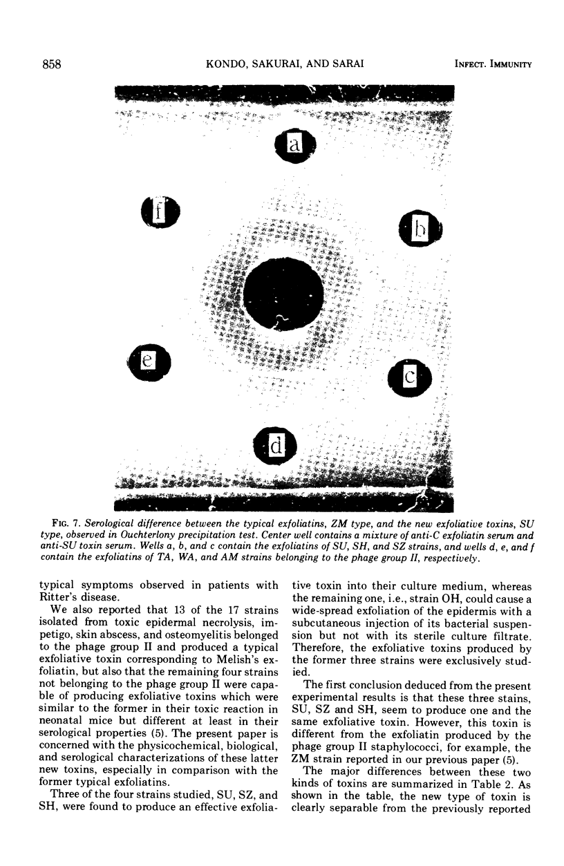
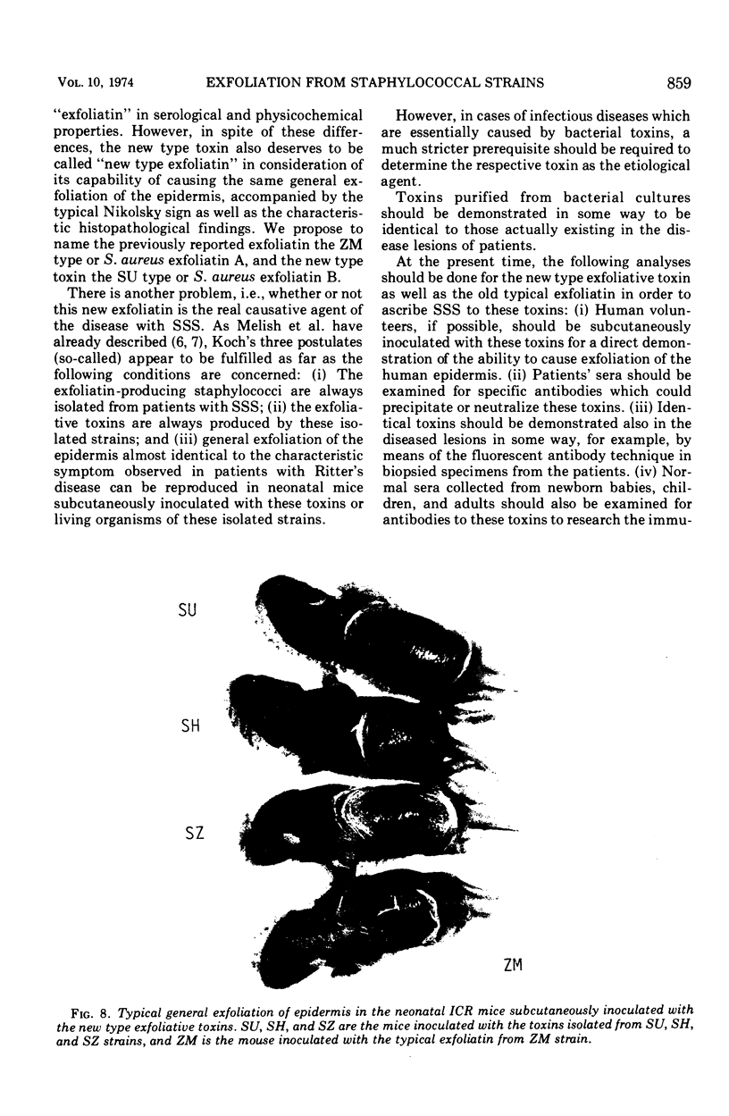
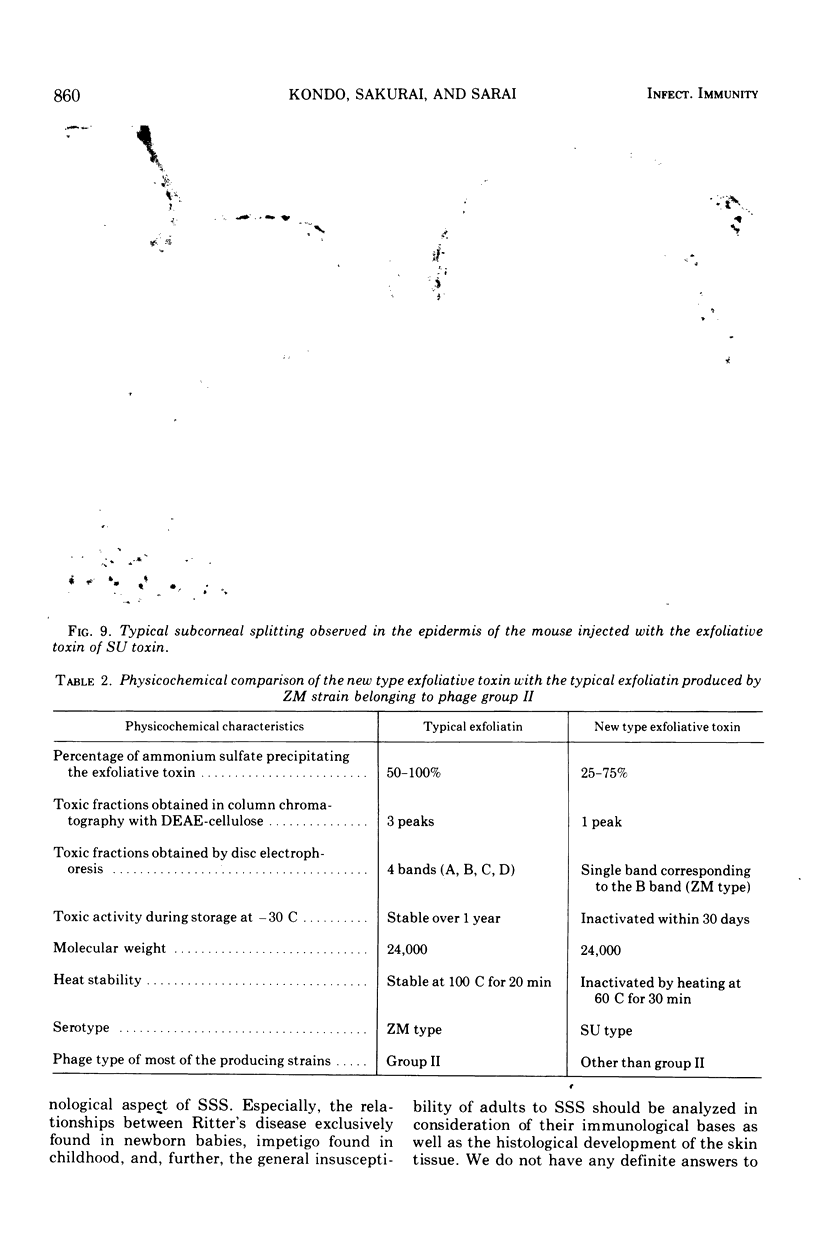
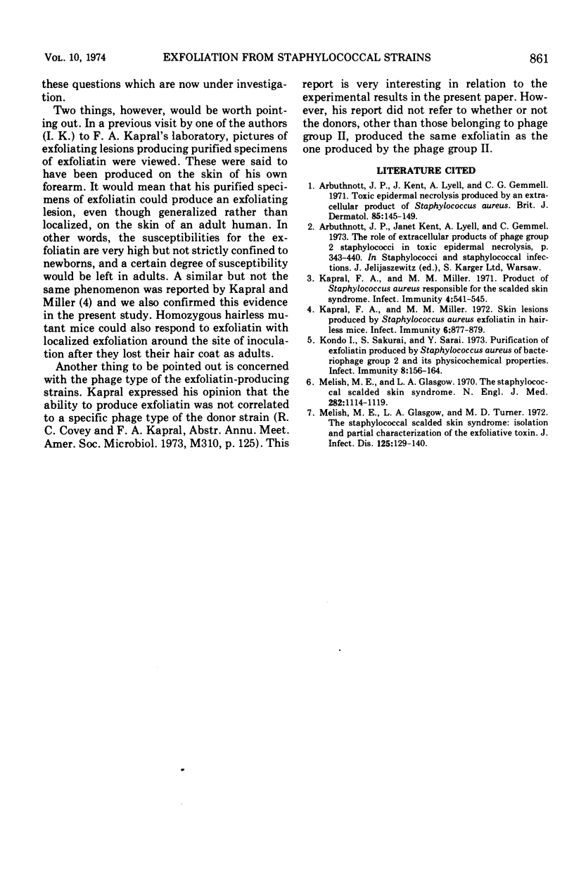
Four strains of Staphylococcus aureus of a phage type other than the second group, isolated from patients with impetigo and Ritter's disease, were found to produce an exotoxin similar to those reported by Melish et al. (1972), Kapral and Miller (1971), and Arbuthnott et al. (1973). This toxin could elicit a general exfoliation of the epidermis with the so-called Nikolsky sign when subcutaneously inoculated into neonatal mice within 4 days after birth. The new toxin was serologically different from exfoliatin produced by the phage group II staphylococci previously reported (Kondo et al., 1973) and showed an electrophoretic pattern corresponding to that of the B-type toxin of the latter in acrylamide disc electrophoresis. It had the same molecular weight as that of the latter, which was estimated to be about 24,000. It was thermolabile and lost its toxic activity by heating at 60 C for 30 min; in addition, most of the toxicity was lost within 1 month of storage even at −30 C. We propose to designate the old typical heat-stable exfoliatin as S. aureus exfoliatin A and the new heat-susceptible exfoliatin as S. aureus exfoliatin B.
Full text
PDF

Images in this article


Selected References
These references are in PubMed. This may not be the complete list of references from this article.
- Arbuthnott J. P., Kent J., Lyell A., Gemmel C. G. The role of extracellular products of 'phage group 2 staphylococci in toxic epidermal necrolysis. Contrib Microbiol Immunol. 1973;1:434–440. [PubMed] [Google Scholar]
- Arbuthnott J. P., Kent J., Lyell A., Gemmell C. G. Toxic epidermal necrolysis produced by an extracellular product of Staphylococcus aureus. Br J Dermatol. 1971 Aug;85(2):145–149. doi: 10.1111/j.1365-2133.1971.tb07200.x. [DOI] [PubMed] [Google Scholar]
- Kapral F. A., Miller M. M. Product of Staphylococcus aureus responsible for the scalded-skin syndrome. Infect Immun. 1971 Nov;4(5):541–545. doi: 10.1128/iai.4.5.541-545.1971. [DOI] [PMC free article] [PubMed] [Google Scholar]
- Kapral F. A., Miller M. M. Skin lesions produced by Staphylococcus aureus exfoliatin in hairless mice. Infect Immun. 1972 Nov;6(5):877–879. doi: 10.1128/iai.6.5.877-879.1972. [DOI] [PMC free article] [PubMed] [Google Scholar]
- Kondo I., Sakurai S., Sarai Y. Purification of exfoliatin produced by Staphylococcus aureus of bacteriophage group 2 and its physicochemical properties. Infect Immun. 1973 Aug;8(2):156–164. doi: 10.1128/iai.8.2.156-164.1973. [DOI] [PMC free article] [PubMed] [Google Scholar]
- Melish M. E., Glasgow L. A. The staphylococcal scalded-skin syndrome. N Engl J Med. 1970 May 14;282(20):1114–1119. doi: 10.1056/NEJM197005142822002. [DOI] [PubMed] [Google Scholar]
- Melish M. E., Glasgow L. A., Turner M. D. The staphylococcal scalded-skin syndrome: isolation and partial characterization of the exfoliative toxin. J Infect Dis. 1972 Feb;125(2):129–140. doi: 10.1093/infdis/125.2.129. [DOI] [PubMed] [Google Scholar]