Abstract
The Osler-Weber-Rendu syndrome or Hereditary Hemorrhagic Telangiectasia (HHT) is a systemic fibrovascular dysplasia characterized by defects in the elastic and vascular walls of blood vessels, making them varicose and prone to disruptions. Lesions occur in different organs and can lead to hemorrhage in the lungs, digestive tract and brain. We describe the case of a patient with cutaneous manifestations and severe impairment of the digestive tract. It is important for the dermatologist to recognize this syndrome, since the cutaneous lesions may play a key role in diagnosis.
Keywords: Diagnosis; Epistaxis; Telangiectasia, hereditary hemorrhagic; Telangiectasis
CASE REPORT
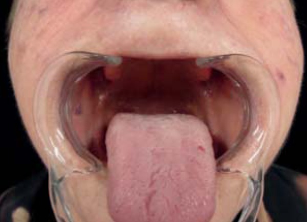
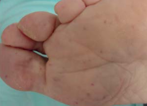
A 73-year-old woman who was hospitalized for hematemesis had multiple, punctiform telangiectasia lesions all over the integument since she was 40 years old. Lesions were most prominent in the oral mucosa, on the tongue, hands and feet (Figures 1 and 2). She also reported recurrent episodes of nasal and intestinal bleeding. The patient underwent hysterectomy at age 39 due to menometrorrhagia. The son and grandson had similar skin lesions.
FIGURE 1.

Telangiectasia on the oral mucosa. Lesions are also observed on the face
FIGURE 2.

Telangiectasia on the foot sole
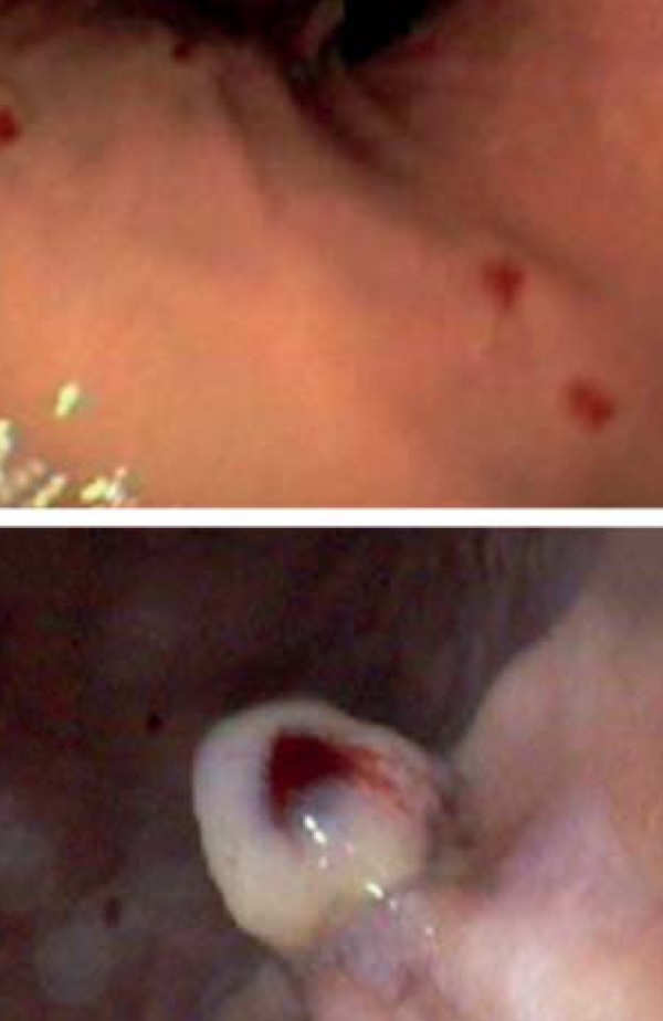
Complementary exams revealed 8.5 g/dL hemoglobin, 79 fl. mean corpuscular volume (MCV), and 35µg/L serum ferritin, which is consistent with iron deficiency anemia. An upper endoscopy revealed angiectases (some were 3 mm in diameter, friable) in the fornix, gastric body and second duodenal portion (Figure 3). CT angiography of the chest and computed cranial tomography showed no alterations.
FIGURE 3.

Upper endoscopy photos: Presence of multiple, friable, shiny telangiectasias in the fornix and gastric body, and tiny angiectases in the second duodenal portion
DISCUSSION
The Osler-Weber-Rendu syndrome or Hereditary Hemorrhagic Telangiectasia (HHT) is a systemic fibrovascular dysplasia characterized by defects in the elastic and vascular walls of blood vessels, making them varicose and prone to disruptions.1,2 It is an underdiagnosed entity.3
HHC has autosomal dominant inheritance, and homozygosity is incompatible with life. There are two subtypes: type 1 is related to a mutation in the endoglin gene (ENG, chromosome 9q34.1) and pulmonary involvement; type 2 is related to a mutation in the activin receptor-like kinase 1 gene (ACVRL 1, ALK1, chromosome 12q31.34), with a mild phenotype and late onset.2,3,4 These two mutations occur in 85% of cases. Both endoglin and ACVRL-1 are expressed in the endothelium, and interfere with the growth-transformation factor β (TGF-β).3,4,5 Mutations have been described in the SMAD4 gene, also related to the TGF-β, in patients with juvenile polyposis and HHT.5,6 TGF-β is important in the differentiation and growth of smooth muscle cells which form the walls of blood vessels. For this reason, its reduction is related to the formation of fragile vessels.7
HHT is characterized by hemorrhagic phenomena and punctiform telangiectasia lesions predominantly located on the face, lips, oral mucosa, hands and feet, with onset usually in the third decade of life.2,3,4,7 The most common manifestation is epistaxis (80-90% of cases) and it regularly precedes cutaneous manifestations. Pulmonary involvement occurs in 30% of patients due to arteriovenous malformations, causing dyspnea, fatigue, and cyanosis. Brain, liver, gastrointestinal and genitourinary involvement may occur.8
The Curaçao diagnostic criteria are: (1) spontaneous, recurrent epistaxis; (2) multiple telangiectases; (3) visceral lesions; (4) positive family history (three first-degree relatives affected.
Treatment is directed to the clinical manifestations of the condition. In the treatment of epistaxis and digestive manifestations, endoscopic or surgical intervention may be indicated.2,3,4,8 Bevacizumab, a monoclonal antibody that blocks the action of VEGF (vascular endothelial growth factor), is being used in cases of epistaxis or gastrointestinal bleeding.9 The Nd YAG and Pulsed Dye lasers have shown to be effective in the treatment of telangiectasias. However, relapses can occur.10
It is important to warn the patient about the possibility of bleeding and provide pre-pregnancy counseling due to a greater risk of complications.11 The case reported here reinforces the importance of knowing this syndrome, which presents skin changes that are often subtle but also decisive for the diagnosis of a condition with severe systemic involvement.
Footnotes
Conflict of interest: None
Financial funding: None
How to cite this article: Boza JC, Dorn T, Oliveira FB, Bakos RM. Case for diagnosis. Hereditary hemorrhagic telangiectasia. An Bras Dermatol. 2014;89(6):999-1001.
Study conducted at the Dermatology Service of the Hospital de Clinicas de Porto Alegre - Universidade Federal do Rio Grande do Sul (HCPA-UFRGS) - Porto Alegre (RS), Brazil.
References
- 1.Fuchizaki U, Miyamori H, Kitagawa S, Kaneko S, Kobayashi K. Hereditary Haemorrhagic Telangiectasia (Rendu-Osler-Weber Disease) Lancet. 2003;362:1490–1494. doi: 10.1016/S0140-6736(03)14696-X. [DOI] [PubMed] [Google Scholar]
- 2.Juares AJC, Dell'Aringa AR, Nardi JC, Kobori K, Rodrigues VLMGM, Filho RMP. Rendu-Osler-Weber Syndrome: case report and literature review. Rev Bras Otorrinolaringol. 2008;74:452–457. doi: 10.1016/S1808-8694(15)30582-6. [DOI] [PMC free article] [PubMed] [Google Scholar]
- 3.Albuquerque G, Terra D, Carvalho C, Quinete S, Oliveira C. Hereditary hemorrhagic telangiectasia: tranexamic acid for plantar ulcer. An Bras Dermatol. 2005;80:S373–S375. [Google Scholar]
- 4.Olitsky SE. Hereditary hemorrhagic telangiectasia: diagnosis and management. Am Fam Physician. 2010;82:785–790. [PubMed] [Google Scholar]
- 5.Tørring P, Brusgaard K, Ousager L, Andersen P, Kjeldsen A. National mutation study among Danish patients with hereditary hemorrhagic telangiectasia. Clin Genet. 2013 doi: 10.1111/cge.12269. [Epub ahead of print] [DOI] [PubMed] [Google Scholar]
- 6.Gallione CJ, Richards JA, Letteboer TG, Rushlow D, Prigoda NL, Leedom TP, et al. SMAD4 mutations found in unselected HHT patients. J Med Genet. 2006;43:793–797. doi: 10.1136/jmg.2006.041517. [DOI] [PMC free article] [PubMed] [Google Scholar]
- 7.Goumans MJ, Liu Z, ten Dijke P. TGF-beta signaling in vascular biology and dysfunction. Cell Res. 2009;19:116–127. doi: 10.1038/cr.2008.326. [DOI] [PubMed] [Google Scholar]
- 8.McDonald J, Bayrak-Toydemir P, Pyeritz RE. Hereditary hemorrhagic telangiectasia: an overview of diagnosis, management, and pathogenesis. Genet Med. 2011;13:607–616. doi: 10.1097/GIM.0b013e3182136d32. [DOI] [PubMed] [Google Scholar]
- 9.Dupuis-Girod S, Ambrun A, Decullier E, Samson G, Roux A, Fargeton AE, et al. ELLIPSE Study: A Phase 1 study evaluating the tolerance of bevacizumab nasal spray in the treatment of epistaxis in hereditary hemorrhagic telangiectasia. MAbs. 2014;6:793–798. doi: 10.4161/mabs.28025. [DOI] [PMC free article] [PubMed] [Google Scholar]
- 10.Halachmi S, Israeli H, Ben-Amitai D, Lapidoth M. Treatment of the skin manifestations of hereditary hemorrhagic telangiectasia with pulsed dye laser. Lasers Med Sci. 2014;29:321–324. doi: 10.1007/s10103-013-1346-x. [DOI] [PubMed] [Google Scholar]
- 11.Shovlin CL, Sodhi V, McCarthy A, Lasjaunias P, Jackson JE, Sheppard MN. Estimates of maternal risks of pregnancy for women with hereditary haemorrhagic telangiectasia (Osler-Weber-Rendu syndrome): suggested approach for obstetric services. BJOG. 2008;115:1108–1115. doi: 10.1111/j.1471-0528.2008.01786.x. [DOI] [PubMed] [Google Scholar]


