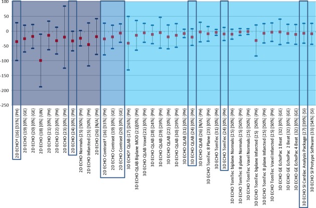
Figure 3.
Comparison of echocardiographic techniques with cardiac magnetic resonance imaging for measurement of end-systolic volume (mL). Red square box indicates bias compared with magnetic resonance imaging. Blue line at each end of the plots indicates the lower and upper limits of agreement calculated by Bland–Altman. MRI = magnetic resonance imaging; 2D ECHO = two-dimensional echocardiography; 3D ECHO = three-dimensional echocardiography; NSR = normal sinus rhythm; MOD = method of disks; QLAB = Philips online and offline LV volume calculation tool; TomTec = offline left ventricular volume calculation tool.

