Abstract
Patient: Male, 29
Final Diagnosis: Traumatic shoulder dislocation
Symptoms: Shoulder pain
Medication: —
Clinical Procedure: Patient-controlled shoulder relocation
Specialty: Orthopedics and Traumatology
Objective:
Management of emergency care
Background:
The glenohumeral joint is the most mobile joint in the human body due to the shallowness of the glenoid socket. This unique anatomy also makes it the most dislocated joint in humans. All the techniques described so far for relocation require operator control and prescription drugs. We describe a technique that is unique, easy, and patient-controlled.
Case Report:
A 29-year-old male patient presented to the Emergency Department after falling from scaffolding at work. He had left shoulder dislocation confirmed by clinical and radiological examination. The patient lay face down on the trolley with trolley being raised with electronic controls. The shoulder was reduced with ease and the patient was discharged home after radiologic confirmation of reduction.
Conclusions:
A new patient-controlled technique for reduction of the glenohumeral joint following dislocation is described. It is simple, safe, and effective to perform in Emergency Departments.
MeSH Keywords: Orthopedic Procedures, Shoulder Dislocation, Stretchers
Background
The glenohumeral joint is the most mobile joint in the human body due to the shallowness of the glenoid socket. This unique anatomy also makes it the most dislocated joint in humans. Males have up to 70% more dislocations compared to females, and 15–35 years age group is the commonest for shoulder dislocation [1]. It is estimated that shoulder dislocation occurs in 1.69 per 1000 person-years in the community [1]. Hippocrates described the oldest known methodology for relocation of the shoulder and a variety of techniques have since then been described to reduce the shoulder. We describe a new technique of shoulder reduction wherein the patient has the control and there is no need for sedation. In our case series we have observed 100% success rate.
Case Report
A 29-year-old presented to the Emergency Department after falling from scaffolding at work. He managed to hold on to the scaffolding rail with his left hand for a brief period of time before falling to the ground. He presented to the Emergency Department with severe pain in his left shoulder. He had no other injury to his body except bruises on the left hip and elbow and was otherwise a healthy young man. He was not taking any medication and had no significant medical or surgical history. He disliked needles and injections. He had no known allergy to any medication. Just before going to work he had his morning breakfast and then he had a cup of tea about 15 minutes prior to arrival to the Emergency Department.
On examination, he was sitting upright with his shoulder in a sling provided by the paramedics. His pulse rate was regular at 98 beats per minute and he was normotensive. He had 97% oxygen saturation on room air. Detailed clinical examination revealed no injury other than the shoulder dislocation. His left shoulder muscles were in spasm. The humeral head was palpable anteriorly. He was holding his arm in slight abduction and external rotation. The deltoid region had normal sensation. His distal radial pulse and capillary refill at all the fingers were within normal range. Any movement of the shoulder was extremely painful, but he was comfortable if there was no shoulder movement.
An urgent x-ray confirmed that the patient had anterior left shoulder dislocation without any fracture to the humerus. His glenoid fossa was intact. As patient had refused to have any needle inserted, he was not given any intravenous or intramuscular analgesics. He was not fasted; hence, immediate sedation had a high risk of aspiration. Clinical staff explained the non-sedating, non-needle technique of shoulder dislocation, for which he gave verbal consent.
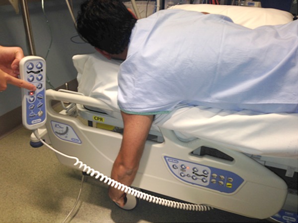
The patient was asked to lie on the trolley with his face down in prone position. His left affected arm was hanging by the side of the trolley (Figure 1). The trolley was lowered so that the patient could hold the brake handle of the trolley. He was asked to continue to hold the handle as long as he could, giving the control to the patient. A doctor then slowly pushed the trolley upwards with the control button as shown in the Figure 1, about 1 inch at a time and within tolerance of the patients’ pain level. As the trolley was lifted about 3–4 inches over a period of 5 minutes, the shoulder was relocated. The patient felt a ‘pop’ and there was immediate pain relief.
Figure 1.

Trolley with electronic control.
Post-reduction clinical examination of the shoulder revealed normal contour of the shoulder and normal distal neurovascular examination. Post-reduction x-ray films revealed a normal left glenohumeral joint. The patient was given a shoulder sling for a week for ligamentous recovery and was discharged home.
Discussion
There are 2 types of anterior shoulder dislocations described in the literature. Subcoracoid dislocations refer to the humeral head slipping anteriorly and medially to the glenoid; this is the commonest type of dislocation. In a subglenoid anterior dislocation, the posterior of the humeral head slips inferior and slightly anterior to the glenoid [2]. More than 90% of cases present with anterior dislocations [3]. Other uncommon types of dislocations are posterior and luxation erecta. Subclavicular and intrathoracic are extremely rare types of shoulder dislocations [4] (Table 1).
Table 1.
Types of shoulder dislocations [10].
| Anterior dislocations |
| Subcoracoid type |
| Subglenoid type |
| Posterior |
| Luxatio erecta |
| Subclavicular |
| Intrathoracic |
A number of techniques have been described in the literature for relocation of a dislocated glenohumeral joint. Kocher’s method and Matson’s traction-counter traction are some of the commonly used techniques. Both these techniques require sedation and there is significant risk of brachial plexus or bone injury [5]. Fares’ technique does not use counter traction but it involves pulling at wrist with a risk of stretching the neural bundles [6]. Stimson’s maneuver involves the patient lying prone on a trolley and pull is provided by weights and sometimes by the assistant as well, but patient does not have control and hence there may even be a risk of falling off the trolley. The scapular maneuver is difficult to master and hence there is high risk of failure. The Milch and Spaso techniques are frequently impossible unless heavy sedation is given, as the positioning of the arm is critical to the success of these procedures [7–9] (Table 2).
Table 2.
Current techniques used for shoulder relocation [11].
| Kocher’s Method |
| Matson’s traction-countertraction method |
| Fare’s technique |
| Stimson’s manoeuvre |
| Scapular manoeuvre |
| Milch Method |
| Spaso technique |
Our technique is a simple to perform. The patient has maximum control and hence the likelihood of nerve or bone injury is very low. The patient is fully awake and there is no need for any sedation. Most developed countries and many developing countries as well have trollies with electronic controls (Figure 1), so there is no need to buy any special equipment for this technique. As the clinician is not pulling any part of patients’ body, there is no need to have a clinician with strong arms for this procedure. One operator is enough to perform our technique. This procedure involves gentle electronic lifting up of the trolley and hence there is gradual stretch and subsequent relocation of the shoulder, which gives satisfaction to the patient and minimizes risk of neurovascular injury. In our cases, we have not seen any complications and all of our reductions have been successful on the first attempt. We have performed a detailed literature search and have not found this technique described anywhere in the literature and hence we refer to this technique as the Doshi-Firke procedure for shoulder relocation.
Limitations of our technique are inability of the patient to lie down in prone position due to respiratory or other problems, in which case this procedure cannot be performed. Patient cooperation and willingness to comply are required for the success of the procedure. Extremely obese, pregnant, and very old patients may not be suitable for this technique. Hospitals without electronic-controlled trolleys may not be able to use our technique.
We suggest that this technique be used for all eligible patients and further case series reports be published in the literature. We conclude that this new and simple technique for shoulder relocation is safe and effective.
Conclusions
A new patient-controlled technique for reduction of the glenohumeral joint following dislocation is described. It is simple, safe, and effective to perform in Emergency Departments.
References:
- 1.Zacchilli MA, Owens BD. Epidemiology of shoulder dislocations presenting to emergency departments in the United States. J Bone Joint Surg Am. 2010;92(3):542–49. doi: 10.2106/JBJS.I.00450. [DOI] [PubMed] [Google Scholar]
- 2.Dodson CC, Cordasco FA. Anterior glenohumeral joint dislocations. Orthop Clin North Am. 2008;39(4):507–18. doi: 10.1016/j.ocl.2008.06.001. [DOI] [PubMed] [Google Scholar]
- 3.Cutts S, Prempeh M, Drew S. Anterior shoulder dislocation. Ann R Coll Surg Engl. 2009;91(1):2–7. doi: 10.1308/003588409X359123. [DOI] [PMC free article] [PubMed] [Google Scholar]
- 4.Yamamoto T, Yoshiya S, Kurosaka M, et al. Luxatio erecta (inferior dislocation of the shoulder): A report of 5 cases and a review of the literature. Am J Orthop (Belle Mead NJ) 2003;32(12):601–3. [PubMed] [Google Scholar]
- 5.Perron AD, Ingerski MS, Brady WJ, et al. Acute complications associated with shoulder dislocation at an academic Emergency Department. J Emerg Med. 2003;24(2):141–45. doi: 10.1016/s0736-4679(02)00717-5. [DOI] [PubMed] [Google Scholar]
- 6.Cleeman E, Flatow EL. Shoulder dislocations in the young patient. Orthop Clin North Am. 2000;31(2):217–29. doi: 10.1016/s0030-5898(05)70142-5. [DOI] [PubMed] [Google Scholar]
- 7.Johnson G, Hulse W, McGowan A. The Milch technique for reduction of anterior shoulder dislocations in an accident and emergency department. Arch Emerg Med. 1992;9(1):40–43. doi: 10.1136/emj.9.1.40. [DOI] [PMC free article] [PubMed] [Google Scholar]
- 8.Yuen MC, Yap PG, Chan YT, Tung WK. An easy method to reduce anterior shoulder dislocation: The Spaso technique. Emerg Med J. 2001;18(5):370–72. doi: 10.1136/emj.18.5.370. [DOI] [PMC free article] [PubMed] [Google Scholar]
- 9.Pasila M, Jaroma H, Kiviluoto I, et al. Early complications of Primary Shoulder dislocations. Acta Orthop Scand. 1978;49:260–63. doi: 10.3109/17453677809005762. [DOI] [PubMed] [Google Scholar]
- 10.Bakal B, Sener S, Turkan H. Scapular manipulation technique for reduction of traumatic anterior shoulder dislocations: Experiences of an Academic Emergency Department. Emerg Med J. 2005;22:336–38. doi: 10.1136/emj.2004.019752. [DOI] [PMC free article] [PubMed] [Google Scholar]
- 11.Manes HR. A New Method of Shoulder Reduction in the Elderly. Clin Orthop. 1980;147:200–2. [PubMed] [Google Scholar]


