Abstract
Purpose: GG-H whole transcriptome array analysis suggested involvement of PININ (PNN) in the alternative splicing of multiple long non-coding RNAs (lncRNAs). To further investigate PNN’s role in regulating the alternative splicing of lncRNAs in a corneal epithelial context, we performed detailed analyses for detecting and identifying alternatively spliced lncRNAs.
Methods: Total RNA was isolated from PNN knockdown human corneal epithelial (HCET) cells or Pnn-deficient mouse corneas, and subjected to real-time–PCR (RT–PCR) assays, and the alternatively spliced lncRNAs were counted. Alternatively spliced lncRNAs were detected with in situ hybridization with variant-specific RNA probes on human cornea sections.
Results: Our analysis uncovered PNN’s impact on the transcript levels of several lncRNAs including Linc00085 and HAS2-AS1. Interestingly, a mouse ortholog of HAS2-AS1, Has2as, clearly exhibited a differential splicing pattern among three major splice variants in the Pnn-deficient mouse cornea. The sequence analyses and quantification of splice variants of candidate lncRNAs, including RP11-295B20.2, RP11–18I14.1, and RP11–322M19.1, demonstrated complex configuration of their splicing changes, with a significant impact of PNN on the process. Knockdown of PNN in HCET cells led to specific changes in the inclusion of multiple cassette exons as well as in the use of alternative splice sites in RP11–322M19.1 and RP11–18I14.1, resulting in considerable net changes in the ratio between the splice variants. Finally, in situ hybridization analyses revealed the presence of RP11–295G20.2 in the nuclei of corneal epithelial cells, but not in the stromal cells of the human cornea, while RP11–322M19.1 was present in epithelial and non-epithelial cells.
Conclusions: The data suggest PNN’s role in the alternative splicing of a specific subset of lncRNAs might have a significant impact on the corneal epithelium.
Introduction
The responsibilities of the specialized surface epithelium of the cornea require it to maintain tightly regulated differentiated properties. There are numerous examples of ocular surface diseases in which the corneal-specific epithelial qualities are not maintained and significant anterior eye physiologic perturbations occur resulting in dramatic loss of vision. Thus, much attention has been focused on the molecular mechanisms central to establishing and maintaining the corneal epithelial phenotype [1-8]. Recent work has focused on the role of the nuclear protein, Pinin (Pnn/DRS/memA), a 140 kDa phosphoprotein associated with splicing apparatus within the nuclei of epithelia, which appears to play a key role in the establishment and maintenance of epithelial phenotypes [9-15]. We previously reported that Pax6-Cre (Le-Cre)–mediated deletion of Pnn in the ocular surface ectoderm resulted in severe malformation of lens placode-derived tissues including the cornea and the lens [11]. Interestingly, deletion of Pnn in the corneal epithelium resulted in the loss of corneal epithelial identity, with downregulation of corneal keratins (K12), enhancement of epidermal keratins (K10 and K14), elevated β-catenin activity, and misregulated p68 levels [11]. These data indicated that Pnn is essential for the activities of major developmental factors of the anterior eye segment.
mRNA splicing assays and nuclear-complex proteomic analyses have revealed that Pnn is involved in transcriptional repression complexes and spliceosomal complexes, specifically the exon junction complex (EJC) and the apoptosis- and splicing-associated protein complex (ASAP) [10,13,16-20]. These data place Pnn at the fulcrum point between chromatin and mRNA splicing. We suggest that Pnn may function through its integral connection between the chromatin and splicing machinery; thus, Pnn may affect crucial alternative splicing (AS) decisions and, in turn, impact cell-type specific gene expression.
Genome-wide studies revealed the astonishing pervasiveness and complexity of eukaryotic alternative splicing [21–28]. It is now appreciated that nearly all human pre-mRNAs undergo alternative splicing, yielding about ten to 12 isoforms per gene in a highly tissue- and stage-specific manner, resulting in tremendous expansion of the transcriptomic repertoire from a limited genome [29-32]. Coordinated control of AS of transcripts allows differential gene expression in specific cell lineages through mRNA isoform switching, resulting in the so-called isoform specialization [33]. Interestingly, many genes that encode critical regulators of eye development (for example Oct4/Oct4a, Foxp1, Fgf4/Fgf4si, FgfR2, and Pax6/Pax6–5a) exhibit mRNA splicing isoform-switching phenomena [33-36].
Our recent efforts focused on the role of Pnn in alternative pre-mRNA splicing in the corneal epithelial context. We created two cell lines of human corneal epithelial cells (HCET) that harbor doxycycline-inducible shRNA against PNN or epithelial splicing regulatory protein 1 (ESRP1), one of PNN’s interaction partners, which has been shown to modulate alternative splicing in an epithelial-specific manner [10,28,35,37-39]. Transcriptome array analysis of ESRP1 or PNN knockdown cells revealed clear and reproducible changes in the transcript profiles and splicing patterns of specific subsets of genes, including PAX6, ENAH, ECT2, FOXJ3, ARHGEF11, NCSTN, and PEN2 [10]. Importantly, the changes in AS do not appear random; instead, they are highly specific and seem to target a subset of genes that would significantly impact the epithelial cell phenotype.
Furthermore, our previous transcriptomic analyses [10] also revealed Pnn-knockdown-induced changes in the alternative splicing of lncRNAs. lncRNAs are a highly diverse family of transcripts of more than 200 nucleotides (NTs), with low protein coding potential. Tens of thousands of lncRNAs have been identified thus far. Some lncRNAs overlap with or are transcribed antisense to protein coding genes, while other lncRNA genes are found within intronic sequences or within the intergenic regions of the genome (lincRNA) [40-45]. It is now well documented that some lncRNAs may exert broad influence over the regulation of stem cell maintenance, cell lineage commitment, and differentiated cellular phenotype [43,46-51]. lncRNAs have been reported to function as molecular scaffolds, site-specific sequence recruiters of transcriptional and epigenetic regulatory factors, decoys that sequester key factors, and signals for integration spatial/developmental stimuli [40]. Intriguingly, lncRNA may regulate the chromatin state [52-56], and some are preferentially located within the large gene deserts that flank transcription factor genes, particularly those with roles in development and differentiation [55,57-62]. The processing of lncRNAs appears to be similar to that of protein-coding mRNAs. lncRNAs are transcribed by RNA polymerase II, exhibit polyadenylation, and undergo splicing through canonical splice sites (GT and AG).
In the present study, we first study lncRNAs of the corneal epithelium by focusing on a small subset of lncRNAs, which exhibited splicing changes in response to PNN knockdown. We demonstrate in vivo expression of lncRNAs, significant alternative splicing of corneal epithelial lncRNAs in primary epithelial explant cultures, and broadening of this AS with the depletion of PNN, both in vitro and in vivo.
Methods
Cells and experimental animals
For doxycycline-inducible PNN or ESRP1 knockdown HCET cells, human TRIPZ shRNAmir clones (V2THS_170187 for PNN and V3THS_400802 for ESRP1) were purchased from Open Biosystems (Lafayette, CO). HCET cells transfected with either shRNAmir clone were then selected with 10 μg/ml of puromycin (Cellgro, Manassas, VA). To induce shRNA expression, the cells were treated with doxycycline at a concentration of 1 μg/ml with daily change of fresh doxycycline medium.
Specific methods for generating Pnn-floxed conditional mice and conditional knockouts were previously reported [11,63]. The Pax6-Cre (Le-Cre) mouse line was kindly provided by Ed Levine (University of Utah) and Peter Gruss (Max-Planck Institute of Biophysical Chemistry). For timed matings, the presence of a vaginal plug was checked in the morning (embryonic day 0.5; E0.5). All mice used in the study were described previously [11]. Animal procedures adhered to the ARVO Statement for the Use of Animals in Ophthalmic and Vision Research and were approved by the Institutional Animal Care and Use Committee at University of Florida.
GG-H human transcriptome array analyses
The transcriptome array analyses of parental HCET, shRNA-PNN HCET, and shRNA-ESRP1 HCET cells, cultured for 3 days with/without doxycycline, are described in Joo et al. [10]. Most relevant to the present investigation, the exon- and transcript-level intensities were determined with APT according to probe set definitions and annotations provided by Affymetrix, and expression levels of junction probes were calculated as the expression of the corresponding probe sets. To detect the alternatively spliced transcripts, the probe sets were filtered out if the signal was near the background noise (detection above background (DABG) <0.01; the DABG p value was used to filter the nonexpressing transcript and exons). A DABG p value below 0.05 was considered expressed. All statistical analyses were performed using the Bioconductor statistical environment [31]. The Junction and Exon array Toolkits for Transcriptome Analysis (JETTA) software was used [64]. Our array data can be accessed at the NCBI Gene Expression Omnibus (GEO) data repository with the accession number GSE41996.
Semiquantitative RT–PCR, sequencing, and quantitative RT–PCR
Total RNA was isolated from corneas, which had been microdissected from embryonic day 17.5 (E17.5) mice, as described previously [11], or cultured HCET cells, and semiquantitative RT–PCR was performed as described previously [10], with NucleoSpin RNA II kit (Clontech, Mountain View, CA) and treated with RNase-free DNase I. One μg of total RNA was reverse transcribed with Superscript III First-Strand Synthesis kit (Invitrogen, Carlsbad, CA) using oligo- dT primers. Subsequent PCR steps were performed with GoTaq Flexi DNA Polymerase (Promega, Madison, WI) according to manufacturer's specifications at 55 °C annealing temperature for 25 PCR cycles. Quantitative real-time RT- PCR assays were carried out by standard curve method with iQ SYBR Green Supermix on MyiQ single color real-time PCR detection system or CFX96 Real-Time PCR Detection System (Bio-Rad, Hercules, CA). Quantification of PCR products of alternatively spliced transcripts was performed with ChemiDoc XRSfl and Image Lab Software Version 4.0 (Bio-Rad) according to the manufacturer’s suggestion. Inclusion of specific exons/introns was determined as a percentage among all PCR products. For sequencing the PCR products, the individual band was gel extracted with the QIAEX II Gel Extraction Kit (Qiagen, Valencia, CA), cloned into pCRII-TOPO vector (Invitrogen, Carlsbad, CA), and sequenced. Quantitative real-time PCR (qRT-PCR) assays were performed by the relative standard curve method with RT2 SYBR Green ROX qPCR Mastermix (Qiagen) on a 7500 Fast Real-Time PCR System (Applied Biosystems, Foster City, CA). Sequences were mapped with the most current genome release for humans (Homo sapiens; Ensembl).
In situ hybridizations
In situ hybridizations were performed using QuantiGene ViewRNA Assays (Affimetrix), which enabled visualization of multiplex mRNA expression. The in situ assays for cultured cells and tissue samples were conducted according to the manufacturer’s protocols.
Results
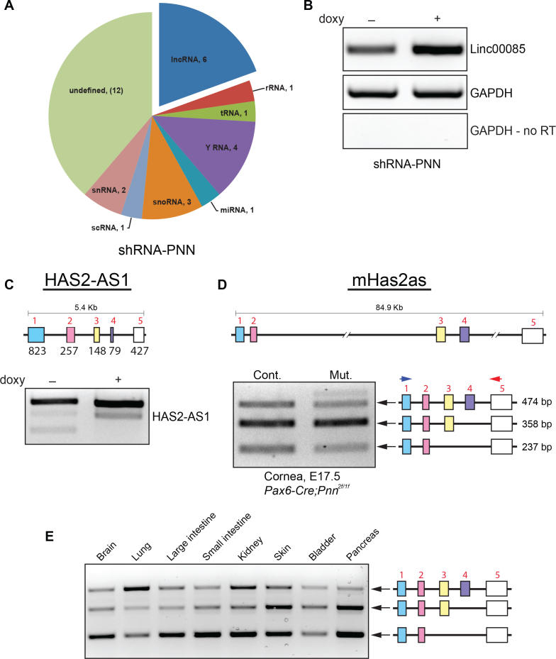
In addition to the profound impact on AS of mRNAs, shRNA-mediated PNN knockdown in HCET cells changed the expression of a collection of ncRNAs (Figure 1A). Interestingly, the doxycycline-inducible shRNA-mediated PNN knockdown in HCET cells resulted in the changed expression and splicing of a specific and small subset of lncRNAs, totaling six, of which we have independently confirmed five (Table 1). One lncRNA, Linc00085, demonstrated an increase in expression subsequent to PNN knockdown in HCET cells; this increase was independently validated with semiquantitative RT–PCR (Figure 1B). These results are consistent with many protein coding genes that change total expression after PNN knockdown [10]. However, other changes in lncRNA expression involved the change in splicing isoforms of lncRNAs. lncRNA, which is located on the opposite DNA strand of hyaluronate synthase 2, HAS2-AS1, demonstrated an obvious change in splice isoforms via transcriptomic arrays. These changes were verified with isoform-specific RT–PCR and sequencing analyses (Figure 1C). The changed observed in Has2-AS1 is of particular interest. HAS2-AS1 is an example of lncRNA that is transcribed from the antisense strand of a protein-coding gene (HA synthase-2), and HAS2-AS1 has been shown to influence the expression of the HAS2 gene [65]. Although lncRNAs may have functional and RNA-structural orthologs, in general, lncRNAs seem to lack clear sequence conservation between species [57,66]. Fortunately, HAS2-AS1 has a clear ortholog in the mouse genome (mHAS2-AS), thus, allowing us to address whether similar changes in the splicing of mHAS2-AS1 exist in the corneal epithelia of conditional (Pax6-cre) PNN-knockouts. Indeed, mHas2as clearly exhibited a differential splicing pattern among three major splice variants in Pnn-deficient mouse cornea. Although the gene organization and intron lengths varied markedly between human HAS2-AS1 and mouse HAS2-AS orthologs, the splicing patterns were similar (Figure 1D and Appendix 1). These data also tempt one to speculate that the change in HAS2-AS1 may be associated with the change in epithelial phenotype seen in the PNN knockout corneal epithelial [11,12,63], since hyaluronic acid (HA) has been closely implicated in the formation of persistent lens stalk, which has been consistently observed in Pnn-deficient mouse embryos [63]. The significant extent tissue-specific isoform-switching of mHAS2-AS1 can be appreciated by examining the splicing pattern of mHas2as in multiple different normal mouse tissues (Figure 1E). The WT mouse tissues tested clearly exhibited a differential splicing pattern among three major splice variants, perhaps suggesting the tissue-specific roles of each splice isoform. It will be of considerable interest to drive Pnn knockout with tissue-specific Cre expression and to then examine if the isoform ratios are shifted after PNN is deleted.
Figure 1.
Induced knockdown of Pnn in HCET cells resulted in altered expression of an array of non-coding RNAs, including long-non-coding RNAs. A: Doxycycline-inducible shRNA-mediated PNN knockdown in human corneal epithelial cells led to the altered expression of specific subsets of ncRNAs, including a subset of lncRNAs, along with snRNA -U RNA, snoRNA, small non-coding components of Ro60 ribonucleoprotein particle (y RNA), rRNA, tRNA, miRNA microRNA, and scRNA small cytoplasmic RNA (7S). B: The increased expression level of Linc00085, seen in the transcriptomic array, was independently validated in RNA from PNN-knockdown human corneal epithelial (HCET) cells with real-time–PCR (RT–PCR). C: The change in splicing isoforms of lncRNA, HAS2-AS1, as demonstrated on the Affimetrix transcriptome-arrays, was verified through isoform-specific RT–PCR and sequencing analyses. D: Conditional knockout of Pnn in developing mouse cornea epithelium led to specific changes in the splicing pattern of mHas2as, the mouse ortholog of human HAS2-AS1. The epithelia from the lens-Cre knockouts demonstrated a decrease in the mHAS2AS1 isoform containing exon 1–5 and isoform containing exons 1, 2, and 5 (see Appendix 1). D: Splicing patterns of mHas2as was examined in multiple different mouse tissues. All tissues tested exhibited a differential splicing pattern across the three major splice variants, suggesting significant normal variations on mHAS2-AS1 across differing tissues and cell types, suggestive of tissue-specific splicing regulation.
Table 1. Long non-coding RNAs with altered expression following PNN knock-down in HCET cells.
| Name | Alias | Ensembl reference | Genomic Location |
|---|---|---|---|
| Linc00085 |
SPACA6P |
ENSG00000182310 |
19:15693340–51712387 (forward strand) |
| HAS2-AS1 |
ENSG00000248690 |
8:121639293–121644693 (forward strand) |
|
| RP11–18I14.1 |
RPARP-AS1 |
ENSG00000269609 |
10:102449817–102461106 (forward strand) |
| RP11–295G20.2 |
ENSC00000233461 |
1:231522388–231528556 (reverse strand) |
|
| RP11–322M19.1 | NUTM2a-AS1 | ENSG00000223482 | 10:87203875–87342612 (reverse strand) |
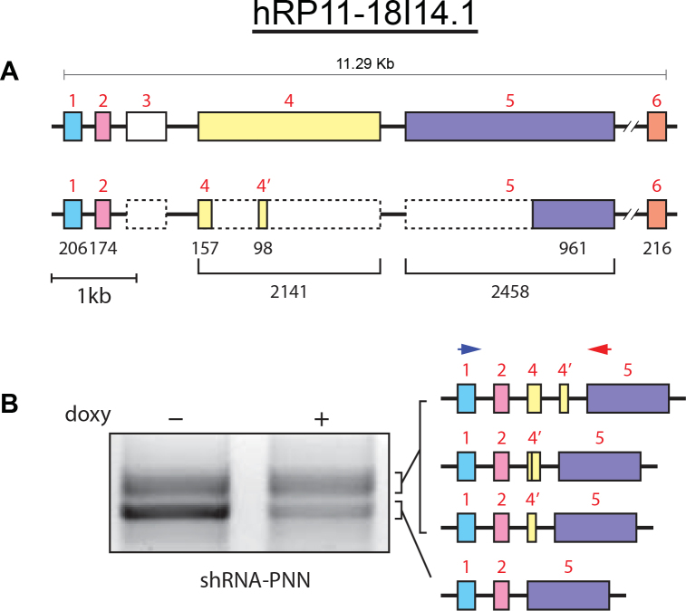
Changes in the RNA splicing of RP11–18I14.1 after Pnn knockdown in HCET cells were also identified through the array analyses. lncRNA RP11–18I14.1, found in the corneal epithelium, contains a distinct pattern of exons, which varies slightly from the predicted full-length lncRNA from lncRNA databases [67]. RT–PCR in combination with sequencing revealed the dominant spliced isoform in WT epithelial contained exons 1,2 and 5, with the exclusion of exon 3, and only the 3′ half of exon 5 (Figure 2A and Appendix 2). In addition, the PCR products included a larger more and more diffuse band, which when sequenced revealed three additional isoforms containing two small segments of exon-4. Pnn knockdown in HCET cells resulted in a shift in RP11–18I14.1 isoforms, with a lower amount of the predominant 1-2-5 isoform and a relative increase in the RP11–18I14.1: 1-2-4-5 isoform.
Figure 2.
Changes in the RNA splicing of RP11–18I14.1 subsequent to Pnn-knockdown in HCET cells. A: RP11–18I14.1 contains six exons spanning 11 kb. RP18I14.1 from corneal epithelia cells contains exons 1–2, partial 4, partial 5, and 6. See Appendix 2. B: Induced knockdown of Pnn resulted in a change in the isoform pattern of RP18I14.1, with a decrease in the predominant exon 1, 2, and 5 isoform and the corresponding increases of isoforms containing one or two stretches of exon 4.
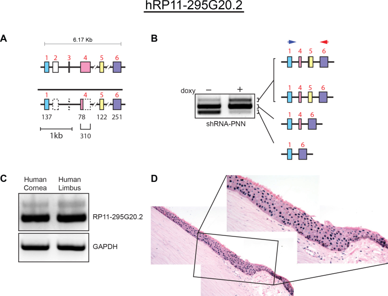
Similar to RP11–18I14.1, the lncRNA RP11–295G20.2 (Figure 3 and Appendix 3) the corneal epithelial cells exhibited a discrete subset of exons that comprise the full length RP11–295G20.2, which include exon 1, only part of exon 4, and full lengths of exon 5 and 6. Exons 2 and 3 of RP11–295G20.2 were not detected from RNA from corneal epithelial cells (Figure 3A). The induction of PNN knockdown in the HCET cells resulted in increased inclusion of exon-5 (Figure 3B). Interestingly, a similar splicing pattern of RP11–295G20.2 was observed in RNA from primary epithelial explant cultures of the central cornea and limbal regions (Figure 3C). In situ hybridizations of corneal sections showed strong nuclear localization of RP11–295G20.2 in the cells comprising the corneal epithelium, with some signal within underlying fibroblasts, and vascular endothelial cells.
Figure 3.
Changes in alternative splicing of RP11–295G20.2 were observed in Pnn-knockdown cells, and RT–PCR and in situ hybridization confirmed in addition to tRP11–295G20.2 expression in the human corneal epithelia. A: RP11–295G20.2 contains six exons spanning just greater than 6 kb. Corneal epithelial cells express RP11–295G20.2 isoforms containing exons 1, 4, 5, and 6. See Appendix 3. B: Knockdown of Pnn resulted in relative decreases in the expression of the two predominant isoforms containing exons 1–6 or 1, 4, and 6, and increased levels of isoforms containing 1, 4, 5, and 6. C: Real-time PCR (RT–PCR) of RNA from explant cultures from central corneal epithelia and limbal epithelia demonstrated robust expression of RP11–295G20.2. D: In situ hybridization analyses on human cornea sections revealed that a lncRNA RP11–295G20.2 is expressed in corneal and limbal epithelium with a nucleus-restricted pattern.
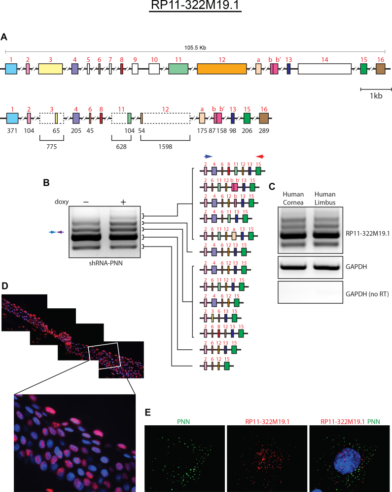
The most complicated corneal epithelial lncRNA examined was RP11–322M19.1 (Figure 4 and Appendix 4). This lncRNA spans more than 100 kb and includes 16 exons and many splice isoforms. Similar to that seen for RP11–18I14.1 and RP11–295G20, the corneal epithelial cells expressed a specific subset of exons from RP11–322M19.1. Of the 16 known exons of RP11–322M19.1, 11 are found within corneal epithelial lncRNA, and many of those are truncated (Figure 4A). The induced depletion of Pnn resulted in the increase on alternate exons, and Pnn depletion also resulted in the inclusion of two transcribed regions within intron 13 (Figure 4B, exons a, b, and bb’), previously unreported. It will be of significant interest to determine whether other tissues and/or other environmental contexts of the corneal epithelia (with or without Pnn depletion) express these alternative exons. Examination of RNA from corneal and limbal epithelial explant cultures revealed a similar pattern of RP11–322M19.1 to the WT HCET cells. Interestingly, fluorescence in situ hybridizations of human cornea revealed strong, albeit heterogeneous labeling of epithelial nuclei, with some labeling of underlying corneal fibroblasts. The most superficial epithelial cells demonstrated significant and consistent labeling. Extending the in situ hybridizations to cultured cells for enhanced resolution revealed predominantly nuclear labeling, as opposed to protein-coding RNA, as shown with Pnn probes. However, in addition, low but significant cytosolic labeling with RP11–322M19.1 probes was observed, which suggests that some RP11–322M19.1 RNA may be exported from the nucleus, at least transiently.
Figure 4.
The long and complex lncRNA, RP11–322M19.1, also showed splicing alterations after Pnn knockdown. A: The reported gene for RP11–322M19.1 spans nearly 106 kb and contains at least 16 exons. Corneal epithelial cells express a complex array of isoforms of RP11–322M19.1 containing 11 of the known exons and two previously unreported exons (labeled a and bb’) were discovered through our RT–PCR and sequencing analyses. See Appendix 4. B: Subsequent to Pnn knockdown, the complexity of RP11–322M19.1isoforms further increased. C: Real-time PCR (RT–PCR) analyses reveal the presence of multiple splice variants of RP11–322M19.1 in corneal and limbal epithelial cells. D: The presence of RNA RP11–322M19.1 in human corneal epithelium is examined through QuantiGene ViewRNA ISH tissue assays. Transcripts of RP11–322M19.1 are visualized with fluorescence in situ hybridization (RNA FISH). RP11–322M19.1 displays a typical nuclear distribution pattern in human corneal epithelial cells. E: RNA distribution pattern of PNN and RP11–322M19.1 transcripts is examined with RNA FISH through QuantiGene ViewRNA ISH cell assay in human corneal epithelial cells (HCET) cells. Although PNN mRNAs are mainly detected in cytoplasm (typical of protein-coding genes), transcripts of lncRNA RP11–322M19.1 are predominantly observed within the nucleus.
An overarching examination of the lncRNA splice isoforms seen for mHAS2-AS, RP11–295G20.2, RP11–295G20, and RP11–322M19.1 in the PNN-depleted cells revealed a pattern of alternate (minor) exon inclusions. Thus, a shift away from the predominant spicing patterns seen in WT cells to a pattern that included a higher proportion of minor isoforms appeared. However, the exon inclusions involve the utilization of previously described exon splice sites, as well as novel splice sites (as observed for RP11–322M19.1 exon a and bb’). Importantly, most of the splice sites conform to the canonical splice site signatures (GT and AG). Therefore, the data from the current study clearly demonstrate that corneal epithelial lncRNAs undergo a complex array of alternative splicing processes as seen in the coding transcripts, and indicate that PNN, in addition to its role in mRNA splicing, plays a role in regulating the alternative splicing of specific lncRNAs.
Discussion
The intensive genomic sequencing efforts, reported by the consortium under the moniker ENCODE [25, 68], changed the way we think about genome organization and gene expression. The outcome of that enormous accomplishment in data collection and integration has significant relevance for this project. First, protein-coding genes account for just 1–2% of the genome; however, more than 90% of the nucleotides that comprise the human genome can be transcribed [68]. Second, in spite of this potential pervasiveness of transcription, at any one time in human cells only 15% of the genome expresses stable transcripts [25]. Finally, genomic regions previously thought to be transcriptionally silent give rise to a wide range of processed transcripts, the lncRNAs, in a tightly regulated fashion [40,69,70]. The current estimates for the total number of lncRNAs, although varied, range between 20,000 and 50,000. The long non-coding RNA transcripts (lncRNAs, ranging from 200 nucleotides to 10,000 residues), although not destined to encode proteins, are alternatively spliced and processed in a manner similar to that for protein-coding genes. The ENCODE project indicated that the annotated lncRNAs exhibit two to 40 isoforms per lncRNA locus. Predictive structural modeling of lncRNA isoform suggests a tremendous range of RNA structures may result from AS of lncRNAs (Lincipedia) [67]. However, further identification and detailed characterization of lncRNA isoforms are needed to fully appreciate the functional relevance of alternative splicing of lncRNAs.
There is significant debate about what proportion of lncRNA represents merely transcriptional noise as opposed to biologic functional moieties. This debate has been fueled by the lack of clear sequence conservation between species [57,66]. However, it is now well documented that some lncRNAs may influence cell lineage commitment and differentiated cellular phenotype [18,23-28]. There is also substantial and growing evidence of cell- and tissue-specific expression of lncRNAs [40,69,70]. Most interestingly, lncRNAs have also been shown to play multiple, fundamental, and profound roles in cancer progression and metastasis in colon and liver cancer [45,71-76]. lncRNAs may represent key regulatory mechanisms for epithelial homeostasis, and abrogation of lncRNAs may be related to loss of key epithelial characteristics associated with carcinoma development. Our early studies revealed a correlation of loss of Pnn expression and cancer progression [77]. It is tempting to speculate that the changes phenotype changes seen in Pnn knockouts and knockdowns may reflect fundamental changes in the lncRNA-mediated regulation of the epithelial program.
Here, we present the first glimpse of the complexity and potential relevance of lncRNAs in the corneal epithelium. We have previously demonstrated that Pnn plays a central role in the establishment and maintenance of epithelia [11,12], and because Pnn is found within transcriptional and spliceosomal complexes, we speculated that Pnn may impact tissue-specific mRNA splicing [10]. However, as the data included in this study demonstrate, that was not the entire story. In addition to RNA processing of protein-coding genes, we noted a small number of lncRNAs that exhibit significant and reproducible changes in expression and RNA processing subsequent to perturbation of PNN expression.
Although the extent of AS and the resultant complexity of lncRNA isoforms within the WT corneal epithelia were intriguing, perturbation of Pnn expression, both in vitro and in the Pax6-Cre animals, resulted in a decrease in the predominant isoforms and an increase in the minor isoforms. These results suggest that normal levels of Pnn may be important for selecting appropriate splice sites and, importantly, the skipping of incorrect splice junctions.
Unlike the impact of Pnn knockdown on mRNA processing, which demonstrated an entire array of changes including exon inclusions, exon exclusions, intron retentions, alternative 5′ or 3′ splice sites, alternative promoters and starts, and an intriguing abundance of alternative splicing changes that lead to alternative 3′UTR [10], the changes in lncRNA splicing were largely restricted to alternative exon usage. These data may indicate a role of Pnn in exon recognition in lncRNAs.
Recent studies have shown that alternative splicing is differentially regulated in many different contexts, such as cell or tissue type, signaling, developmental state, and diseases. The establishment of regulated AS of RNA is achieved through the variable expression/interaction of splicing proteins or the expression of context-specific regulatory factors, or both. Examples of such factors are the cell type-specific splicing proteins, such as Nova, nPTB, MBNL, and ESRPs [28,78–80]. Recent data suggest epigenetics, specifically chromatin markings, have an integral role in regulating AS. Bickmore and colleagues found Pinin in complex with Psip1/Ledgf p52. Psip1, a PWWP protein, binds methylated histone H3K36 and splicing factors and contributes to the regulation of alternative splicing [81]. It is tempting to speculate that loss of Pnn may lead to incorrect exon recognition.
There is of course the possibility that Pnn may be involved in the more rapid turnover of aberrant RNAs, and lower levels of Pnn thus allow the accumulation of these transient RNAs. Pnn has been reported to be present in the ASAP complex, which interacts with the exon-junction complex (EJC), a messenger ribonucleoprotein complex involved in post-transcriptional regulation [20]. Other ASAP subunits (Acinus, RNPS1, and SAP18) have been individually implicated in transcriptional regulation, pre-mRNA splicing, and mRNA quality control. The exon-junction complex itself has fundamental roles in RNA transport and nonsense-mediated decay of aberrant RNAs [82,83].
In situ hybridizations of lncRNAs (RP11-295G20 and RP11–322M19.1) in human corneal epithelia and cultured HCET cells revealed that both exhibited strong nuclear labeling. Interestingly, our first attempts at isoform-specific in situ hybridizations exhibited no substantial difference in tissues or cells. However, these studies are preliminary, and far more isoforms must be examined to make a more definitive statement.
We explored several candidate lncRNAs identified in the corneal epithelium and searched for the biologic significance of these newly emerging key players in a corneal epithelial identity. Our studies, insomuch that they address fundamental processes and mechanisms controlling cell- and tissue-specific splicing decisions on lncRNAs, have relevance for all differentiating systems within the eye.
Acknowledgments
Supported by NIH Grant R01 EY007883, P30 EY021721. We also thank Debra Akin and Todd A. Barnash for their assistance in preparation of the manuscript.
Appendix 1.
To access the data, click or select the words “Appendix 1.” Exon map and transcript sequence of HAS2-AS1 of HCET cells, and exon map and transcript sequence mHas2as of day 17.5 mouse cornea. At the top of human and mouse transcript sequence is a gene diagram depicting relative size and position of the exons of HAS2-AS1 and mHas2-as. The corresponding sequences of each exon are identified. Esembl URls for the reference sequences are provided.
Appendix 2.
To access the data, click or select the words “Appendix 2.” Exon map and transcript sequence of RP11–18I14.1 of HCET cells. At the top is a diagram of the full-length RP11–18I14.1 depicting relative size and position of the exons. The diagram immediately beneath demonstrates the relative size and position of the exons found within the corneal epithelial RP11–18I14.1. Corresponding sequences of each exon are identified. Esembl URls for the reference sequences are provided.
Appendix 3.
To access the data, click or select the words “Appendix 3.” Exon map and transcript sequence of RP11–295G20.2 of HCET cells. At the top is a diagram of the full-length RP11–295G20.2 depicting relative size and position of the exons. The diagram immediately beneath demonstrates the relative size and position of the exons found within the corneal epithelial RP11–295G20.2. The corresponding sequences of each exon are identified. Esembl URls for the reference sequences are provided.
Appendix 4.
To access the data, click or select the words “Appendix 4.” Exon map and transcript sequence of RP11–322M19.1 of HCET cells. At the top is a diagram of the full length RP11–322M19.1 depicting relative size and position of the exons. The diagram immediately beneath demonstrates the relative size and position of the exons found within the corneal epithelial RP11–322M19.1. The corresponding sequences of each exon are identified. In addition to the previously identified exons, we have identified two exons in the corneal epithelial transcripts, labeled; Exon a and Exon bb’. Esembl URls for the reference sequences are provided.
References:
- 1.Pearton DJ, Yang Y, Dhouailly D. Transdifferentiation of corneal epithelium into epidermis occurs by means of a multistep process triggered by dermal developmental signals. Proc Natl Acad Sci USA. 2005;102:3714–9. doi: 10.1073/pnas.0500344102. [DOI] [PMC free article] [PubMed] [Google Scholar]
- 2.Davis J, Duncan MK, Robison WG, Piatigorsky J. Requirement for Pax6 in corneal morphogenesis: a role in adhesion. J Cell Sci. 2003;116:2157–67. doi: 10.1242/jcs.00441. [DOI] [PubMed] [Google Scholar]
- 3.Baulmann DC, Ohlmann A, Flugel-Koch C, Goswami S, Cvekl A, Tamm ER. Pax6 heterozygous eyes show defects in chamber angle differentiation that are associated with a wide spectrum of other anterior eye segment abnormalities. Mech Dev. 2002;118:3–17. doi: 10.1016/s0925-4773(02)00260-5. [DOI] [PubMed] [Google Scholar]
- 4.Wolosin JM, Budak MT, Akinci MAM. Ocular surface epithelial and stem cell development. Int J Dev Biol. 2004;48:981–91. doi: 10.1387/ijdb.041876jw. [DOI] [PubMed] [Google Scholar]
- 5.Xiong L, Woodward AM, Argüeso P. Notch signaling modulates MUC16 biosynthesis in an in vitro model of human corneal and conjunctival epithelial cell differentiation. IOVS. 2011;52:5641–6. doi: 10.1167/iovs.11-7196. [DOI] [PMC free article] [PubMed] [Google Scholar]
- 6.Li W, Chen Y-T, Hayashida Y, Blanco G, Kheirkah A, He H, Chen S-Y, Liu C-Y, Tseng SCG. Down-regulation of Pax6 is associated with abnormal differentiation of corneal epithelial cells in severe ocular surface diseases. J Pathol. 2008;214:114–22. doi: 10.1002/path.2256. [DOI] [PMC free article] [PubMed] [Google Scholar]
- 7.Kao WW. Ocular surface tissue morphogenesis in normal and disease states revealed by genetically modified mice. Cornea. 2006;25(Suppl 1):S7–19. doi: 10.1097/01.ico.0000247207.55520.a4. [DOI] [PubMed] [Google Scholar]
- 8.Kao WWY. Corneal morphogenesis during development and diseases. Eye Contact Lens. 2010;36:265–8. doi: 10.1097/ICL.0b013e3181ef0e00. [DOI] [PubMed] [Google Scholar]
- 9.Shi Y, Tabesh M, Sugrue SP. Role of cell adhesion-associated protein, pinin (DRS/memA), in corneal epithelial migration. IOVS. 2000;41:1337–45. [PubMed] [Google Scholar]
- 10.Joo J-H, Correia GP, Li J-L, Lopez M-C, Baker HV, Sugrue SP. Transcriptomic Analysis of PNN- and ESRP1-regulated Alternative pre-mRNA Splicing in Human Corneal Epithelial Cells. IOVS. 2013. [DOI] [PMC free article] [PubMed] [Google Scholar]
- 11.Joo J-H, Kim YH, Dunn NW, Sugrue SP. Disruption of mouse corneal epithelial differentiation by conditional inactivation of pnn. IOVS. 2010;51:1927–34. doi: 10.1167/iovs.09-4591. [DOI] [PMC free article] [PubMed] [Google Scholar]
- 12.Joo J-H, Taxter TJ, Munguba GC, Kim YH, Dhaduvai K, Dunn NW, Degan WJ, Oh SP, Sugrue SP. Pinin modulates expression of an intestinal homeobox gene, Cdx2, and plays an essential role for small intestinal morphogenesis. Dev Biol. 2010;345:191–203. doi: 10.1016/j.ydbio.2010.07.009. [DOI] [PMC free article] [PubMed] [Google Scholar]
- 13.Alpatov R, Munguba GC, Caton P, Joo J-H, Shi Y, Shi Y, Hunt ME, Sugrue SP. Nuclear speckle-associated protein Pnn/DRS binds to the transcriptional corepressor CtBP and relieves CtBP-mediated repression of the E-cadherin gene. Mol Cell Biol. 2004;24:10223–35. doi: 10.1128/MCB.24.23.10223-10235.2004. [DOI] [PMC free article] [PubMed] [Google Scholar]
- 14.Alpatov R, Shi Y, Munguba GC, Moghimi B, Joo J-H, Bungert J, Sugrue SP. Corepressor CtBP and nuclear speckle protein Pnn/DRS differentially modulate transcription and splicing of the E-cadherin gene. Mol Cell Biol. 2008;28:1584–95. doi: 10.1128/MCB.00421-07. [DOI] [PMC free article] [PubMed] [Google Scholar]
- 15.Joo J-H, Alpatov R, Munguba GC, Jackson MR, Hunt ME, Sugrue SP. Reduction of Pnn by RNAi induces loss of cell-cell adhesion between human corneal epithelial cells. Mol Vis. 2005;11:133–42. [PubMed] [Google Scholar]
- 16.Costa E, Canudas S, Garcia-Bassets I, Perez S, Fernandez I, Giralt E, Azorin F, Espinas ML. Drosophila dSAP18 is a nuclear protein that associates with chromosomes and the nuclear matrix, and interacts with pinin, a protein factor involved in RNA splicing. Chromosome Res. 2006;14:515–26. doi: 10.1007/s10577-006-1046-5. [DOI] [PubMed] [Google Scholar]
- 17.Dellaire G, Makarov EM, Cowger JJ, Longman D, Sutherland HG, Luhrmann R, Torchia J, Bickmore WA. Mammalian PRP4 kinase copurifies and interacts with components of both the U5 snRNP and the N-CoR deacetylase complexes. Mol Cell Biol. 2002;22:5141–56. doi: 10.1128/MCB.22.14.5141-5156.2002. [DOI] [PMC free article] [PubMed] [Google Scholar]
- 18.Zimowska G, Shi J, Munguba G, Jackson MR, Alpatov R, Simmons MN, Shi Y, Sugrue SP. Pinin/DRS/memA interacts with SRp75, SRm300 and SRrp130 in corneal epithelial cells. IOVS. 2003;44:4715–23. doi: 10.1167/iovs.03-0240. [DOI] [PubMed] [Google Scholar]
- 19.Wang P, Lou P-J, Leu S, Ouyang P. Modulation of alternative pre-mRNA splicing in vivo by pinin. Biochem Biophys Res Commun. 2002;294:448–55. doi: 10.1016/S0006-291X(02)00495-3. [DOI] [PubMed] [Google Scholar]
- 20.Murachelli AG, Ebert J, Basquin C, Le Hir H, Conti E. The structure of the ASAP core complex reveals the existence of a Pinin-containing PSAP complex. Nat Struct Mol Biol. 2012;19:378–86. doi: 10.1038/nsmb.2242. [DOI] [PubMed] [Google Scholar]
- 21.Sie CPG, Kuchka M. RNA editing adds flavor to complexity. Biochemistry Biokhimii͡a. 2011;76:869–81. doi: 10.1134/S0006297911080025. [DOI] [PubMed] [Google Scholar]
- 22.de Almeida SF, Carmo-Fonseca M. Design principles of interconnections between chromatin and pre-mRNA splicing. Trends Biochem Sci. 2012;37:248–53. doi: 10.1016/j.tibs.2012.02.002. [DOI] [PubMed] [Google Scholar]
- 23.Pan Q, Shai O, Lee LJ, Frey BJ, Blencowe BJ. Deep surveying of alternative splicing complexity in the human transcriptome by high-throughput sequencing. Nat Genet. 2008;40:1413–5. doi: 10.1038/ng.259. [DOI] [PubMed] [Google Scholar]
- 24.Chen F-C. Are all of the human exons alternatively spliced? Brief Bioinform. 2013 doi: 10.1093/bib/bbt025. [DOI] [PubMed] [Google Scholar]
- 25.A user's guide to the encyclopedia of DNA elements (ENCODE). PLoS Biol. 2011;9:e1001046. doi: 10.1371/journal.pbio.1001046. [DOI] [PMC free article] [PubMed] [Google Scholar]
- 26.Douglas AG, Wood MJ. RNA splicing: disease and therapy. Briefings in Functional Genomics. 2011;10:151–64. doi: 10.1093/bfgp/elr020. [DOI] [PubMed] [Google Scholar]
- 27.Ecker JR, Bickmore WA, Barroso I, Pritchard JK, Gilad Y, Segal E. Genomics: ENCODE explained. Nature. 2012;489:52–5. doi: 10.1038/489052a. [DOI] [PubMed] [Google Scholar]
- 28.Warzecha CC, Shen S, Xing Y, Carstens RP. The epithelial splicing factors ESRP1 and ESRP2 positively and negatively regulate diverse types of alternative splicing events. RNA Biol. 2009;6:546–62. doi: 10.4161/rna.6.5.9606. [DOI] [PMC free article] [PubMed] [Google Scholar]
- 29.Johnson TL, Vilardell J. Regulated pre-mRNA splicing: the ghostwriter of the eukaryotic genome. Biochim Biophys Acta. 2012;1819:538–45. doi: 10.1016/j.bbagrm.2011.12.011. [DOI] [PMC free article] [PubMed] [Google Scholar]
- 30.Kwan T, Benovoy D, Dias C, Gurd S, Provencher C, Beaulieu P, Hudson TJ, Sladek R, Majewski J. Genome-wide analysis of transcript isoform variation in humans. Nat Genet. 2008;40:225–31. doi: 10.1038/ng.2007.57. [DOI] [PubMed] [Google Scholar]
- 31.Anders S, Reyes A, Huber W. Detecting differential usage of exons from RNA-seq data. Genome Res. 2012;22:2008–17. doi: 10.1101/gr.133744.111. [DOI] [PMC free article] [PubMed] [Google Scholar]
- 32.Chen L, Tovar-Corona JM, Urrutia AO. Alternative splicing: a potential source of functional innovation in the eukaryotic genome. Int J Evol Biol. 2012;2012:596274. doi: 10.1155/2012/596274. [DOI] [PMC free article] [PubMed] [Google Scholar]
- 33.Chepelev I, Chen X. Alternative splicing switching in stem cell lineages. Front Biol. 2013;8:50–9. doi: 10.1007/s11515-012-1198-y. [DOI] [PMC free article] [PubMed] [Google Scholar]
- 34.Pinson J, Mason JO, Simpson TI, Price DJ. Regulation of the Pax6 : Pax6(5a) mRNA ratio in the developing mammalian brain. BMC Dev Biol. 2005;5:13. doi: 10.1186/1471-213X-5-13. [DOI] [PMC free article] [PubMed] [Google Scholar]
- 35.Warzecha CC, Sato TK, Nabet B, Hogenesch JB, Carstens RP. ESRP1 and ESRP2 are epithelial cell-type-specific regulators of FGFR2 splicing. Mol Cell. 2009;33:591–601. doi: 10.1016/j.molcel.2009.01.025. [DOI] [PMC free article] [PubMed] [Google Scholar]
- 36.Chauhan BK, Yang Y, Cveklova K, Cvekl A. Functional properties of natural human PAX6 and PAX6(5a) mutants. Invest Ophthalmol Vis Sci. 2004;45:385–92. doi: 10.1167/iovs.03-0968. [DOI] [PMC free article] [PubMed] [Google Scholar]
- 37.Dittmar KA, Jiang P, Park JW, Amirikian K, Wan J, Shen S, Xing Y, Carstens RP. Genome-wide determination of a broad ESRP-regulated posttranscriptional network by high-throughput sequencing. Mol Cell Biol. 2012;32:1468–82. doi: 10.1128/MCB.06536-11. [DOI] [PMC free article] [PubMed] [Google Scholar]
- 38.Lekva T, Berg JP, Fougner SL, Olstad OK, Ueland T, Bollerslev J. Gene Expression Profiling Identifies ESRP1 as a Potential Regulator of Epithelial Mesenchymal Transition in Somatotroph Adenomas from a Large Cohort of Patients with Acromegaly. J Clin Endocrinol Metab. 2012;97:E1506–14. doi: 10.1210/jc.2012-1760. [DOI] [PubMed] [Google Scholar]
- 39.Yae T, Tsuchihashi K, Ishimoto T, Motohara T, Yoshikawa M, Yoshida GJ, Wada T, Masuko T, Mogushi K, Tanaka H, Osawa T, Kanki Y, Minami T, Aburatani H, Ohmura M, Kubo A, Suematsu M, Takahashi K, Saya H, Nagano O. Alternative splicing of CD44 mRNA by ESRP1 enhances lung colonization of metastatic cancer cell. Nature Communications. 2012;3:883. doi: 10.1038/ncomms1892. [DOI] [PubMed] [Google Scholar]
- 40.Ernst C, Morton CC. Identification and function of long non-coding RNA. Frontiers in Cellular Neuroscience. 2013;7:168. doi: 10.3389/fncel.2013.00168. [DOI] [PMC free article] [PubMed] [Google Scholar]
- 41.Fatica A, Bozzoni I. Long non-coding RNAs: new players in cell differentiation and development. Nat Rev Genet. 2014;15:7–21. doi: 10.1038/nrg3606. [DOI] [PubMed] [Google Scholar]
- 42.Ilott NE, Ponting CP. Predicting long non-coding RNAs using RNA sequencing. Methods. 2013 doi: 10.1016/j.ymeth.2013.03.019. [DOI] [PubMed] [Google Scholar]
- 43.Iyengar BR, Choudhary A, Sarangdhar MA, Venkatesh KV, Gadgil CJ, Pillai B. Non-coding RNA interact to regulate neuronal development and function. Frontiers in Cellular Neuroscience. 2014;8:47. doi: 10.3389/fncel.2014.00047. [DOI] [PMC free article] [PubMed] [Google Scholar]
- 44.Moran VA, Perera RJ, Khalil AM. Emerging functional and mechanistic paradigms of mammalian long non-coding RNAs. Nucleic Acids Res. 2012;40:6391–400. doi: 10.1093/nar/gks296. [DOI] [PMC free article] [PubMed] [Google Scholar]
- 45.Niland CN, Merry CR, Khalil AM. Emerging Roles for Long Non-Coding RNAs in Cancer and Neurological Disorders. Frontiers in Genetics. 2012;3:25. doi: 10.3389/fgene.2012.00025. [DOI] [PMC free article] [PubMed] [Google Scholar]
- 46.Aprea J, Prenninger S, Dori M, Ghosh T, Monasor LS, Wessendorf E, Zocher S, Massalini S, Alexopoulou D, Lesche M, Dahl A, Groszer M, Hiller M, Calegari F. Transcriptome sequencing during mouse brain development identifies long non-coding RNAs functionally involved in neurogenic commitment. EMBO J. 2013;32:3145–60. doi: 10.1038/emboj.2013.245. [DOI] [PMC free article] [PubMed] [Google Scholar]
- 47.Rapicavoli NA, Poth EM, Blackshaw S. The long noncoding RNA RNCR2 directs mouse retinal cell specification. BMC Dev Biol. 2010;10:49. doi: 10.1186/1471-213X-10-49. [DOI] [PMC free article] [PubMed] [Google Scholar]
- 48.Guttman M, Donaghey J, Carey BW, Garber M, Grenier JK, Munson G, Young G, Lucas AB, Ach R, Bruhn L, Yang X, Amit I, Meissner A, Regev A, Rinn JL, Root DE, Lander ES. lincRNAs act in the circuitry controlling pluripotency and differentiation. Nature. 2011;477:295–300. doi: 10.1038/nature10398. [DOI] [PMC free article] [PubMed] [Google Scholar]
- 49.Ramos AD, Diaz A, Nellore A, Delgado RN, Park K-Y, Gonzales-Roybal G, Oldham MC, Song JS, Lim DA. Integration of genome-wide approaches identifies lncRNAs of adult neural stem cells and their progeny in vivo. Cell Stem Cell. 2013;12:616–28. doi: 10.1016/j.stem.2013.03.003. [DOI] [PMC free article] [PubMed] [Google Scholar]
- 50.Lin N, Chang K-Y, Li Z, Gates K, Rana ZA, Dang J, Zhang D, Han T, Yang C-S, Cunningham TJ, Head SR, Duester G, Si Dong PD, Rana TM. An Evolutionarily Conserved Long Noncoding RNA TUNA Controls Pluripotency and Neural Lineage Commitment. Mol Cell. 2014 doi: 10.1016/j.molcel.2014.01.021. [DOI] [PMC free article] [PubMed] [Google Scholar]
- 51.Paralkar VR, Mishra T, Luan J, Yao Y, Kossenkov AV, Anderson SM, Dunagin M, Pimkin M, Gore M, Sun D, Konuthula N, Raj A, An X, Mohandas N, Bodine DM, Hardison RC, Weiss MJ. Lineage and species-specific long noncoding RNAs during erythro-megakaryocytic development. Blood. 2014;123:1927–37. doi: 10.1182/blood-2013-12-544494. [DOI] [PMC free article] [PubMed] [Google Scholar]
- 52.Chu C, Qu K, Zhong FL, Artandi SE, Chang HY. Genomic maps of long noncoding RNA occupancy reveal principles of RNA-chromatin interactions. Mol Cell. 2011;44:667–78. doi: 10.1016/j.molcel.2011.08.027. [DOI] [PMC free article] [PubMed] [Google Scholar]
- 53.Flynn RA, Flynn RA, Chang HY, Chang HY. Active chromatin and noncoding RNAs: an intimate relationship. Curr Opin Genet Dev. 2012;22:172–8. doi: 10.1016/j.gde.2011.11.002. [DOI] [PMC free article] [PubMed] [Google Scholar]
- 54.Marchese FP, Huarte M. Long non-coding RNAs and chromatin modifiers: Their place in the epigenetic code. Epigenetics. Official Journal of the DNA Methylation Society. 2013;9:1. doi: 10.4161/epi.27472. [DOI] [PMC free article] [PubMed] [Google Scholar]
- 55.Mercer TR, Mattick JS. Structure and function of long noncoding RNAs in epigenetic regulation. Nat Struct Mol Biol. 2013;20:300–7. doi: 10.1038/nsmb.2480. [DOI] [PubMed] [Google Scholar]
- 56.Saxena A, Carninci P. Long non-coding RNA modifies chromatin: epigenetic silencing by long non-coding RNAs. BioEssays: News and Reviews in Molecular. Cellular and Developmental Biology. 2011;33:830–9. doi: 10.1002/bies.201100084. [DOI] [PMC free article] [PubMed] [Google Scholar]
- 57.Ulitsky I, Bartel DP. lincRNAs: genomics, evolution, and mechanisms. Cell. 2013;154:26–46. doi: 10.1016/j.cell.2013.06.020. [DOI] [PMC free article] [PubMed] [Google Scholar]
- 58.Guttman M, Amit I, Garber M, French C, Lin MF, Feldser D, Huarte M, Zuk O, Carey BW, Cassady JP, Cabili MN, Jaenisch R, Mikkelsen TS, Jacks T, Hacohen N, Bernstein BE, Kellis M, Regev A, Rinn JL, Lander ES. Chromatin signature reveals over a thousand highly conserved large non-coding RNAs in mammals. Nature. 2009;458:223–7. doi: 10.1038/nature07672. [DOI] [PMC free article] [PubMed] [Google Scholar]
- 59.Pauli A, Rinn JL, Schier AF. Non-coding RNAs as regulators of embryogenesis. Nat Rev Genet. 2011;12:136–49. doi: 10.1038/nrg2904. [DOI] [PMC free article] [PubMed] [Google Scholar]
- 60.Burgess DJ. Non-coding RNA: HOTTIP goes the distance. Nat Rev Genet. 2011;12:300. doi: 10.1038/nrg2992. [DOI] [PubMed] [Google Scholar]
- 61.Bertani S, Sauer S, Bolotin E, Sauer F. The noncoding RNA Mistral activates Hoxa6 and Hoxa7 expression and stem cell differentiation by recruiting MLL1 to chromatin. Mol Cell. 2011;43:1040–6. doi: 10.1016/j.molcel.2011.08.019. [DOI] [PMC free article] [PubMed] [Google Scholar] [Retracted]
- 62.Rapicavoli NA, Poth EM, Zhu H, Blackshaw S. The long noncoding RNA Six3OS acts in trans to regulate retinal development by modulating Six3 activity. Neural Dev. 2011;6:32. doi: 10.1186/1749-8104-6-32. [DOI] [PMC free article] [PubMed] [Google Scholar]
- 63.Joo J-H, Lee YJ, Munguba GC, Park S, Taxter TJ, Elsagga MY, Jackson MR, Oh SP, Sugrue SP. Role of Pinin in neural crest, dorsal dermis, and axial skeleton development and its involvement in the regulation of Tcf/Lef activity in mice. Developmental Dynamics of the American Association of Anatomists. 2007;236:2147–58. doi: 10.1002/dvdy.21243. [DOI] [PubMed] [Google Scholar]
- 64.Seok J, Xu W, Gao H, Davis RW, Xiao W. JETTA: junction and exon toolkits for transcriptome analysis. Bioinformatics. 2012;28:1274–5. doi: 10.1093/bioinformatics/bts134. [DOI] [PMC free article] [PubMed] [Google Scholar]
- 65.Michael DR, Phillips AO, Krupa A, Martin J, Redman JE, Altaher A, Neville RD, Webber J, Kim MY, Bowen T. The human hyaluronan synthase 2 (HAS2) gene and its natural antisense RNA exhibit coordinated expression in the renal proximal tubular epithelial cell. J Biol Chem. 2011;286:19523–32. doi: 10.1074/jbc.M111.233916. [DOI] [PMC free article] [PubMed] [Google Scholar]
- 66.Diederichs S. The four dimensions of noncoding RNA conservation. Trends in genetics. TIG. 2014;30:121–3. doi: 10.1016/j.tig.2014.01.004. [DOI] [PubMed] [Google Scholar]
- 67.Volders PJ, Helsens K, Wang X, Menten B, Martens L, Gevaert K, Vandesompele J, Mestdagh P. LNCipedia: a database for annotated human lncRNA transcript sequences and structures. Nucleic Acids Res. 2013;41(Database issue):D246–51. doi: 10.1093/nar/gks915. [DOI] [PMC free article] [PubMed] [Google Scholar]
- 68.Consortium EP, Dunham I, Kundaje A, Aldred SF, Collins PJ, Davis CA, Doyle F, Epstein CB, Frietze S, Harrow J, Kaul R, Khatun J, Lajoie BR, Landt SG, Lee BK, Pauli F, Rosenbloom KR, Sabo P, Safi A, Sanyal A, Shoresh N, Simon JM, Song L, Trinklein ND, Altshuler RC, Birney E, Brown JB, Cheng C, Djebali S, Dong X, Dunham I, Ernst J, Furey TS, Gerstein M, Giardine B, Greven M, Hardison RC, Harris RS, Herrero J, Hoffman MM, Iyer S, Kelllis M, Khatun J, Kheradpour P, Kundaje A, Lassmann T, Li Q, Lin X, Marinov GK, Merkel A, Mortazavi A, Parker SC, Reddy TE, Rozowsky J, Schlesinger F, Thurman RE, Wang J, Ward LD, Whitfield TW, Wilder SP, Wu W, Xi HS, Yip KY, Zhuang J, Bernstein BE, Birney E, Dunham I, Green ED, Gunter C, Snyder M, Pazin MJ, Lowdon RF, Dillon LA, Adams LB, Kelly CJ, Zhang J, Wexler JR, Green ED, Good PJ, Feingold EA, Bernstein BE, Birney E, Crawford GE, Dekker J, Elinitski L, Farnham PJ, Gerstein M, Giddings MC, Gingeras TR, Green ED, Guigo R, Hardison RC, Hubbard TJ, Kellis M, Kent WJ, Lieb JD, Margulies EH, Myers RM, Snyder M, Starnatoyannopoulos JA, Tennebaum SA, Weng Z, White KP, Wold B, Khatun J, Yu Y, Wrobel J, Risk BA, Gunawardena HP, Kuiper HC, Maier CW, Xie L, Chen X, Giddings MC, Bernstein BE, Epstein CB, Shoresh N, Ernst J, Kheradpour P, Mikkelsen TS, Gillespie S, Goren A, Ram O, Zhang X, Wang L, Issner R, Coyne MJ, Durham T, Ku M, Truong T, Ward LD, Altshuler RC, Eaton ML, Kellis M, Djebali S, Davis CA, Merkel A, Dobin A, Lassmann T, Mortazavi A, Tanzer A, Lagarde J, Lin W, Schlesinger F, Xue C, Marinov GK, Khatun J, Williams BA, Zaleski C, Rozowsky J, Roder M, Kokocinski F, Abdelhamid RF, Alioto T, Antoshechkin I, Baer MT, Batut P, Bell I, Bell K, Chakrabortty S, Chen X, Chrast J, Curado J, Derrien T, Drenkow J, Dumais E, Dumais J, Duttagupta R, Fastuca M, Fejes-Toth K, Ferreira P, Foissac S, Fullwood MJ, Gao H, Gonzalez D, Gordon A, Gunawardena HP, Howald C, Jha S, Johnson R, Kapranov P, King B, Kingswood C, Li G, Luo OJ, Park E, Preall JB, Presaud K, Ribeca P, Risk BA, Robyr D, Ruan X, Sammeth M, Sandu KS, Schaeffer L, See LH, Shahab A, Skancke J, Suzuki AM, Takahashi H, Tilgner H, Trout D, Walters N, Wang H, Wrobel J, Yu Y, Hayashizaki Y, Harrow J, Gerstein M, Hubbard TJ, Reymond A, Antonarakis SE, Hannon GJ, Giddings MC, Ruan Y, Wold B, Carninci P, Guigo R, Gingeras TR, Rosenbloom KR, Sloan CA, Learned K, Malladi VS, Wong MC, Barber GP, Cline MS, Dreszer TR, Heitner SG, Karolchik D, Kent WJ, Kirkup VM, Meyer LR, Long JC, Maddren M, Raney BJ, Furey TS, Song L, Grasfeder LL, Giresi PG, Lee BK, Battenhouse A, Sheffield NC, Simon JM, Showers KA, Safi A, London D, Bhinge AA, Shestak C, Schaner MR, Kim SK, Zhang ZZ, Mieczkowski PA, Mieczkowska JO, Liu Z, McDaniell RM, Ni Y, Rashid NU, Kim MJ, Adar S, Zhang Z, Wang T, Winter D, Keefe D, Birney E, Iyer VR, Lieb JD, Crawford GE, Li G, Sandhu KS, Zheng M, Wang P, Luo OJ, Shahab A, Fullwood MJ, Ruan X, Ruan Y, Myers RM, Pauli F, Williams BA, Gertz J, Marinov GK, Reddy TE, Vielmetter J, Partridge EC, Trout D, Varley KE, Gasper C, Bansal A, Pepke S, Jain P, Amrhein H, Bowling KM, Anaya M, Cross MK, King B, Muratet MA, Antoshechkin I, Newberry KM, McCue K, Nesmith AS, Fisher-Aylor KI, Pusey B, DeSalvo G, Parker SL, Balasubramanian S, Davis NS, Meadows SK, Eggleston T, Gunter C, Newberry JS, Levy SE, Absher DM, Mortazavi A, Wong WH, Wold B, Blow MJ, Visel A, Pennachio LA, Elnitski L, Margulies EH, Parker SC, Petrykowska HM, Abyzov A, Aken B, Barrell D, Barson G, Berry A, Bignell A, Boychenko V, Bussotti G, Chrast J, Davidson C, Derrien T, Despacio-Reyes G, Diekhans M, Ezkurdia I, Frankish A, Gilbert J, Gonzalez JM, Griffiths E, Harte R, Hendrix DA, Howald C, Hunt T, Jungreis I, Kay M, Khurana E, Kokocinski F, Leng J, Lin MF, Loveland J, Lu Z, Manthravadi D, Mariotti M, Mudge J, Mukherjee G, Notredame C, Pei B, Rodriguez JM, Saunders G, Sboner A, Searle S, Sisu C, Snow C, Steward C, Tanzer A, Tapanari E, Tress ML, van Baren MJ, Walters N, Washieti S, Wilming L, Zadissa A, Zhengdong Z, Brent M, Haussler D, Kellis M, Valencia A, Gerstein M, Raymond A, Guigo R, Harrow J, Hubbard TJ, Landt SG, Frietze S, Abyzov A, Addleman N, Alexander RP, Auerbach RK, Balasubramanian S, Bettinger K, Bhardwaj N, Boyle AP, Cao AR, Cayting P, Charos A, Cheng Y, Cheng C, Eastman C, Euskirchen G, Fleming JD, Grubert F, Habegger L, Hariharan M, Harmanci A, Iyenger S, Jin VX, Karczewski KJ, Kasowski M, Lacroute P, Lam H, Larnarre-Vincent N, Leng J, Lian J, Lindahl-Allen M, Min R, Miotto B, Monahan H, Moqtaderi Z, Mu XJ, O'Geen H, Ouyang Z, Patacsil D, Pei B, Raha D, Ramirez L, Reed B, Rozowsky J, Sboner A, Shi M, Sisu C, Slifer T, Witt H, Wu L, Xu X, Yan KK, Yang X, Yip KY, Zhang Z, Struhl K, Weissman SM, Gerstein M, Farnham PJ, Snyder M, Tenebaum SA, Penalva LO, Doyle F, Karmakar S, Landt SG, Bhanvadia RR, Choudhury A, Domanus M, Ma L, Moran J, Patacsil D, Slifer T, Victorsen A, Yang X, Snyder M, White KP, Auer T, Centarin L, Eichenlaub M, Gruhl F, Heerman S, Hoeckendorf B, Inoue D, Kellner T, Kirchmaier S, Mueller C, Reinhardt R, Schertel L, Schneider S, Sinn R, Wittbrodt B, Wittbrodt J, Weng Z, Whitfield TW, Wang J, Collins PJ, Aldred SF, Trinklein ND, Partridge EC, Myers RM, Dekker J, Jain G, Lajoie BR, Sanyal A, Balasundaram G, Bates DL, Byron R, Canfield TK, Diegel MJ, Dunn D, Ebersol AK, Ebersol AK, Frum T, Garg K, Gist E, Hansen RS, Boatman L, Haugen E, Humbert R, Jain G, Johnson AK, Johnson EM, Kutyavin TM, Lajoie BR, Lee K, Lotakis D, Maurano MT, Neph SJ, Neri FV, Nguyen ED, Qu H, Reynolds AP, Roach V, Rynes E, Sabo P, Sanchez ME, Sandstrom RS, Sanyal A, Shafer AO, Stergachis AB, Thomas S, Thurman RE, Vernot B, Vierstra J, Vong S, Wang H, Weaver MA, Yan Y, Zhang M, Akey JA, Bender M, Dorschner MO, Groudine M, MacCoss MJ, Navas P, Stamatoyannopoulos G, Kaul R, Dekker J, Stamatoyannopoulos JA, Dunham I, Beal K, Brazma A, Flicek P, Herrero J, Johnson N, Keefe D, Lukk M, Luscombe NM, Sobral D, Vaquerizas JM, Wilder SP, Batzoglou S, Sidow A, Hussami N, Kyriazopoulou-Panagiotopoulou S, Libbrecht MW, Schaub MA, Kundaje A, Hardison RC, Miller W, Giardine B, Harris RS, Wu W, Bickel PJ, Banfai B, Boley NP, Brown JB, Huang H, Li Q, Li JJ, Noble WS, Bilmes JA, Buske OJ, Hoffman MM, Sahu AO, Kharchenko PV, Park PJ, Baker D, Taylor J, Weng Z, Iyer S, Dong X, Greven M, Lin X, Wang J, Xi HS, Zhuang J, Gerstein M, Alexander RP, Balasubramanian S, Cheng C, Harmanci A, Lochovsky L, Min R, Mu XJ, Rozowsky J, Yan KK, Yip KY, Birney E. An integrated encyclopedia of DNA elements in the human genome. Nature. 2012;489:57–74. doi: 10.1038/nature11247. [DOI] [PMC free article] [PubMed] [Google Scholar]
- 69.Niazi F, Valadkhan S. Computational analysis of functional long noncoding RNAs reveals lack of peptide-coding capacity and parallels with 3' UTRs. RNA. 2012;18:825–43. doi: 10.1261/rna.029520.111. [DOI] [PMC free article] [PubMed] [Google Scholar]
- 70.Rose D, Hiller M, Schutt K, Hackermuller J, Backofen R, Stadler PF. Computational discovery of human coding and non-coding transcripts with conserved splice sites. Bioinformatics. 2011;27:1894–900. doi: 10.1093/bioinformatics/btr314. [DOI] [PubMed] [Google Scholar]
- 71.Crea F, Clermont PL, Parolia A, Wang Y, Helgason CD. The non-coding transcriptome as a dynamic regulator of cancer metastasis. Cancer Metastasis Rev. 2013;33:1–16. doi: 10.1007/s10555-013-9455-3. [DOI] [PMC free article] [PubMed] [Google Scholar]
- 72.Wang J, Wang H, Zhang Y, Zhen N, Zhang L, Qiao Y, Weng W, Liu X, Ma L, Xiao W, Yu W, Chu Q, Pan Q, Sun F. Mutual inhibition between YAP and SRSF1 maintains long non-coding RNA, Malat1-induced tumourigenesis in liver cancer. Cell Signal. 2014;26:1048–59. doi: 10.1016/j.cellsig.2014.01.022. [DOI] [PubMed] [Google Scholar]
- 73.Quagliata L, Matter MS, Piscuoglio S, Arabi L, Ruiz C, Procino A, Kovac M, Moretti F, Makowska Z, Boldanova T, Andersen JB, Hammerle M, Tornillo L, Heim MH, Diederichs S, Cillo C, Terracciano LM. Long noncoding RNA HOTTIP/HOXA13 expression is associated with disease progression and predicts outcome in hepatocellular carcinoma patients. Hepatology. 2014;59:911–23. doi: 10.1002/hep.26740. [DOI] [PMC free article] [PubMed] [Google Scholar]
- 74.Gibb EA, Brown CJ, Lam WL. The functional role of long non-coding RNA in human carcinomas. Mol Cancer. 2011;10:38. doi: 10.1186/1476-4598-10-38. [DOI] [PMC free article] [PubMed] [Google Scholar]
- 75.Huang J-L, Zheng L, Hu Y-W, Wang Q. Characteristics of long non-coding RNA and its relation to hepatocellular carcinoma. Carcinogenesis. 2014;35:507–14. doi: 10.1093/carcin/bgt405. [DOI] [PubMed] [Google Scholar]
- 76.Xue B. An expanding universe of the non-coding genome in cancer biology. Carcinogenesis. 2014;35:1209–16. doi: 10.1093/carcin/bgu099. [DOI] [PMC free article] [PubMed] [Google Scholar]
- 77.Shi Y, Shi Y, Ouyang P, Ouyang P, Sugrue SP, Sugrue SP. Characterization of the gene encoding pinin/DRS/memA and evidence for its potential tumor suppressor function. Oncogene. 2000;19:289–97. doi: 10.1038/sj.onc.1203328. [DOI] [PubMed] [Google Scholar]
- 78.Kim S, Kim H, Fong N, Erickson B, Bentley DL. Pre-mRNA splicing is a determinant of histone H3K36 methylation. Proc Natl Acad Sci USA. 2011;108:13564–9. doi: 10.1073/pnas.1109475108. [DOI] [PMC free article] [PubMed] [Google Scholar]
- 79.Han H, Irimia M, Ross PJ, Sung H-K, Alipanahi B, David L, Golipour A, Gabut M, Michael IP, Nachman EN, Wang E, Trcka D, Thompson T. O'Hanlon D, Slobodeniuc V, Barbosa-Morais NL, Burge CB, Moffat J, Frey BJ, Nagy A, Ellis J, Wrana JL, Blencowe BJ. MBNL proteins repress ES-cell-specific alternative splicing and reprogramming. Nature. 2013;498:241–5. doi: 10.1038/nature12270. [DOI] [PMC free article] [PubMed] [Google Scholar]
- 80.Bitel CL, Perrone-Bizzozero NI, Frederikse PH. HuB/C/D, nPTB, REST4, and miR-124 regulators of neuronal cell identity are also utilized in the lens. Mol Vis. 2010;16:2301–16. [PMC free article] [PubMed] [Google Scholar]
- 81.Pradeepa MM, Sutherland HG, Ule J, Grimes GR, Bickmore WA. Psip1/Ledgf p52 binds methylated histone H3K36 and splicing factors and contributes to the regulation of alternative splicing. PLoS Genet. 2012;8:e1002717. doi: 10.1371/journal.pgen.1002717. [DOI] [PMC free article] [PubMed] [Google Scholar]
- 82.Michelle L, Cloutier A, Toutant J, Shkreta L, Thibault P, Durand M, Garneau D, Gendron D, Lapointe E, Couture S, Le Hir H, Klinck R, Elela SA, Prinos P, Chabot B. Proteins associated with the exon junction complex also control the alternative splicing of apoptotic regulators. Mol Cell Biol. 2012;32:954–67. doi: 10.1128/MCB.06130-11. [DOI] [PMC free article] [PubMed] [Google Scholar]
- 83.Chen L. A global comparison between nuclear and cytosolic transcriptomes reveals differential compartmentalization of alternative transcript isoforms. Nucleic Acids Res. 2010;38:1086–97. doi: 10.1093/nar/gkp1136. [DOI] [PMC free article] [PubMed] [Google Scholar]


