Abstract
Cellular prion protein (PrPC) plays an important role in the cellular defense against oxidative stress. However, the exact protective mechanism of PrPC is unclear. Autophagy is essential for survival, differentiation, development, and homeostasis in several organisms. Although the role that autophagy plays in neurodegenerative disease has yet to be established, it is clear that autophagy-induced cell death is observed in neurodegenerative disorders that exhibit protein aggregations. Moreover, autophagy can promote cell survival and cell death under various conditions. In this review, we describe the involvement of autophagy in prion disease and the effects of PrPC.
Keywords: prion, Prnp-deficient, prion diseases, autophagy, autophagic flux, oxidative stress
1. Introduction
Normal cellular prion protein (PrPC) is a glycosylphosphatidylinositol (GPI)-anchored glycoprotein on the extracellular surface that is highly expressed in the central nervous system (CNS), in particular by neurons [1,2,3,4]. Normal PrPC is converted into the abnormal scrapie isoform, PrPSc, when a portion of its α-helical coil structure is refolded into a β-sheet [3,4]. This structural change confers partial resistance to proteolytic degradation and detergent insolubility to PrPSc [5,6]. Prion diseases, such as scrapie and bovine spongiform encephalopathy (BSE) in animals and Creutzfeldt-Jakob disease (CJD) in humans, are neurodegenerative conditions characterized by the accumulation of this altered PrP isoform, PrPSc [7,8].
A certain degree of neurodegeneration in these diseases is induced by autophagic cell death, which is characterized by the accumulation of autophagic vacuoles including preautophagosomes, autophagosomes, and autophagolysosomes [9,10,11]. During this process, the cytoplasmic form of microtubule-associated light chain 3 (LC3-I, 18 kDa) is converted into the preautophagosomal and autophagosomal membrane-bound form of LC3 (LC3-II, 16 kDa), which is the most reliable marker for the activation of autophagy [12]. For normal cell growth and development, protein synthesis and organelle biogenesis are balanced against protein degradation and organelle turnover [9]. The major pathways for the degradation of cellular constituents are autophagy and cytosolic turnover by the proteasome [9]. Autophagy plays an important role in cellular homeostasis, i.e., the turnover of intracellular organelles and long-lived protein; however, excessive autophagy has been proposed to cause cellular destruction [9,10]. Autophagy is observed in all nucleated cell types that have been analyzed, and the process is essentially the same in yeast, plant, and animal cells [13,14,15]. However, the functions of autophagy, particularly in neurons, are still largely unknown. In this review, we focus on the possible role that PrPC play in the autophagy pathway.
2. The Functional Role of PrPC on Autophagy in vitro
The PrPC is encoded by the Prnp gene, which is highly conserved in a wide range of mammalian species. PrPC is highly expressed throughout the CNS but has also been observed in other organs [1,2,16]. The exact physiological functions of PrPC in the CNS are unclear, but several reports demonstrated that this protein is involved in various biological processes.
Autophagy is a lysosomal degradation process that is associated with the intracellular turnover of cytoplasmic proteins and organelles [9]. Autophagy is involved in cell survival in response to nutrient deprivation and is also associated with various diseases [17,18].
In prion diseases, the appearance of autophagic vacuoles was first observed in neurons in experimental rodent models affected by transmissible spongiform encephalopathies (TSEs) and in scrapie prion-infected cultured cells [19,20,21]. Autophagic vacuoles were identified in neurons in experimentally induced scrapie and human transmissible encephalopathies [22,23]. It was therefore proposed by Liberski that autophagy could contribute to the spongiform degeneration that is a pathological hallmark in brains affected by prion diseases [22]. In neurons from scrapie-infected mice, increased levels of stimulator of chondrogenesis 1/scrapie responsive gene 1 (SCRG1) are detected in autophagic vacuoles [24]. More recently, it was reported that the pharmacological induction of autophagy by treatment with trehalose or lithium can decrease pathogenic and infectious PrPSc expression in persistently prion-infected neurons [25,26]. Moreover, GSS transgenic mice that were treated with rapamycin, an autophagy inducer, exhibited a dose-related delay in disease onset, a reduction in clinical sign severity, and an extension of survival [27]. These results suggest that the administration of an autophagy inducer may be a therapeutically tenable option for treating prion diseases. In addition, an enhanced macroautophagic response was observed in scrapie-infected (strain 263K) hamsters and in human genetic prion diseases [28], indicating that there is a correlation between the up-regulation of autophagy activation and the pathogenesis of prion diseases.
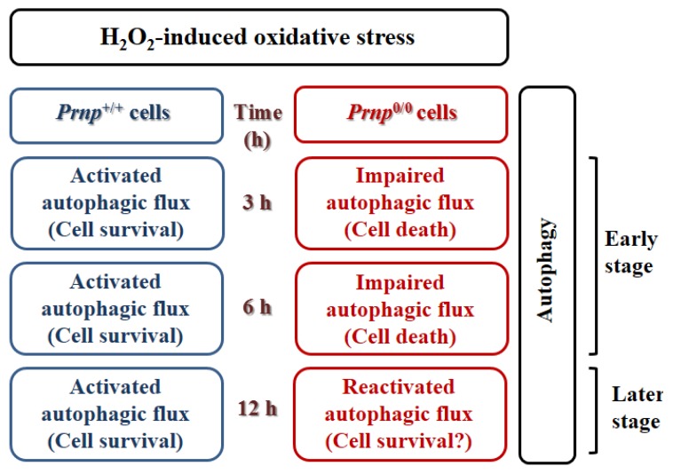
In addition to autophagy in prion diseases, a correlation between PrPC and autophagy was recently described. Increased expression of LC3-II, autophagy marker protein and autophagosomes were observed in Zürich I Prnp0/0 hippocampal neurons compared to wild-type control cells under serum starvation [29]. This increased LC3-II was inhibited by the transfection of the wild-type Prnp gene into Prnp0/0 hippocampal neurons, but not by the introduction of PrPC lacking the octapeptide repeat region. Thus, the octapeptide repeat region of PrPC might play a crucial role in the control of autophagy in neurons. Although the autophagic responses of wild-type and Prnp0/0 hippocampal neurons were clearly different, no definitive association between PrPC and the autophagy pathway was demonstrated in this previous report. In wild-type cells, decreased autophagy induction may be due to the effect that PrPC has on anti-oxidant activity. A recent report revealed a novel protective mechanism that PrPC-associated autophagy has against hydrogen peroxide (H2O2)-induced oxidative stress [30]. Interestingly, autophagy was oppositely regulated in Prnp+/+ and Prnp0/0 hippocampal neurons under oxidative stress. In this study, increased autophagy following H2O2 treatment was due to enhanced and impaired autophagic flux in Prnp+/+ and Prnp0/0 hippocampal neurons, respectively. Inhibition of autophagy by siRNA knockdown of Atg7, which is essential for the formation of autophagosomes, supports the suggestion that enhanced autophagy led to cell survival in H2O2-treated Prnp+/+ cells and that impaired autophagic flux contributed to cell death in H2O2-treated Prnp0/0 cells. Moreover, PrPC itself may not be directly involved in autophagic flux in these cells given that a PrPC deficiency did not affect the basal autophagic flux under normal culture conditions without H2O2 treatment (Figure 1) [30].
Figure 1.
Schematic representation of time-dependent H2O2-induced cell death in Prnp+/+ and Prnp0/0 cells [30].
It was recently reported that the ectopic overexpression of PrP-like protein doppel (PRND) in addition to PrPC deficiency in Ngsk (NP0/0) mice provokes the impairment of autophagic flux in central nervous system neurons, an effect that is potentially associated with progressive cerebellar Purkinje cell death in these animals [31,32]. PRND alone can cause neurotoxicity, and PRND toxicity is involved in the upregulation of both heme oxygenase 1 (HMOX1) and nitric oxide synthase (nNOS and iNOS) systems, which suggests that there is increased oxidative stress in the brains of the NP0/0 mice [33,34]. Thus, the defective autophagic flux exhibited by NP0/0 mice may be due to PRND toxicity-induced oxidative stress.
In human malignant glioma cell lines and non-glial tumor cells, the knockdown of PrPC using antisense oligonucleotides targeting the Prnp transcript induces autophagic cell death without the presence of apoptosis markers [35]. This evidence suggests that PrPC may directly modulate the autophagy-dependent cell death pathway.
3. The functional role of PrPC on autophagy in vivo
Normal PrPC is highly expressed in the neurons of CNS and especially in their synaptic plasma membrane [3,4,36]. PrPC has several roles in cellular metabolism and maintenance, including neurotransmitter metabolism, signal transduction, copper metabolism, cell adhesion, neuritogenesis, and anti-oxidant activity. Furthermore, many studies have demonstrated that PrPC has neuroprotective and anti-apoptotic functions [37,38,39,40,41,42].
Mice in which Prnp is ablated have been used in several studies that examined behavior and cognition [43]. More recent studies have noted certain differences between PrPC knockout and wild-type mice. Several important facts have emerged from these studies. For example, PrP knockout mice exhibit increased susceptibility to neuronal damage by oxidative stress and cerebral ischemia. Additionally, neurotoxicity is caused by the expression of Doppel and N-terminally truncated PrP [31,44,45,46]. The multiple effects of PrP deficiency in the same transgenic mouse line suggest its essential function and has broad implications [47].
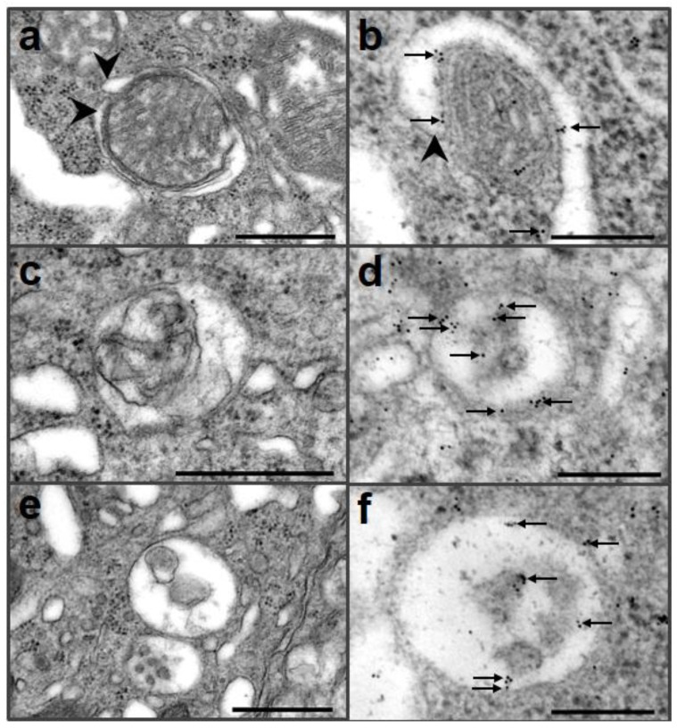
Large deletions in Prnp, (e.g., in the Ngsk PrP-deficient mouse line) have neurodegenerative effects in Purkinje cells, which may induce neuronal autophagy [31,32]. Ultrastructural analysis of the Purkinje cells of Ngsk PrP-deficient mouse revealed multivesicular bodies and mitochondria around the autophagic membrane in the soma (Figure 2). Dystrophic axons were also observed that exhibited features of acute autophagy, numerous reticular phagophores, and autophagosomes containing axoplasmic material [32] (Table 1).
Figure 2.
Immunogold electron microscopic analysis of autophagy in Purkinje cells of Ngsk mice using an anti-LC3 antibody. Autophagosomal membranes (arrowheads) began to form vacuoles that contained mitochondria (a, b), and gold particles (arrows) were located in isolated membranes (b). Autophagolysosome showed that single membrane surrounding mitochondrion (c). Immunogold particles (arrows) were located in single membrane and residues (d). Autophagolysosomes were bounded by a single membrane and contained residues (e), and gold particles (arrows) were detected in single membrane and residues (f). Scale bar = 500 nm (a, c, e), 200 nm (b, d, f).
Table 1.
A summary of autophagic alteration with aging of Ngsk PrP-deficient mouse line [32].
| 3-4 months of age | 6-8 months of age | |
|---|---|---|
| PrP-deficient mice |
|
|
Autophagy is a natural process that removes misfolded proteins, dysfunctional mitochondria, and other potentially toxic proteins or organelles [9]. Although the role of autophagy in neurodegeneration has yet to be established, it is clear that cell death is induced by autophagy in neurodegenerative disorders, such as Alzheimer’s, Huntington’s and Parkinson’s disease (PD), that exhibit protein aggregation [10,48,49]. The results from experiments where autophagy is either inhibited or stimulated indicates that altered protein clearance and organelle clearance, either increased or decreased, are involved in the onset of PD [48]. Furthermore, in the degenerated hippocampus of an early-onset Alzheimer’s mouse model, there are increased protein levels of the autophagy formation marker LC3-II. In addition, actin cytoskeletal and molecular motor defects lead to transport abnormalities and the accumulation of autophagosomes in dystrophic neurites in the hippocampus of these mice [49].
The mechanisms of neuronal death have been examined intensively to gain insight into the pathological processes that are associated with acute and chronic neurological illnesses. Prion diseases belong to the family of neurodegenerative disorders that affect both humans and animals. It is known that one of the fundamental steps in the pathogenesis of these diseases is the conversion of the host’s cellular prion protein, PrPC, into the disease-associated form, PrPSc [6,50]. Membrane-anchored PrPC is required to transduce the neurotoxic signals that are elicited by the pathogenic forms of PrP [51]. However, neuronal death is also induced in PrP-knockout mice, and this toxicity is dose-dependently suppressed by the coexpression of full-length PrP [52]. These results suggest that the normal biological activity of PrPC may be altered during the disease process. However, the cellular pathway and molecular components that are involved in this mechanism have yet to be identified.
Various mechanisms have been proposed to explain neuronal death in prion diseases, with apoptosis and autophagy being the most probable types of cell death involved [3,18,22]. Recently, evidence of apoptosis, such as morphologically apoptotic nuclei or cells immunostained with antibody against the activated form of caspase-3, was not detected in prion disease [4]. In vivo investigations show contradictory results, especially regarding the function of Bax in neuronal cell death in prion disease [21,26,53]. Dong and coworkers demonstrated that deletion of the proapoptotic protein Bax does not alter either the clinical signs or the Purkinje cell degeneration in Dpl transgenic mice [54].
Finally, many studies have reported that a defective autophagy pathway is directly involved in other neurodegenerative disorders, such as Alzheimer’s, Huntington’s, PD, frontotemporal dementia and acute brain injuries [10,48,49,55,56]. However, the evidence for defective autophagy is unclear with respect to prion diseases. Hence, determining the exact role of autophagy in the context of PrPC loss-of-function and prion diseases is likely to contribute to elucidating the pathogenesis of these conditions.
4. Conclusions
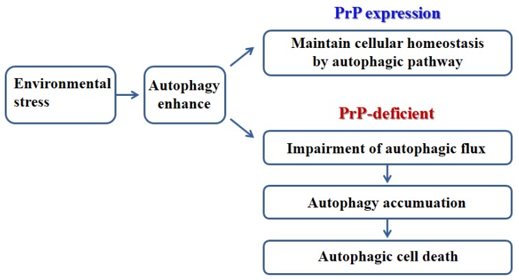
Decreased autophagy induction may be due to the influence that PrPC has on anti-stress activity in normal cells. The role of PrPC-associated autophagy is revealed a novel protective mechanism against oxidative stress [30]. However, in PrP-deficient cells, enhanced autophagy leads to impaired autophagic flux, which contributes to cell death by oxidative stress [30]. The impairment of autophagic flux in CNS neurons is potentially associated with progressive cerebellar Purkinje cell death in the Ngsk PrP-deficient mouse line [30,31,32]. Taken together, the deficiency of PrPC contributes to autophagic neuronal cell death via impaired autophagic flux (Figure 3).
Figure 3.
Schematic representation of environmental stress induced autophagic pathway in PrP-expression and PrP-deficient models.
Acknowledgments
This work was supported by the National Research Foundation of Korea Grant funded by the Korean Government (NRF-2011-619-E0001) and Basic Science Research Program through the National Research Foundation of Korea (NRF) funded by the Ministry of Education, Science and Technology (2012-0000308).
References
- 1.Basler K., Oesch B., Scott M., Westaway D., Wälchli M., Groth D.F., McKinley M.P., Prusiner S.B., Weissmann C. Scrapie and cellular PrP isoforms are encoded by the same chromosomal gene. Cell. 1986;46:417–428. doi: 10.1016/0092-8674(86)90662-8. [DOI] [PubMed] [Google Scholar]
- 2.Kretzschmar H.A., Prusiner S.B., Stowring L.E., DeArmond S.J. Scrapie prion proteins are synthesized in neurons. Am. J. Pathol. 1986;122:1–5. [PMC free article] [PubMed] [Google Scholar]
- 3.Prusiner S.B. Molecular biology of prion diseases. Science. 1991;252:1515–1522. doi: 10.1126/science.1675487. [DOI] [PubMed] [Google Scholar]
- 4.Sales N., Rodolfo K., Hassig R., Faucheux B., Di Giamberardino L., Moya K.L. Cellular prion protein localization in rodent and primate brain. Eur. J. Neurosci. 1998;10:2464–2471. doi: 10.1046/j.1460-9568.1998.00258.x. [DOI] [PubMed] [Google Scholar]
- 5.Bolton D.C., McKinley M.P., Prusiner S.B. Identification of a protein that purifies with the scrapie prion. Science. 1982;218:1309–1311. doi: 10.1126/science.6815801. [DOI] [PubMed] [Google Scholar]
- 6.Prusiner S.B. Prions. Proc. Natl. Acad. Sci. USA. 1998;95:13363–13383. doi: 10.1073/pnas.95.23.13363. [DOI] [PMC free article] [PubMed] [Google Scholar]
- 7.Aguzzi A., Weissmann C. Spongiform encephalopathies: a suspicious signature. Nature. 1996;383:666–667. doi: 10.1038/383666a0. [DOI] [PubMed] [Google Scholar]
- 8.Weissmann C. The Ninth Datta Lecture. Molecular biology of transmissible spongiform encephalopathies. FEBS Lett. 1996;389:3–11. doi: 10.1016/0014-5793(96)00610-2. [DOI] [PubMed] [Google Scholar]
- 9.Klionsky D.J., Emr S.D. Autophagy as a regulated pathway of cellular degradation. Science. 2000;290:1717–1721. doi: 10.1126/science.290.5497.1717. [DOI] [PMC free article] [PubMed] [Google Scholar]
- 10.Petersen A., Larsen K.E., Behr G.G., Romero N., Przedborski S., Brundin P., Sulzer D. Expanded CAG repeats in exon 1 of the Huntington's disease gene stimulate dopamine-mediated striatal neuron autophagy and degeneration. Hum. Mol. Genet. 2001;10:1243–1254. doi: 10.1093/hmg/10.12.1243. [DOI] [PubMed] [Google Scholar]
- 11.Selimi F., Lohof A.M., Heitz S., Lalouette A., Jarvis C.I., Bailly Y., Mariani J. Lurcher GRID2-induced death and depolarization can be dissociated in cerebellar Purkinje cells. Neuron. 2003;37:813–819. doi: 10.1016/S0896-6273(03)00093-X. [DOI] [PubMed] [Google Scholar]
- 12.Kabeya Y., Mizushima N., Ueno T., Yamamoto A., Kirisako T., Noda T., Kominami E., Ohsumi Y., Yoshimori T. LC3, a mammalian homologue of yeast Apg8p, is localized in autophagosome membranes after processing. EMBO J. 2000;19:5720–5728. doi: 10.1093/emboj/19.21.5720. [DOI] [PMC free article] [PubMed] [Google Scholar]
- 13.Noda T., Matsuura A., Wada Y., Ohsumi Y. Novel system for monitoring autophagy in the yeast Saccharomyces cerevisiae. Biochem. Biophys. Res. Commun. 1995;210:126–132. doi: 10.1006/bbrc.1995.1636. [DOI] [PubMed] [Google Scholar]
- 14.Aubert S., Gout E., Bligny R., Marty-Mazars D., Barrieu F., Alabouvette J., Marty F., Douce R. Ultrastructural and biochemical characterization of autophagy in higher plant cells subjected to carbon deprivation: control by the supply of mitochondria with respiratory substrates. Cell. Biol. 1996;133:1251–1263. doi: 10.1083/jcb.133.6.1251. [DOI] [PMC free article] [PubMed] [Google Scholar]
- 15.Reunanen H., Punnonen E.L., Hirsimäki P. Studies on vinblastine-induced autophagocytosis in mouse liver. V. A cytochemical study on the origin of membranes. Histochemistry. 1985;83:513–517. doi: 10.1007/BF00492453. [DOI] [PubMed] [Google Scholar]
- 16.Manson J., West J.D., Thomson V., McBride P., Kaufman M.H., Hope J. The prion protein gene: a role in mouse embryogenesis? Development. 1992;115:117–122. doi: 10.1242/dev.115.1.117. [DOI] [PubMed] [Google Scholar]
- 17.Cuervo A.M. Autophagy: in sickness and in health. Trends Cell. Biol. 2004;14:70–77. doi: 10.1016/j.tcb.2003.12.002. [DOI] [PubMed] [Google Scholar]
- 18.Shintani T., Klionsky D.J. Autophagy in health and disease: a double-edged sword. Science. 2004;306:990–995. doi: 10.1126/science.1099993. [DOI] [PMC free article] [PubMed] [Google Scholar]
- 19.Boellaard J.W., Schlote W., Tateishi J. Neuronal autophagy in experimental Creutzfeldt-Jakob's disease. Acta Neuropathol. 1989;78:410–418. doi: 10.1007/BF00688178. [DOI] [PubMed] [Google Scholar]
- 20.Boellaard J.W., Kao M., Schlote W., Diringer H. Neuronal autophagy in experimental scrapie. Acta Neuropathol. 1991;82:225–228. doi: 10.1007/BF00294449. [DOI] [PubMed] [Google Scholar]
- 21.Schätzl H.M., Laszlo L., Holtzman D.M., Tatzelt J., DeArmond S.J., Weiner R.I., Mobley W.C., Prusiner S.B. A hypothalamic neuronal cell line persistently infected with scrapie prions exhibits apoptosis. J. Virol. 1997;71:8821–8831. doi: 10.1128/jvi.71.11.8821-8831.1997. [DOI] [PMC free article] [PubMed] [Google Scholar]
- 22.Liberski P.P., Sikorska B., Bratosiewicz-Wasik J., Gajdusek D.C., Brown P. Neuronal cell death in transmissible spongiform encephalopathies (prion diseases) revisited: from apoptosis to autophagy. Int. J. Biochem. Cell. Biol. 2004;36:2473–2490. doi: 10.1016/j.biocel.2004.04.016. [DOI] [PubMed] [Google Scholar]
- 23.Sikorska B., Liberski P.P., Giraud P., Kopp N., Brown P. Autophagy is a part of ultrastructural synaptic pathology in Creutzfeldt-Jakob disease: a brain biopsy study. Int. J. Biochem. Cell. Biol. 2004;36:2563–2573. doi: 10.1016/j.biocel.2004.04.014. [DOI] [PubMed] [Google Scholar]
- 24.Dron M., Bailly Y., Beringue V., Haeberlé A.M., Griffond B., Risold P.Y., Tovey M.G., Laude H., Dandoy-Dron F. Scrg1 is induced in TSE and brain injuries, and associated with autophagy. Eur. J. Neurosci. 2005;22:133–146. doi: 10.1111/j.1460-9568.2005.04172.x. [DOI] [PubMed] [Google Scholar]
- 25.Aguib Y., Heiseke A., Gilch S., Riemer C., Baier M., Schätzl H.M., Ertmer A. Autophagy induction by trehalose counteracts cellular prion infection. Autophagy. 2009;5:361–369. doi: 10.4161/auto.5.3.7662. [DOI] [PubMed] [Google Scholar]
- 26.Heiseke A., Aguib Y., Riemer C., Baier M., Schätzl H.M. Lithium induces clearance of protease resistant prion protein in prion-infected cells by induction of autophagy. J. Neurochem. 2009;109:25–34. doi: 10.1111/j.1471-4159.2009.05906.x. [DOI] [PubMed] [Google Scholar]
- 27.Cortes C.J., Qin K., Cook J., Solanki A., Mastrianni J.A. Rapamycin delays disease onset and prevents PrP plaque deposition in a mouse model of Gerstmann-Sträussler-Scheinker disease. J. Neurosci. 2012;32:12396–12405. doi: 10.1523/JNEUROSCI.6189-11.2012. [DOI] [PMC free article] [PubMed] [Google Scholar]
- 28.Xu Y., Tian C., Wang S.B., Xie W.L., Guo Y., Zhang J., Shi Q., Chen C., Dong X.P. Activation of the macroautophagic system in scrapie-infected experimental animals and human genetic prion diseases. Autophagy. 2012;8:1604–1620. doi: 10.4161/auto.21482. [DOI] [PMC free article] [PubMed] [Google Scholar]
- 29.Oh J.M., Shin H.Y., Park S.J., Kim B.H., Choi J.K., Choi E.K., Carp R.I., Kim Y.S. The involvement of cellular prion protein in the autophagy pathway in neuronal cells. Mol. Cell. Neurosci. 2008;39:238–247. doi: 10.1016/j.mcn.2008.07.003. [DOI] [PubMed] [Google Scholar]
- 30.Oh J.M., Choi E.K., Carp R.I., Kim Y.S. Oxidative stress impairs autophagic flux in prion protein-deficient hippocampal cells. Autophagy. 2012;8:1448–1461. doi: 10.4161/auto.21164. [DOI] [PubMed] [Google Scholar]
- 31.Heitz S., Grant N.J., Bailly Y. Doppel induces autophagic stress in prion protein-deficient Purkinje cells. Autophagy. 2009;5:422–424. doi: 10.4161/auto.5.3.7882. [DOI] [PubMed] [Google Scholar]
- 32.Heitz S., Grant N.J., Leschiera R., Haeberlé A.M., Demais V., Bombarde G., Bailly Y. Autophagy and cell death of Purkinje cells overexpressing Doppel in Ngsk Prnp-deficient mice. Brain. Pathol. 2010;20:119–132. doi: 10.1111/j.1750-3639.2008.00245.x. [DOI] [PMC free article] [PubMed] [Google Scholar]
- 33.Wong B.S., Liu T., Paisley D., Li R., Pan T., Chen S.G., Perry G., Petersen R.B., Smith M.A., Melton D.W., Gambetti P., Brown D.R., Sy M.S. Induction of HO-1 and NOS in doppel-expressing mice devoid of PrP: implications for doppel function. Mol. Cell. Neurosci. 2001;17:768–775. doi: 10.1006/mcne.2001.0963. [DOI] [PubMed] [Google Scholar]
- 34.Cui T., Holme A., Sassoon J., Brown D.R. Analysis of doppel protein toxicity. Mol. Cell. Neurosci. 2003;23:144–155. doi: 10.1016/S1044-7431(03)00017-4. [DOI] [PubMed] [Google Scholar]
- 35.Barbieri G., Palumbo S., Gabrusiewicz K., Azzalin A., Marchesi N., Spedito A., Biggiogera M., Sbalchiero E., Mazzini G., Miracco C., Pirtoli L., Kaminska B., Comincini S. Silencing of cellular prion protein (PrPC) expression by DNA-antisense oligonucleotides induces autophagy-dependent cell death in glioma cells. Autophagy. 2011;7:840–853. doi: 10.4161/auto.7.8.15615. [DOI] [PubMed] [Google Scholar]
- 36.Herms J., Tings T., Gall S., Madlung A., Giese A., Siebert H., Schürmann P., Windl O., Brose N., Kretzschmar H. Evidence of presynapticlocation and function of the prion protein. J. Neurosci. 1999;19:8866–8875. doi: 10.1523/JNEUROSCI.19-20-08866.1999. [DOI] [PMC free article] [PubMed] [Google Scholar]
- 37.Brown D.R., Wong B.S., Hafiz F., Clive C., Haswell S.J., Jones I.M. Normal prion protein has an activity like that of superoxide dismutase. Biochem. J. 1999;344:1–5. doi: 10.1042/0264-6021:3440001. [DOI] [PMC free article] [PubMed] [Google Scholar]
- 38.Lee H.G., Park S.J., Choi E.K., Carp R.I., Kim Y.S. Increased expression of prion protein is associated with changes in dopamine metabolism and MAO activity in PC12 cells. J. Mol. Neurosci. 1999;13:121–126. doi: 10.1385/JMN:13:1-2:121. [DOI] [PubMed] [Google Scholar]
- 39.Graner E., Mercadante A.F., Zanata S.M., Forlenza O.V., Cabral A.L., Veiga S.S., Juliano M.A., Roesler R., Walz R., Minetti A., Izquierdo I., Martins V.R., Brentani R.R. Cellular prion protein binds laminin and mediates neuritogenesis. Brain. Res. Mol. Brain. Res. 2000;76:85–92. doi: 10.1016/S0169-328X(99)00334-4. [DOI] [PubMed] [Google Scholar]
- 40.Mouillet-Richard S., Ermonval M., Chebassier C., Laplanche J.L., Lehmann S., Launay J.M., Kellermann O. Signal transduction through prion protein. Science. 2000;289:1925–1928. doi: 10.1126/science.289.5486.1925. [DOI] [PubMed] [Google Scholar]
- 41.Mange A., Milhavet O., Umlauf D., Harris D., Lehmann S. PrP-dependent cell adhesion in N2a neuroblastoma cells. FEBS. Lett. 2002;514:159–162. doi: 10.1016/S0014-5793(02)02338-4. [DOI] [PubMed] [Google Scholar]
- 42.Li A., Harris D.A. Mammalian prion protein suppresses Bax-induced cell death in yeast. J. Biol. Chem. 2005;280:17430–17434. doi: 10.1074/jbc.C500058200. [DOI] [PubMed] [Google Scholar]
- 43.Bueler H., Fischer M., Lang Y., Bluethmann H., Lipp H.P., DeArmond S.J., Prusiner S.B., Aguet M., Weissmann C. Normal development and behaviour of mice lacking the neuronal cell-surface PrP protein. Nature. 1992;356:577–582. doi: 10.1038/356577a0. [DOI] [PubMed] [Google Scholar]
- 44.Criado J.R., Sa´nchez-Alavez M., Conti B., Giacchino J.L., Wills D.N., Henriksen S.J., Race R., Manson J.C., Chesebro B., Oldstone M.B. Mice devoid of prion protein have cognitive deficits that are rescued by reconstitution of PrP in neurons. Neurobiol. Dis. 2005;19:255–265. doi: 10.1016/j.nbd.2005.01.001. [DOI] [PubMed] [Google Scholar]
- 45.Weissmann C., Flechsig E. PrP knock-out and PrP transgenic mice in prion research. Br. Med. Bull. 2003;66:43–60. doi: 10.1093/bmb/66.1.43. [DOI] [PubMed] [Google Scholar]
- 46.Prestori F., Rossi P., Bearzatto B., Laine´ J., Necchi D., Diwakar S., Schiffmann S.N., Axelrad H., D'Angelo E. Altered Neuron excitability and synaptic plasticity in the cerebellar granular layer of juvenile prion protein knock-out mice with impaired motor control. J. Neurosci. 2008;28:7091–7103. doi: 10.1523/JNEUROSCI.0409-08.2008. [DOI] [PMC free article] [PubMed] [Google Scholar]
- 47.Singh A., Kong Q., Luo X., Petersen R.B., Meyerson H., Singh N. Prion protein (PrP) knock-out mice show altered iron metabolism: A functional role for PrP in iron uptake and transport. PLoS ONE. 2009;4:1–14. doi: 10.1371/journal.pone.0006115. [DOI] [PMC free article] [PubMed] [Google Scholar]
- 48.Sánchez-Pérez A.M., Claramonte-Clausell B., Sánchez-Andrés J.V., Herrero M.T. Parkinson's disease and autophagy. Parkinsons. Dis. 2012;2012 doi: 10.1155/2012/429524. [DOI] [PMC free article] [PubMed] [Google Scholar]
- 49.Sanchez-Varo R., Trujillo-Estrada L., Sanchez-Mejias E., Torres M., Baglietto-Vargas D., Moreno-Gonzalez I., De Castro V., Jimenez S., Ruano D., Vizuete M., Davila J.C., Garcia-Verdugo J.M., Jimenez A.J., Vitorica J., Gutierrez A. Abnormal accumulation of autophagic vesicles correlates with axonal and synaptic pathology in young Alzheimer's mice hippocampus. Acta. Neuropathol. 2012;123:53–70. doi: 10.1007/s00401-011-0896-x. [DOI] [PMC free article] [PubMed] [Google Scholar]
- 50.Jackson G.S., Clarke A.R. Mammalian prion proteins. Curr. Opin. Struct. Biol. 2000;10:69–74. doi: 10.1016/S0959-440X(99)00051-2. [DOI] [PubMed] [Google Scholar]
- 51.Brandner S., Isenmann S., Raeber A., Fischer M., Sailer A., Kobayashi Y., Marino S., Weissmann C., Aguzzi A. Normal host prion protein necessary for scrapie-induced neurotoxicity. Nature. 1996;379:339–343. doi: 10.1038/379339a0. [DOI] [PubMed] [Google Scholar]
- 52.Chu C.T., Plowey E.D., Dagda R.K., Hickey R.W., Cherra S.J., Clark R.S. Autophagy in neurite injury and neurodegeneration: in vitro and in vivo models. Methods. Enzymol. 2009;453:217–249. doi: 10.1016/S0076-6879(08)04011-1. [DOI] [PMC free article] [PubMed] [Google Scholar]
- 53.Bursch W., Ellinger A., Gerner C., Frohwein U., Schulte-Hermann R. Programmed cell death (PCD). Apoptosis, autophagic PCD, or others? Ann. N. Y. Acad. Sci. 2000;926:1–12. doi: 10.1111/j.1749-6632.2000.tb05594.x. [DOI] [PubMed] [Google Scholar]
- 54.Dong J., Li A., Yamaguchi N., Sakaguchi S., Harris D.A. Doppel induces degeneration of cerebellar Purkinje cells independently of Bax. Am. J. Pathol. 2007;171:599–607. doi: 10.2353/ajpath.2007.070262. [DOI] [PMC free article] [PubMed] [Google Scholar]
- 55.Lee J.A. Autophagy in neurodegeneration: two sides of the same coin. BMB Rep. 2009;42:324–330. doi: 10.5483/BMBRep.2009.42.6.324. [DOI] [PubMed] [Google Scholar]
- 56.Wong E., Cuervo A.M. Autophagy gone awry in neurodegenerative diseases. Nat. Neurosci. 2010;13:805–811. doi: 10.1038/nn.2575. [DOI] [PMC free article] [PubMed] [Google Scholar]


