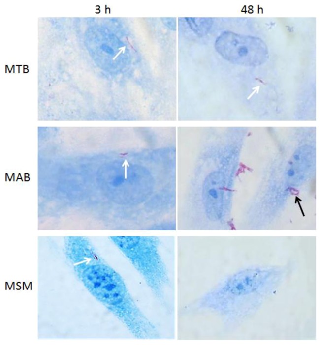
Figure 2.

Optic microscopy images of limbo-corneal fibroblasts infected with mycobacteria. The limbo-corneal fibroblasts were infected for 3 h with MTB, MAB, or MSM, and the infection was followed for 48 hours post-infection. At each time point, the monolayers were stained using the Ziehl-Neelsen technique. In the cells infected with MTB, the presence of intracellular bacilli is observed (white arrows) at 3 h and 48 h, although there is no evidence of intracellular replication. At 3 h, intracellular bacilli are detected (white arrow) in the cells infected with MAB, and at 48 h, there is evidence of the intracellular replication of the bacilli (black arrow). In the cells infected with MSM, the presence of intracellular bacilli is only evident at 3 h post-infection (white arrow); at 48 h post-infection, there is no evidence of intracellular bacilli. 1000× magnification.
