Summary
This pictorial review describes the spectrum of CT/MR imaging findings of solitary extramedullary and bone plasmacytomas in different locations related to neuroradiology. Plasmacytoma is considered a counterpart of multiple myeloma that is described as a solitary and discrete mass of monoclonal neoplastic plasma cells. It may arise from osseous (medullary) or extramedullary sites. Isolated extramedullary plasmacytoma is very rare and comprises less than 4% of all plasma cellular diseases of which more than 80% are localized to the submucosal lymphoid tissue of the nasopharynx, nasal cavity and paranasal sinuses. We will demonstrate imaging findings in ten histopathologically proven plasmacytomas in different locations related to neuroradiology. Extramedullary and osseous plasmacytoma show nonspecific CT and MR imaging findings. MR is the preferred modality for evaluation due to better soft tissue contrast. Features that may suggest the diagnosis of plasmacytoma are bulky soft tissue mass and relatively isointense signal on T2-weighted MR images due to high cellularity.
Keywords: plasmacytoma, CT and MR imaging
Introduction
Plasmacytoma, a form of plasma cell dyscrasia, is described as a solitary and discrete mass of monoclonal neoplastic plasma cells. It may arise from osseous (medullary) or extramedullary sites. Plasmacytoma can be solitary or multiple. Extramedullary plasmacytoma (EMP) is extremely rare accounting for less than 4% of all plasma cell tumors of which more than 80% are localized to the upper aerodigestive tract. Solitary plasmacytoma remains a diagnostic challenge with limited literature available on imaging of this entity.
This pictorial review demonstrates neuroimaging findings of solitary extramedullary and bone plasmacytomas.
Figure 1.
A 62-year-old man with a solitary plasmacytoma of the right maxillary alveolar process. Axial (A, bone window) and coronal (B, soft tissue window) contrast-enhanced CT images demonstrate a bony destructive lesion (arrow) involving the right maxillary alveolar process with soft tissue component (arrowhead).
Figure 2.
A 55-year-old woman with a solitary plasmacytoma of the nasal cavities. Sagittal T1 precontrast (A) and axial T1 postcontrast (B) MR images demonstrate a large avidly enhancing soft tissue mass in bilateral nasal cavities (arrow) with intracranial extension (arrowhead).
Figure 3.
A 61-year-old woman with a solitary plasmacytoma of the left nasal cavity. Coronal (A, soft tissue window) and axial CT (B, soft tissue window) images demonstrate a soft tissue mass (arrows) completely filling the left nasal cavity posteriorly projecting into the nasopharynx.
Figure 4.
A 51-year-old man with a solitary plasmacytoma of the nasopharynx. Axial (A) and coronal (B) contrast-enhanced CT images demonstrate a large enhancing submucosal mass in the nasopharynx on the left (arrow) with posterior extension into the perivertebral space (arrowhead).
Figure 5.
A 60-year-old man with a solitary plasmacytoma of the left orbit. Coronal CT (A, soft tissue window) and coronal T1 post contrast MR (B) images demonstrate a large intensely enhancing extraconal mass (arrows) in the left orbit eroding through the roof with intracranial extradural extension (arrowhead).
Figure 6.
A 49-year-old woman with a solitary plasmacytoma of the spinal epidural space. Sagittal T2 (A) and sagittal T1 postcontrast (B) MR images demonstrate a large, T2 isointense, primarily extradural mildly enhancing mass ( arrows) in the lumbar spinal canal causing severe thecal sac compression.
Figure 7.
A 70-year-old man with a solitary plasmacyotma of the sacrum. Sagittal T1 precontrast (A) and axial T1 postcontrast (B) MR images demonstrate a partially visualized large heterogeneously enhancing sacral mass (arrows).
Figure 8.
A 69-year-old woman with a solitary plasmacytoma of the lumbar spine. Axial T1 precontrast (A) and axial T1 postcontrast (B) MR images show a large enhancing left dorsal paraspinal soft tissue mass (arrows) with epidural extension causing thecal sac compression (arrowhead). There was also osseous involvement of the posterior elements of the L2 vertebra (not shown).
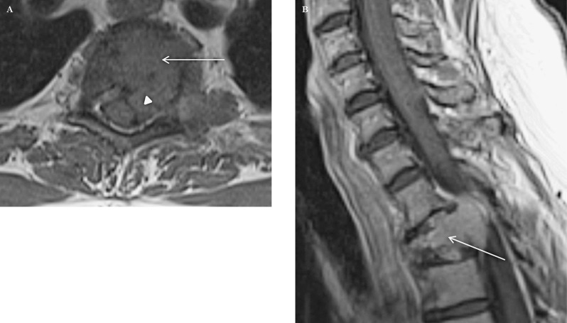
Figure 9.
A 65-year-old woman with a solitary plasmacytoma of the T2 vertebra. Axial T1 precontrast (A) and sagittal T1 postcontrast (B) MR images demonstrate a heterogeneously enhancing mass involving the T2 vertebra (arrows) with extraosseous enhancing epidural soft tissue (arrowheads) causing mild cord compression. There is a pathological fracture of T2.
Figure 10.
A 60-year-old man with a solitary plasmacytoma of the T9 vertebra. Axial CT (A, soft tissue window) and axial T1 post contrast MR (B) images demonstrate a large destructive osseous lesion involving the T9 vertebra (arrow) with an enhancing extraosseous epidural soft tissue component (arrowheads) causing moderate cord compression.
Discussion
Plasmacytoma refers to a malignant plasma cell tumor growing within soft tissue or within the axial skeleton. There are three distinct groups of plasmacytoma defined by the International Myeloma Working Group: solitary plasmacytoma of bone (SPB), extramedullary plasmacytoma (EMP) and multiple plasmacytomas that are either primary or recurrent 1,2. The most common of these is SPB, accounting for 3-5% of all plasma cell malignancies. SPBs usually present as lytic lesions within the axial skeleton with or without an extraosseous soft tissue component. EMPs most often occur in the upper respiratory tract (85%), but can occur in any soft tissue including gastrointestinal tract, skin, chest wall, breast and retroperitoneum 2,3. Plasmacytoma is distinguished from multiple myeloma by the presence of only one lesion (either in bone or soft tissue), normal bone marrow (<5% plasma cells), normal skeletal survey, absent or low paraprotein and no end organ damage. The osseous form of plasmacytoma usually progresses to multiple myeloma in three to five years. The incidence of plasmacytoma is more common in males and the 55-75 year age group 1,3. Clinical presentation of patients depends on the location of the lesion and mass effect. SEPs and EMPs are usually treated with radiotherapy. Wide surgical excision is also an option. Prognosis is usually good with more than 70% patients surviving longer than five years 2,4.
Imaging with CT and MRI is important in these patients for assessing local disease and for staging. Evaluation of apparently solitary plasmacytomas with imaging may reveal multiple soft tissue lesions or additional bone lesions, thereby upstaging the disease. Cross-sectional imaging also helps to better depict regional lymphadenopathy which may be present in up to 25-50% of patients. However, imaging alone cannot distinguish from other malignancies and requires histologic evidence for diagnosis 3,5. These tumors are usually locally aggressive with involvement and destruction of adjacent structures. CT and MR imaging are complementary to each other. CT is the modality of choice to assess the bony lesion while MRI provides a better assessment of soft tissue lesions. EMP generally presents as a bulky soft tissue mass, isointense to muscle on T1-weighted MR images, isointense to slightly hyperintense to muscle on T2-weighted MR images with heterogeneous enhancement. Large tumors may show area of necrosis, infiltration of adjacent structures and vascular encasement. Regional lymphadenopathy may be present, particularly in the abdomen and thorax 2,5.
This review demonstrates imaging findings in ten cases of histopathologically confirmed plasmacytoma, related to neuroradiology (5 SPB and 5 EMP). One patient presented primarily with a large epidural mass on MRI of the lumbar spine. Two patients presented primarily as a nasal cavity soft tissue mass. One patient presented as an isolated nasopharyngeal mass with parapharyngeal extension.
Conclusion
Solitary extramedullary and osseous plasmacytoma show nonspecific CT and MR imaging findings. MR is the modality of choice for soft tissue evaluation. CT is the modality of choice for assessment of the bony abnormality. Features that may suggest the diagnosis of plasmacytoma are a bulky soft tissue mass and relatively isointense signal on T2-weighted MR images due to high cellularity. Imaging helps to detect additional lesions and regional lymphadenopathy.
References
- 1.International Myeloma Working Group. Criteria for the classification of monoclonal gammopathies, multiple myeloma and related disorders: a report of the International Myeloma Working Group. Br J Haematol. 2003;121(5):749–757. doi: 10.1046/j.1365-2141.2003.04355.x. [PubMed] [Google Scholar]
- 2.Ooi GC, Chim JC, Au WY, et al. Radiologic manifestations of primary solitary extramedullary and multiple solitary plasmacytomas. Am J Roentgenol. 2006;186(3):821–827. doi: 10.2214/AJR.04.1787. doi: 10.2214/AJR.04.1787. [DOI] [PubMed] [Google Scholar]
- 3.Ching AS, Khoo JB, Chong VF. CT and MR imaging of solitary extramedullary plasmacytoma of the nasal tract. Am J Neuroradiol. 2002;23(10):1632–1636. [PMC free article] [PubMed] [Google Scholar]
- 4.Vogl TJ, Steger W, Grevers G, et al. MR characteristics of primary extramedullary plasmacytoma in the head and neck. Am J Neuroradiol. 1996;17(7):1349–1354. [PMC free article] [PubMed] [Google Scholar]
- 5.Susnerwala SS, Shanks JH, Banerjee SS, et al. Extramedullary plasmacytoma of the head and neck region: clinicopathological correlation in 25 cases. Br J Cancer. 1997;75(6):921–927. doi: 10.1038/bjc.1997.162. doi: 10.1038/bjc.1997.162. [DOI] [PMC free article] [PubMed] [Google Scholar]


