Abstract
Summary
Brain death is defined as the irreversible cessation of functioning of the entire brain, including the brainstem. Brain death is principally established using clinical criteria including coma, absence of brainstem reflexes and loss of central drive to breathe assessed with apnea test. In situations in which clinical testing cannot be performed or when uncertainty exists about the reliability of its parts due to confounding conditions ancillary tests (i.a. imaging studies) may be useful. The objective of ancillary tests in the diagnosis of brain death is to demonstrate the absence of cerebral electrical activity (EEG and evoked potentials) or cerebral circulatory arrest. In clinical practice catheter cerebral angiography, perfusion scintigraphy, transcranial Doppler sonography, CT angiography and MR angiography are used. Other methods, like perfusion CT, xenon CT, MR spectroscopy, diffusion weighted MRI and functional MRI are being studied as potentially useful in the diagnosis of brain death. CT angiography has recently attracted attention as a promising alternative to catheter angiography – a reference test in the diagnosis of brain death. Since 1998 several major studies were published and national guidelines were introduced in several countries (e.g. in France, Austria, Switzerland, the Netherlands and Canada). This paper reviews technique, characteristic findings and criteria for the diagnosis of cerebral circulatory arrest in CT angiography.
MeSH eywords: Brain Death, Cerebral Angiography, Multidetector Computed Tomography
Background
The diagnosis of brain death (BD) is of particular importance in view of the contemporary medical and technological achievements in supporting, repairing and replacing failing human organs. The declaration of BD determines the termination of futile life support and potential procurement of organs for transplantation.
Brain death is defined as the irreversible cessation of functioning of the entire brain, including the brainstem with two pathways to reach that point: (i) permanent absence of circulation or (ii) subsequent to a catastrophic brain injury [1]. To determine BD physicians must:
determine the presence of unresponsive coma,
determine the absence of brainstem reflexes,
determine the absence of respiratory drive after a CO2 challenge – apnea testing.
To ensure that the cessation of brain function is “irreversible” physicians must:
determine the cause of coma,
exclude mimicking medical conditions,
observe the patient for a period of time to exclude the possibility of recovery [2].
Confounding conditions that may invalidate testing for cessation of brain function include: hypothermia; the use of therapeutic hypothermia; the presence of central nervous system depressing drugs that may explain or contribute to coma; high cervical spine injury; acquired or therapeutic neuromuscular paralysis; and severe acid-base, electrolyte, endocrine abnormality that may explain or contribute to coma [3].
When uncertainty exists about the reliability of parts of the clinical examination or when the apnea test cannot be performed ancillary tests may be useful. The objective of ancillary tests is to demonstrate the absence of cerebral electrical activity (EEG and evoked potentials) or cerebral circulatory arrest. A number of blood flow tests were introduced to the diagnosis of BD including: catheter cerebral angiography, nuclear scan, transcranial Doppler sonography (TCD), computed tomographic angiography (CTA), magnetic resonance angiography (MRA), perfusion CT (PCT), xenon CT, magnetic resonance perfusion (MRP), magnetic resonance spectroscopy (MRS), diffusion-weighted MRI (DWI) and functional MRI (fMRI).
Although catheter angiography is considered as a reference test in the diagnosis of BD the method has major drawbacks: invasiveness, technical complexity, inability to be performed at the bedside, and limited accessibility of the angiographic labs. Therefore modern, noninvasive and ubiquitous modality like CTA has recently attracted attention. CTA to the diagnosis of BD was introduced in 1998 by Dupas et al. [4].
CT Angiography Findings in Cerebral Circulatory Arrest
Cerebral blood flow (CBF) is continuously regulated by cerebral perfusion pressure (CPP) and cerebrovascular resistance (CVR). CPP is measured as a difference between the entry pressure - mean arterial pressure (MAP) and the exit pressure - intracranial pressure (ICP). According to Poiseuille equation:
Normal CBF at rest is maintained within a range of 45–60 mL/100 g of brain tissue/min. A brain injury is accompanied by cerebral edema. Because the skull bones limit the intracranial volume this “mass effect” leads to the intracranial hypertension. The process spreads from the region of primary injury gradually involving the whole brain and the intracranial vessels become compressed but preserve their patency. Initially CVR is kept almost constant by the activated autoregulation mechanisms releasing blood vessel tone. However, this phenomenon is capable to preserve a sufficient blood supply as long as CPP exceeds approx. 50 mmHg [5]. Further elevation of ICP results in an increase of CVR. The deep cerebral veins and capillaries as the most susceptible vessels are primarily affected; then the process spreads proximally involving bigger arteries progressively.
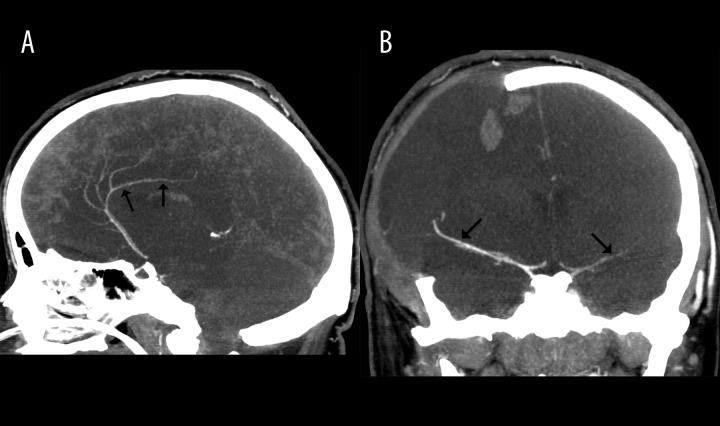
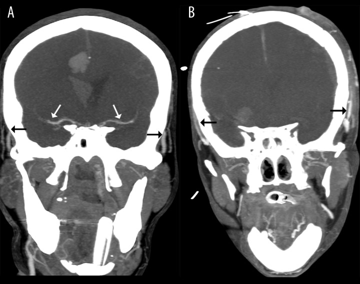
Unfortunately, CTA findings have not been studied in non-brain-dead patients with intracranial hypertension yet. The earliest sign of cerebral circulatory arrest in CTA is a lack of opacification of the deep veins – the internal cerebral veins (ICV) and the great cerebral vein (GCV, vein of Galen). The sensitivity of this finding in the diagnosis of cerebral circulatory arrest in CTA is 98–100% [4,6–8]. A lack of opacification of cortical branches of the middle cerebral arteries (MCA-M4) is a slightly less sensitive indicator of cerebral circulatory arrest with a sensitivity of 86–100% [6,7]. The basilar artery (BA) and cortical branches of the posterior cerebral arteries (PCA-P2) are more frequently opacified in cerebral circulatory arrest than MCA-M4. Their sensitivities are 83–94% and 79%, respectively [4,6,9]. The least sensitive finding of cerebral circulatory arrest is a lack of opacification of cortical branches of the anterior cerebral artery (ACA-A3, pericallosal artery) with a sensitivity of 64% [6]. Such a sequence is explained by the highest susceptibility of cortical branches of the MCA to the intracranial hypertension and the lowest cerebral perfusion pressure (CPP) in these arteries – see Figure 1. In the course of cerebral circulatory arrest, proximal segments of the cerebral arteries may show opacification for some time – Figure 2A. These arteries appear as thin and their filling is delayed and weak [10]. Intracranial stasis of contrast is observed with its extremely slow elimination through arteriovenous shunts and extravasation due to the interrupted blood-brain barrier. The last sign of cerebral circulatory arrest is intracranial non-filling – see Figure 2B.
Figure 1.
The case of a 22-year-old woman with brain stem ischemic stroke and right-sided craniectomy presenting signs of BD on clinical examination; (A) – 10-mm MIP in the sagittal plane in CTA shows opacification of the right pericallosal artery (thin arrows); (B) – 10-mm MIP in the coronal plane in CTA shows opacification of M1 segments of MCAs (thin arrows).
Figure 2.
Positive results of CTA in the diagnosis of BD: (A) – 10-mm MIP in the coronal plane shows stasis filling with delayed opacification of proximal MCAs (white arrows); please note the simultaneous opacification of the superficial temporal arteries (black arrows) (B) – 10-mm MIP in the coronal plane shows no intracranial filling; these findings confirm the diagnosis of BD.
Technique of CT Angiography in the Diagnosis of Cerebral Circulatory Arrest
The technique of CTA involves rapid, intravenous administration of iodinated contrast followed by volume scanning of the whole brain. For the diagnosis of BD, at least 3 acquisitions should be performed:
Non-enhanced scanning as a reference for assessing vascular opacification.
Early post-contrast scanning for assessing intra- and, which is more important, extracranial vascular opacification. Usually this phase is started 20 sec. after the beginning of injection. Opacification of branches of external carotid arteries – the superficial temporal or the facial arteries indicate that the contrast was injected correctly and there are no hemodynamic abnormalities causing a delay of contrast delivery to the vessels of the head and neck.
Late post-contrast scanning for assessing intracranial vascular opacification. This phase is started 60 sec. after the beginning of injection, with a delay of 40 sec. to the early phase. The necessity of performing the late phase of CTA in the diagnosis of cerebral circulatory arrest is motivated by a possible delayed vascular opacification in intracranial hypertension. Termination of the study on the early phase may result in a failure to recognize delayed intracranial filling giving a false positive result.
Criteria for the Diagnosis of Cerebral Circulatory Arrest in CT Angiography
There are no widely accepted criteria of CTA in the diagnosis of cerebral circulatory arrest. Four methods of evaluation have been proposed so far; they are summarized in Table 1.
Table 1.
CTA evaluation scales in the diagnosis of BD.
| Criteria | Lack of opacification of |
|---|---|
| Intracranial non-filling |
|
| 10-point* |
|
| 7-point* |
|
| 4-point* |
|
One point is noted for each nonopacified vessel in the late phase. Cerebral circulatory arrest is diagnosed with the score of 10, 7, or 4 points, accordingly;
according to the 4-point scale, opacification of 1 or 2 cortical branches of MCA on the same side does not exclude the diagnosis of cerebral circulatory arrest provided there is no opacification of ICVs.
Usefulness of CT Angiography in the Diagnosis of Cerebral Circulatory Arrest
CTA is far more widely available than any other blood flow test. The study is noninvasive, technically uncomplicated and non-time consuming. However, CTA cannot be applied at the bedside, while the transport of ICU patients is always insecure. The potential risk of damage of transplantable organs caused by iodinated contrast, although postulated, has not been confirmed yet. Moreover, it was revealed that contrast medium administration to donors does not affect kidney graft function after transplantation [11].
The accuracy of CTA in the diagnosis of cerebral circulatory arrest has not been reliably studied yet. In particular, specificity of the method has not been evaluated with non-brain-dead patients with intracranial hypertension as controls. However there are no reports on false positive CTAs involving multiphase scanning. Skull decompression and ischemic-hypoxic injury after cardiac arrest are the predisposing factors to false negative results of CTA [12–14] – see Table 2.
Table 2.
Sensitivity of CTA in the diagnosis of BD.
| Study authors and year | No of cases | Sensitivity (%) | ||
|---|---|---|---|---|
| 10-point | 7-point | 4-point | ||
| Combes et al. 2007 [15] | 43 | 70 | ||
| Welschehold et al. 2013 [16] | 63 | 54 * | ||
| Dupas et al. 1998 [4] | 14 | 100 | ||
| Quesnel et al. 2007 [13] | 21 | 52** | ||
| Frampas et al. 2009 [6] | 105 | 63 | 86 | |
| Rieke et al. 2011 [14] | 29 | 76 | 93 | |
| Leclerc et al. 2006 [7] | 15 | 87 | ||
| Sawicki et al. 2014 [9] | 82 | 67 | 74 | 96 |
GCV was not assessed,
the study included 5 out of 21 patients with anoxic brain injury.
Limitations of CT Angiography in the Diagnosis of Cerebral Circulatory Arrest
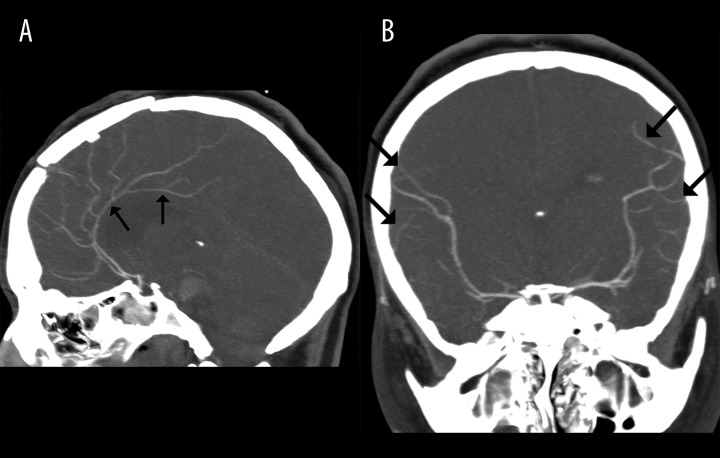
The limitations of CTA result most of all from uneven distribution of intracranial hypertension in some brain-dead patients due to local decompression of the cranial fossa – see Figure 3. There are several mechanisms causing a local decrease of ICP: craniectomy, ventricular drainage, skull fracture, open fontanelles and unfused sutures in neonates and infants. The regionally decreased ICP may result in preservation or restoration of residual cerebral blood flow at the site of decompression while in the remaining parts of the brain the cessation of blood flow is detected [15,16]. A similar problem can be met in some cases of infratentorial injuries in which the increase of ICP in the subtentorial compartment precedes its increase in the supratentorial region due to the protective function of the cerebellar tentorium [14]. In such cases the blood flow in cerebral hemispheres ceases with a marked delay in comparison to the posterior circulation. An opposite sequence is known as well, when cessation of blood flow in the supratentorial region precedes cerebral circulatory arrest in the basilar artery [17]. Diagnostic difficulties are also encountered in case of hypoxic-ischemic brain injuries after cardiac arrest when the ineffective restoration of cerebral blood flow is observed [12,13].
Figure 3.
The case of a 30-year-old woman with brain stem hematoma and frontal craniotomy presenting signs of BD on clinical examination; (A) – 10-mm MIP in the sagittal plane in CTA shows opacification of both pericallosal arteries (thin arrows); (B) – 10-mm MIP in the coronal plane in CTA shows opacification of cortical segments of MCAs (thin arrows); these findings exclude the diagnosis of BD.
Future Trends
CTA is the most promising ancillary test considering the recent introduction of ultrafast, wide detector scanners. Such machines are capable of performing time-resolved dynamic CTA (4D CTA). Combination of 4D CTA with whole-brain PCT using a single contrast injection seems to be the most promising alternative for the future. The problem of portability of CT may be resolved in next years. First portable CT scanners are already on the market.
References
- 1.Shemie SD, Hornby L, Baker A, et al. International guideline development for the determination of death. Intensive Care Med. 2014;40(6):788–97. doi: 10.1007/s00134-014-3242-7. [DOI] [PMC free article] [PubMed] [Google Scholar]
- 2.Wijdicks EF, Varelas PN, Gronseth GS, et al. Evidence-based guideline update: determining brain death in adults: report of the Quality Standards Subcommittee of the American Academy of Neurology. Neurology. 2010;74:1911–18. doi: 10.1212/WNL.0b013e3181e242a8. [DOI] [PubMed] [Google Scholar]
- 3.Shemie SD, Doig C, Dickens B, et al. Severe brain injury to neurological determination of death: Canadian forum recommendations. CMAJ. 2006;174(6):S1–13. doi: 10.1503/cmaj.045142. [DOI] [PMC free article] [PubMed] [Google Scholar]
- 4.Dupas B, Gayet-Delacroix M, Villers D, et al. Diagnosis of brain death using two-phase spiral CT. AJNR Am J Neuroradiol. 1998;19(4):641–47. [PMC free article] [PubMed] [Google Scholar]
- 5.Greenberg MS. Handbook of neurosurgery. Tampa, Fl, New York, NY: Greenberg Graphics, Thieme Medical Publishers; 2010. [Google Scholar]
- 6.Frampas E, Videcoq M, de Kerviler E, et al. CT angiography for brain death diagnosis. AJNR Am J Neuroradiol. 2009;30:1566–70. doi: 10.3174/ajnr.A1614. [DOI] [PMC free article] [PubMed] [Google Scholar]
- 7.Leclerc X, Taschner CA, Vidal A, et al. The role of spiral CT for the assessment of the intracranial circulation in suspected brain-death. J Neuroradiol. 2006;33:90–95. doi: 10.1016/s0150-9861(06)77237-6. [DOI] [PubMed] [Google Scholar]
- 8.Bohatyrewicz R, Sawicki M, Walecka A, et al. Computed tomographic angiography and perfusion in the diagnosis of brain death. Transplant Proc. 2010;42:3941–46. doi: 10.1016/j.transproceed.2010.09.143. [DOI] [PubMed] [Google Scholar]
- 9.Sawicki M, Bohatyrewicz R, Safranow K, et al. Computed tomographic angiography criteria in the diagnosis of brain death-comparison of sensitivity and interobserver reliability of different evaluation scales. Neuroradiology. 2014 doi: 10.1007/s00234-014-1364-9. [Epub ahead of print] [DOI] [PMC free article] [PubMed] [Google Scholar]
- 10.Sawicki M, Bohatyrewicz R, Safranow K, et al. Dynamic evaluation of stasis filling phenomenon with computed tomography in diagnosis of brain death. Neuroradiology. 2013;55:1061–69. doi: 10.1007/s00234-013-1210-5. [DOI] [PMC free article] [PubMed] [Google Scholar]
- 11.Grosse K, Brauer B, Kucuk O, et al. Does contrast medium administration in organ donors affect early kidney graft function? Transplant Proc. 2006;38:668–69. doi: 10.1016/j.transproceed.2006.01.034. [DOI] [PubMed] [Google Scholar]
- 12.Escudero D, Otero J, Marques L, et al. Diagnosing brain death by CT perfusion and multislice CT angiography. Neurocrit Care. 2009;11:261–71. doi: 10.1007/s12028-009-9243-7. [DOI] [PubMed] [Google Scholar]
- 13.Quesnel C, Fulgencio JP, Adrie C, et al. Limitations of computed tomographic angiography in the diagnosis of brain death. Intensive Care Med. 2007;33:2129–35. doi: 10.1007/s00134-007-0789-6. [DOI] [PubMed] [Google Scholar]
- 14.Rieke A, Regli B, Mattle HP, et al. Computed tomography angiography (CTA) to prove circulatory arrest for the diagnosis of brain death in the context of organ transplantation. Swiss Med Wkly. 2011;141:w13261. doi: 10.4414/smw.2011.13261. [DOI] [PubMed] [Google Scholar]
- 15.Combes JC, Chomel A, Ricolfi F, et al. Reliability of computed tomographic angiography in the diagnosis of brain death. Transplant Proc. 2007;39:16–20. doi: 10.1016/j.transproceed.2006.10.204. [DOI] [PubMed] [Google Scholar]
- 16.Welschehold S, Kerz T, Boor S, et al. Detection of intracranial circulatory arrest in brain death using cranial CT-angiography. Eur J Neurol. 2013;20(1):173–79. doi: 10.1111/j.1468-1331.2012.03826.x. [DOI] [PubMed] [Google Scholar]
- 17.Braun M, Ducrocq X, Huot JC, et al. Intravenous angiography in brain death: report of 140 patients. Neuroradiology. 1997;39:400–5. doi: 10.1007/s002340050432. [DOI] [PubMed] [Google Scholar]


