

An 81-year-old cognitively normal man (APOE ε3/ε3) without risk factors had amyloid-related imaging abnormalities with sulcal effusions and edema (ARIA-E), siderosis, and microhemorrhages (ARIA-H) on MRI (figure 1).1 He was identified from 1,006 participants followed with 3,385 MRIs in 2010–2013 in Alzheimer's Disease Neuroimaging Initiative (ADNI) 2/ADNI Grand Opportunities. Amyloid standard uptake value ratio on PET was 1.85 (positive) (figure 2). ARIA-E and associated ARIA-H can be observed in cognitively normal elderly without the APOE ε4 risk allele, who have no prior microhemorrhages, and who are not receiving amyloid-modifying treatments. Focal amyloid deposits around the region of ARIA-H suggest that cerebral amyloid angiopathy may be responsible for the occurrence of ARIA in this case.2
Figure 1. MRI of amyloid-related imaging abnormalities with microhemorrhages and amyloid-related imaging abnormalities with sulcal effusions and edema.
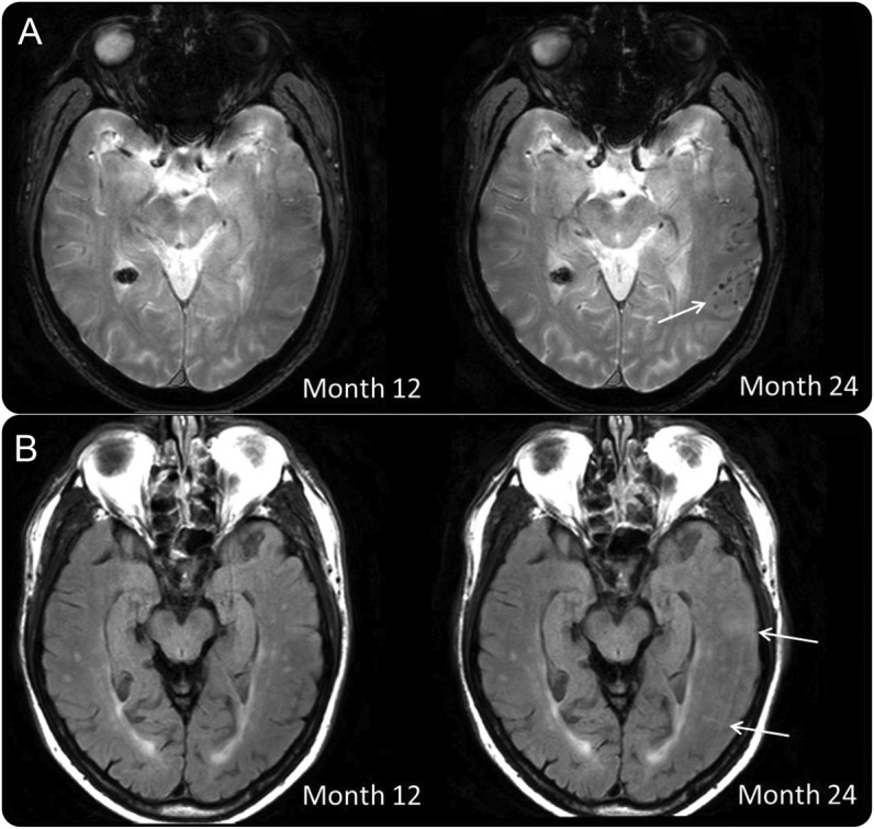
Amyloid-related imaging abnormalities with microhemorrhages (arrow) on T2* gradient recalled echo (A), amyloid-related imaging abnormalities with sulcal effusions and edema (arrows) on fluid-attenuated inversion recovery MRI (B) at month 24, which were not present on the 12-month MRI.
Figure 2. Amyloid PET of a patient with spontaneous amyloid-related imaging abnormalities with microhemorrhages and amyloid-related imaging abnormalities with sulcal effusions and edema.
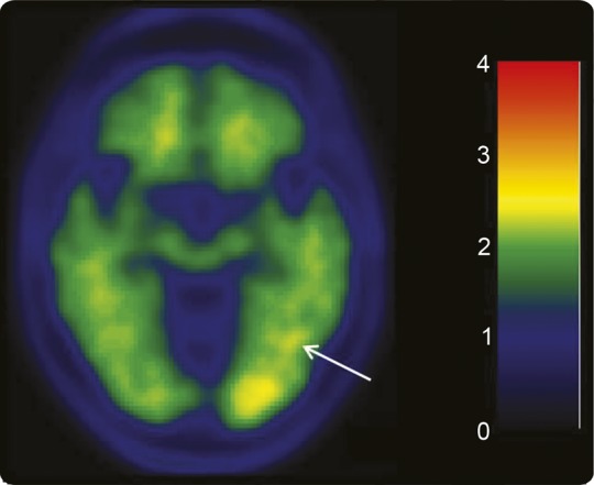
C-11 Pittsburgh compound B PET (month 24) shows focal amyloid deposits in the left temporal and occipital lobe. Arrow indicates increased uptake in the location of the microhemorrhages.
Footnotes
Author contributions: Mekala R. Raman: drafting/revising the manuscript, study concept or design, analysis or interpretation of data, accepts responsibility for conduct of research and final approval. Heather J. Wiste: analysis or interpretation of data, accepts responsibility for conduct of research and final approval, statistical analysis. Matthew L. Senjem: drafting/revising the manuscript, analysis or interpretation of data, accepts responsibility for conduct of research and final approval, contribution of vital reagents/tools/patients, acquisition of data. Chadwick P. Ward: analysis or interpretation of data, accepts responsibility for conduct of research and final approval, acquisition of data. Clifford R. Jack: drafting/revising the manuscript, study concept or design, analysis or interpretation of data, accepts responsibility for conduct of research and final approval, acquisition of data, study supervision, obtaining funding. Kejal Kantarci: drafting/revising the manuscript, study concept or design, analysis or interpretation of data, accepts responsibility for conduct of research and final approval, study supervision, obtaining funding.
Study funding: No targeted funding reported.
Disclosure: M. Raman, H. Wiste, M. Senjem, and C. Ward report no disclosures relevant to the manuscript. C. Jack provides consulting services for Janssen. He receives research funding from the NIH (R01-AG011378, R01-AG041851, R01-AG037551, U01-HL096917, U01-AG032438, U01-AG024904) and the Alexander Family Alzheimer's Disease Research Professorship of the Mayo Foundation. K. Kantarci serves on the data safety monitoring board for Pfizer Inc., Janssen Alzheimer Immunotherapy, Takeda Global Research & Development Center, Inc., and is funded by the NIH (R01AG040042 [PI], Mayo Clinic Alzheimer's Disease Research Center/Project 1 P50 AG16574/P1 [PI], P50 AG44170/Project 2 [PI], and R01 AG11378 [Co-I]). Go to Neurology.org for full disclosures.
References
- 1.Sperling RA, Jack CR, Jr, Black SE, et al. Amyloid-related imaging abnormalities in amyloid-modifying therapeutic trials: recommendations from the Alzheimer's Association Research Roundtable Workgroup. Alzheimers Dement 2011;7:367–385. [DOI] [PMC free article] [PubMed] [Google Scholar]
- 2.Greenberg SM, Grabowski T, Gurol ME, et al. Detection of isolated cerebrovascular beta-amyloid with Pittsburgh compound B. Ann Neurol 2008;64:587–591. [DOI] [PMC free article] [PubMed] [Google Scholar]