Abstract
The aim of this study was to identify the developmental stages of Rubus alceifolius and to determine one or more characteristic morphological markers for each stage. The developmental reconstitution method used involved a detailed description of many individuals throughout the different stages of growth, from germination to the development of an adult shoot capable of fruiting. Results revealed that R. alceifolius passes through five developmental stages that can be distinguished by changes in several morphological markers such as internode length and diameter, pith diameter and plant shape. This analysis indicated that R. alceifolius has a heteroblastic developmental pattern, midway between that of a bush and a liana. Moreover, results showed that this species taps environmental resources early in its development, i.e. foliarization is high (the foliar component overrides the caulinary component) and an autotrophic stage is rapidly reached, whereas it ‘explores’ the environment during the adult stage, i.e. axialization is substantial (the caulinary component overrides the foliar component) and autotrophy occurs at a later stage. The morphological markers identified could benefit land‐use managers attempting to control this species before it reaches its optimum developmental stage.
Key words: Rubus alceifolius, architecture, morphometric markers, metamer and primary structure approach, foliarization, axialization, invasive bramble, bush, liana, Réunion island
INTRODUCTION
Plants respond sensitively to the environment not by moving their bodies but by varying their physiology and growth (Waller, 1986). Architectural analysis provides access to the global architecture of a plant and its spatiotemporal development (Hallé and Oldeman, 1970; Edelin, 1977, 1984; Hallé et al., 1978; Barthélémy et al., 1989). It consists of a structural description of individuals that have reached various stages of development in differing environments. This approach involves an a posteriori reconstitution of the development of each individual and is based on different morphological markers, thus highlighting the chronological developmental patterns of each structure assessed (e.g. Day et al., 1997; Nicolini, 1998; Nicolini and Chanson, 1999; Sabatier and Barthélémy, 1999; Heuret et al., 2000; Lauri and Kelner, 2001; Nicolini et al., 2001).
A recent morphometric approach used by Lauri (1988, 1991; Lauri and Térouanne, 1991, 1995) operates on another level, focusing on the structure of the caulinary axis components, the metamer (White, 1979, 1984), and variations in the balance between the different components (leaves, nodes and internodes). Considering the site of leaf projection and its subjacent basal extension, Lauri (1988, 1991) and Lauri and Térouanne (1991, 1995) suggested that the foliar component may override the caulinary component (= foliarization), whereas during development towards a more axialized metamer the caulinary component overrides the foliar component (= axialization). In these studies, internode diameter was used as the stem parameter (Lauri, 1988, 1991; Lauri and Térouanne, 1991, 1995; Lauri and Kelner, 2001). However, the shoot axis is represented by the central pith region (primary structure) that is corticated by surrounding leaf tissues (secondary structure) (Troll and Rauh, 1950; Kaplan, 2001). Both structures must therefore be considered when studying stem parameters, as demonstrated successfully for Fagus sylvatica, a temperate tree (Nicolini and Chanson, 1999). Therefore, pith has been used like the main stem parameters in this study.
Architectural analysis can be more meaningful when quantitative measurements are combined with morphological description. Trees of economic importance have been studied often (e.g. Heuret, 2000; Lauri and Kelner, 2001; Nicolini et al., 2001; Sabatier and Barthélémy, 2001), but other plant groups are comparatively poorly studied, especially those with life forms intermediate between those of trees and herbaceous perennial plants.
Weakly lignified brambles belonging to the genus Rubus constitute a unique biological model, having high specific diversity throughout the world and a unique growth habit and architecture. Their development and growth capacity are such that many species, when translocated by man to new areas, become highly invasive.
Some studies have documented the developmental stages of different species of Rubus: R. fruticosus (Barnola, 1971a, b; Amor, 1974; Amor and Richardson, 1980), R. ulmifolius (Heslop‐Harrison, 1959) and R. idaeus (Hudson, 1959; Barnola, 1970). However, these descriptive studies have generally focused on one architectural parameter (for example, rooting of the stem apex).
Among the invasive Rubus species in the world, R. alceifolius, native to eastern Asia (northern Vietnam to Java), became a major weed in Queensland, Australia, and the Indian Ocean Islands, Réunion–Mauritius–Mayotte–Madagascar, after its recent introduction by humans. A recent genetic study of R. alceifolius has shown that the plant invaded Réunion Island from a single clone (Amsellem et al., 2000).
To gain a better understanding of the invasiveness of this plant, growth and developmental stages of this plant were documented using architectural and morphological analyses by addressing the following questions: (1) does Rubus alceifolius have a particular growth habit; (2) does the morphometric approach, particularly of the primary structures, provide markers to characterize the developmental stages of this plant; and (3) can these markers be used to describe the development of each individual and thus predict its spatial invasive potential?
MATERIALS AND METHODS
Study site
The study was carried out on Réunion, a volcanic island in the Indian Ocean (21°06′S, 55°32′E). Rubus alceifolius is a species that thrives in wet habitats. It proliferates on the eastern and southeastern coasts, where rainfall is heaviest, from sea level to 1700 m a.s.l., whereas it is only found at 500 m a.s.l. and above in gullies along the western coast. Measurements were carried out at two sites: (1) Grand Etang, located in the eastern region, between 500 and 550 m a.s.l., where the annual average rainfall is high (6306 mm year–1) and the annual average temperature is 20·4 °C (source France Météo, year 2000); and (2) Mare Longue, located in the southern region at 500 m a.s.l. Precipitation is also abundant (approx. 4000 mm y–1) and the annual average temperature ranges from 19 to 20 °C.
For the purposes of our analysis, we did not differentiate between the two study sites.
Biological material
All descriptions were based on individual plants that had been harvested near gaps in the forest, and which had reached the following specific height stages: stage a, 1–10 cm seedlings; stage b, 30–50 cm individuals; stage c, 90–120 cm individuals; stage d, 170–220 cm individuals; and stage e, individuals whose height ranged from 5 m to the top of the canopy. Five plants were studied for each stage. One hundred other individuals were also assessed for more general features.
Observation methods and parameters studied
An individual of R. alceifolius grown from seed can be regarded as stock (also called rootstock) that bears shoots of various sizes, which are emitted successively during plant development. On each of these axes, the total number of nodes was counted, and each component of the successive metamers (stem part and leaf) was measured. Data presented are: (1) the global trend for each descriptor (internode length, pith and internode areas, leaf form, etc.) of the most representative individual per stage, to give an overall view; and (2) tables presenting mean values with statistical comparisons for all individuals. In these tables, we do not present all metamer data: according to architectural descriptions, the main plant axis was divided into four different parts (1–4) and only data recorded for four metamers selected at a median position in each part are presented.
Description of stem components
On each of the axes, the total number of nodes was counted and internode lengths were measured from base to tip. Internode length may be considered to be a good marker of the growth rate and internal rhythm of plants (Pieters, 1983, 1985; Lauri, 1988; Nicolini, 1998). The diameter of the axis at each internode level was measured. The internode diameter measurement takes into account the primary (pith) and secondary structures (surrounding tissues), especially for the oldest individuals. To assess the intrinsic potential of the primary developing meristem on the basis of the structure of its products (leaves, stems), it is essential to be able to determine the share of primary stem tissues that results from its functioning. Although the structure of each individual was analysed at a macroscopic level, a posteriori we decided to retain only the primary structure part. The pith of Rubus alceifolius stems was clearly visible, so, using the naked eye or with the aid of a dissecting microscope, it was possible to measure the pith diameter using a small ruler. Based on pith diameter measured in the middle of each internode (d) and internode length (l), pith volume at each internode was calculated from the formula πd2l.
Leaf description
After numbering and dehydrating the leaves located at each node of the axis, leaf area was measured using an image analysis software program (CorelDRAW 9). Leaf thickness was also measured using a calliper (0·01 cm accuracy). Using these two parameters, leaf volume was calculated (leaf area × thickness). The length of the midrib (RL1) and of a secondary rib (RL2) was also measured, and the ratio RL2 : RL1 calculated. This ratio accounts for the shape of R. alceifolius leaves and their developmental patterns.
RESULTS
Individual plant architecture
Seeds germinated along forest paths and in clearings. About 3 months after germination, seedlings (stage a, Fig. 1A), which were 1–10 cm high, had a non‐ramified thin main axis (2·39 ± 0·33 mm diameter) that was vertical for smaller plants and slightly inclined for larger ones. This axis comprised successive nodes, each having a simple leaf, separated by internodes of variable length. Leaves were arranged around the stem according to an alternate‐spiral phyllotaxis arrangement with a 2/5 index. At each leaf axil there was a small lateral bud whose length depended on the associated leaf position: the largest buds were located at the base of the epicotyl and were 2–3 mm long, whereas those located on the rest of the axis were less than 1 mm long.
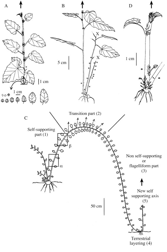
Fig. 1. Architecture of different R. alceifolius individuals at different developmental stages. A, Young stage a. The axis shown was derived from a seed. B, Intermediate stage b. C, Adult stage e; the lianescent axis tip can root and form a new axis (D).
Larger individuals (30–50 cm height) comprised several axes of different heights. Assessment of one individual revealed the organization described below (stage b, Fig 1B). The smallest axis had stopped developing and was usually dry. This was often the axis that issued from the seed; it had less‐developed internodes (1–5 cm). At its base, it had a more developed axis that was still alive and comprised longer internodes (2–8 cm) whose development ceased following necrosis of the terminal meristem. This axis had stopped developing, was dry, like the axis that issued from the seed, and bore a higher axis with longer internodes (4–11 cm) that were still growing at the time of examination. It had the overall appearance of a stock bearing vertical axes at different stages of development. The leaves of the live axes were in an alternate‐distichous arrangement on a plane on the stem. This is the only foliar arrangement that occurred thereafter.
This general organization was also noted in larger individuals (stage c, 90–120 cm; stage d, 170–220 cm). Larger‐sized structures appeared at this stage (basal diameter: stage c, 5·2 ± 0·44 mm; stage d, 8·5 ± 1·70 mm). The mean basal axis diameter differed significantly (Mann–Whitney U test) among developmental stages.
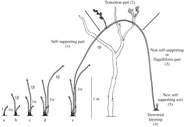
Stage e individuals were organized differently (Fig. 1C). Examination of a representative individual of this class showed that it had several axes of different heights starting at a stock of greater dimensions than that of the previous stage. All axes except one were poorly developed (around 2 m) and were confined to within the understorey. There was a major vertical axis at the basal part of the plant with a horizontal axis at the distal part. Some axes had stopped growing, whereas others continued to grow a little. They were all sterile at the time of examination, except the most highly developed axis that had reached the canopy. This 14‐m axis was organized differently to the other axes. Based on the organization of the main axis (growth direction and branching) from the base to the apical part, it could be divided into five parts as described in Fig. 1C. The first part was vertical and rigid from the stock up to the lower level of the canopy (part 1α and β: corresponding to the unique part observed in the younger stages, Fig. 1C); the second part was curved and grew within the canopy with lateral flowering and fruiting branches (part 2); the third part was supple and hung from canopy to the ground (part 3); the fourth part was rooted in the ground (part 4, Fig. 1D); and the fifth part was vertical, still growing, and constituted a new vertical axis (part 1 of a new axis, Fig. 1D). As part 1 varied between the first developmental stages, we distinguished two zones, α and β (called part 1α and part 1β).
Development of the metameric caulinary and foliar components
Internode lengths increased markedly from the axis base to its tip (Table 1). For all individuals described in stages a–d, this pattern could be divided into two stages (Fig. 2A): in the first (part 1α), internode lengths increased from the base to the median part of the axis, whereas in the second stage (part 1β) an inverse pattern was noted from the median to the distal part of the axis. Stage e individuals also showed this pattern in the first part. However, these individuals had shorter internodes until the median part of part 2, then the distance increased again until the beginning of part 3. On this part of the axis, the size of the internodes remained constant before decreasing suddenly. Internodes were very short on the rooted part (part 4). Longer internodes were then found on a new vertically growing axis. A comparison of the mean longest internodes (part α) revealed a significant increase in length from one developmental stage to another (Table 1; Fig. 2A).
Table 1.
Mean internodes, pith diameters of metamers and ratio of secondary rib length : midrib length (RL2/RL1) of each leaf corresponding to these metamers, located in different axis parts for individuals at different developmental stages
| Stage | Part | Internode length (mm) | Pith diameter (mm) | RL2 : RL1 |
| a | 1 | 10·1 ± 4·13a | 0·89 ± 0·10a | 0·35 ± 0·05a |
| b | 1α | 54·6 ± 8·96b | 1·19 ± 0·15b | 0·43 ± 0·02bc |
| 1β | 37·3 ± 9·24c | 1·00 ± 0·16ab | 0·41 ± 0·02b | |
| c | 1α | 127·8 ± 18·31d | 1·57 ± 0·23c | 0·48 ± 0·02d |
| 1β | 57·4 ± 12·01b | 1·09 ± 0·12b | 0·45 ± 0·03c | |
| d | 1α | 164·6 ± 22·73ef | 4·51 ± 0·53d | 0·56 ± 0·01e |
| 1β | 102·4 ± 11·25g | 2·54 ± 0·30e | 0·53 ± 0·03f | |
| e | 1α | 266·0 ± 22·66h | 9·73 ± 0·96f | 0·62 ± 0·02g |
| 1β | 178·0 ± 9·31f | 8·58 ± 1·13f | 0·62 ± 0·03g | |
| 2 | 140·3 ± 15·08de | 6·45 ± 1·23g | 0·60 ± 0·04eg | |
| 3 | 232·3 ± 8·97i | 3·02 ± 0·31h | 0·62 ± 0·03g | |
| 4 | 3·6 ± 0·47j | 6·09 ± 0·55g |
a–e Represent the five developmental stages analysed. One axis can be divided into four distinct parts: an ascending or self‐supporting part (1), a transition part (2), a bending or lianescent part (3) and a terrestrial layering part (4). Only part 1 was subdivided further into a median part, α, and a part located at the end of part 1, β. In each part (1α, 1β, 2, 3, 4), four metamers found in a median position were selected. Means of the parameters were then calculated on the basis of these metamers. A 95 % confidence interval was associated with each mean. The metamer means for the five plant groups (Mann–Whitney U test) are represented by the letters a, b, c, d and e. Different superscript letters within a column indicate that the corresponding distribution is significantly different at the 95 % threshold.
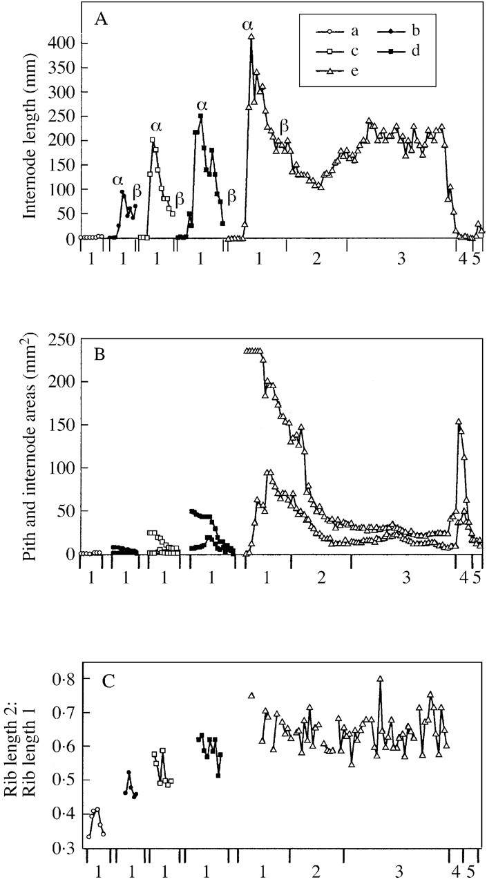
Fig. 2. Variations in characters analysed on successive metamer‐forming axes for individuals at different developmental stages. The characters are: internode length (A), pith and total internode area (B), and the ratio of secondary rib length : midrib length (C). Variations occurred from the base towards the axis tip. Each line represents a different developmental stage: stage a (open circles), stage b (closed circles), stage c (open square), stage d (closed square) and stage e (triangle). In B, the lower curve represents the cross‐sectional area of the pith (primary structure), whereas the upper curve represents the total cross‐sectional area for an internode (primary and secondary structure). The area between the two curves gives an approximation of the stem tissue area (conductance, support and storage), mainly of secondary origin. The different axis parts are represented on the abscissa. The y‐axis corresponds to five distinct parts: an ascending or self‐supporting part (1), a transition part (2), a bending or flagelliform part (3) and a terrestrial layering part (4). Part 5 corresponds to the base of a new ascending or self‐supporting part. Stages a–d have axes formed only with part 1 (ascending or self‐supporting part).
Changes in pith diameter at successive internodes showed the same pattern as that for internode length along the axis and also between developmental stages (Table 1). However, qualitative differences were sometimes noted with respect to individuals at stage e. The pith diameter decreased in part 3 whereas it thickened substantially in the rooted part (part 4).
Two curves (Fig. 2B) provided information about the tissue area (conductance, support and storage) in the stem. This tissue area was greater in the self‐supporting part at each developmental stage. The tissue area increased progressively between stages. At the adult stage (stage e), the minimum value was obtained in the lianescent part.
The ratio between the length of rib 2 (LN2) and the length of rib 1 (LN1) (an indicator of leaf shape) increased significantly between stages (Table 1; Fig. 2C), but it varied little along an axis.
Comparative development of the metameric caulinary and leaf volumes
Changes in the metameric structure can also be considered through a comparison of the development of different volume components (Table 2; Fig. 3). Pith and leaf metameric volumes in part 1α significantly increased between developmental stages (Table 2). Figure 3A–E also highlights the fact that the pith volume increased more rapidly than the leaf volume between developmental stages. The leaf volume : pith volume ratio gradually changed from 829 ± 373 at stage a to 0·45 ± 0·2 at stage e.
Table 2.
Mean values for leaf volume, pith volume and the leaf volume : pith volume ratio for metamers located in different axis parts for individuals at different developmental stages
| Stage | Part | Pith volume (mm3) | Leaf volume (mm3) | Leaf volume: internode pith volume |
| a | 1 | 0·8 ± 0·35a | 281 ± 70a | 829·23 ± 373·07a |
| b | 1α | 68·2 ± 21·4b | 1074 ± 202b | 21·90 ± 6·60b |
| 1β | 31·3 ± 15·1c | 689 ± 108c | 30·49 ± 8·67c | |
| c | 1α | 297·4 ± 141·1d | 2046 ± 320d | 12·03 ± 3·76d |
| 1β | 59·8 ± 14·7b | 2210 ± 466d | 41·30 ± 11·24c | |
| d | 1α | 2913·5 ± 956e | 5592 ± 854e | 2·96 ± 0·94e |
| 1β | 569·4 ± 203f | 4016 ± 703f | 8·74 ± 2·07d | |
| e | 1α | 21 129·0 ± 4360g | 6299 ± 1475e | 0·45 ± 0·20f |
| 1β | 11 085·7 ± 2617h | 9284 ± 1392g | 1·21 ± 0·41g | |
| 2 | 5908·7 ± 2066e | 7760 ± 1020g | 3·49 ± 1·80e | |
| 3 | 1694·3 ± 303i | 1461 ± 356b | 0·90 ± 0·18g | |
| 4 | 105·9 ± 21·6j | →0 | →0 |
a, b, c, d and e represent the five developmental stages analysed (see Table 1 for part codes and for explanation of statistical analysis).
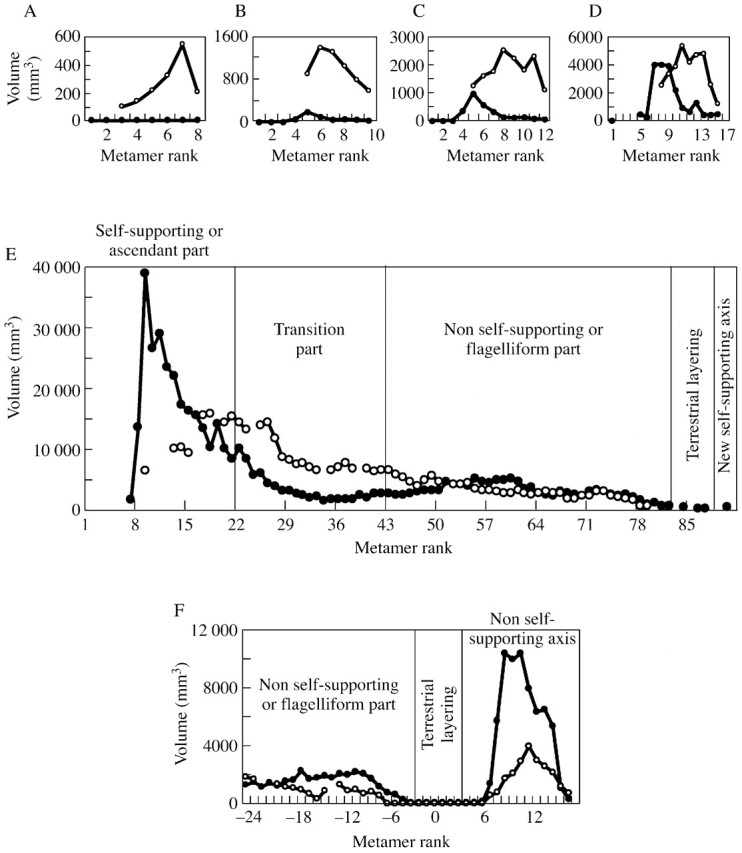
Fig. 3. Patterns of variation in pith volume and leaf volume for successive metamer‐forming axes of individuals at different developmental stages. Variations occurred from the base towards the axis tip. Each graph represents a developmental stage: stage a (A), stage b (B), stage c (C), stage d (D) and stage e (E). F, Variations at the end of part 3 (flagelliform part) for an individual at stage e. Closed circles, pith volume variations; open circles, variations in leaf volume.
The pith volume, leaf volume and the leaf volume : pith volume ratio varied significantly (Table 2; Fig. 3) along an axis at all developmental stages. Considering, for instance, an individual at stage e (Fig. 3E), pith volume predominated over leaf volume (ratio approx. 0·2) in part 1α, whereas in part 1β pith volume decreased while successive leaf volumes increased. This pattern continued in part 2 where the leaf volume finally predominated over the pith volume (ratio approx. 4). The pattern was then reversed, i.e. pith volume increased substantially, whereas leaf volume decreased gradually. In part 3, medullary and leaf volumes were relatively similar and the leaf volume : pith volume ratio oscillated around 1.
Figure 3F shows medullar and foliar patterns at the end of part 3, in part 4 and at the beginning of the new part 1. This new part corresponds to the creation of a new vertical axis that was growing during the observations; patterns noted in this part were similar to those of part 1 of the main axis.
DISCUSSION
All plants go through a series of developmental changes from non‐reproductive to reproductive stages. In many plants, shifts in development are subtle, involving small changes in leaf size and shape. In others, the changes are so marked that early and late stages have been identified as separate species. Goebel (1900) recognized these differences in the extent of developmental change, designating the former species as homoblastic and the latter as heteroblastic. Since Goebel’s treatment, the heteroblastic developmental concept has been extended to species in which the juvenile‐to‐adult transition is more gradual, which is true of the majority of plants (Allsopp, 1967), with more developmental traits being considered: leaf anatomy, phyllotaxis, internode length, stem thickness, shoot apex structure and zonation, trophic response, regenerative capacity, physiology and reproductive status (Troll, 1939; Doorenbos, 1965; Richards, 1983; Lee and Richards, 1991).
Development of Rubus alceifolius, a species with an organization midway between a bush and a liana
The development of R. alceifolius can be summarized as follows. After seed germination in an opening in the forest canopy, the plant forms a small vertical leafy axis with reduced development. The development of this first axis is followed shortly by the formation of a new vertical axis from a lateral meristem located at the basal part of the axis (basitonic ramification). This new axis has larger dimensions than the preceding one, and so on. While the older shoots dry out, new self‐supporting shoots, with a vertical part and a more or less oblique part, are formed from an increasingly larger stock. This understorey developmental phase, based on the basitonic ramification phenomenon, is characteristic of bush development (Champagnat, 1947; Crabbé, 1976), and has previously been observed in other Rubus species (Heslop‐Harrison, 1959; Barnola, 1970).
At a certain developmental stage, characterized by a relatively large stock containing sufficient reserves with a well‐developed root system able to extract essential nutrient reserves from the soil, a new shoot is formed by the plant and, after a self‐supporting phase (part 1, self‐supporting part), it reaches the top of the canopy. From there its growth direction changes, switching gradually from vertical to horizontal, forming an arch (part 2, transition part), with the formation of flowering twigs. When the shoot reaches the mature stage, it expresses a new whole‐organism potential, represented by a positive geotropic growth phase during which the apical meristem produces a supple axis (part 3, flagelliform part) which reaches the ground and forms roots (part 4, terrestrial layering). This stage, similar to that observed in other Rubus species (Heslop‐Harrison, 1959; Barnola, 1971a, b), is followed by the formation of a new self‐supporting axis from the same terminal meristem.
The change from a self‐supporting developmental phase to a non‐self‐supporting phase is a key feature of liana development (Caballé, 1998), and also of R. alceifolius. This was confirmed by our observation of a growing axis during the descending phase: the terminal meristem produced an axis with long and fine internodes, which Madison (1977) and Blanc (1980) referred to as a flagelliform axis for tropical liana development in the Araceae family. A vegetative mode of propagation and a capacity to grow roots from flagelliform axes with or without soil contact are two characteristics of R. alceifolius that are generally specific to lianas (Caballé, 1980), thus confirming that this species belongs to the liana biotype.
The lianescent propagation mode is acquired gradually, which is also the case with respect to the acquisition of maturity and other lower profile potentials that are not expressed during the initial stages of plant development. The mature reproductive stage is reached and the lianescent character is expressed after an installation phase, a phenomenon described in many plant species (e.g. Heuret et al., 2000; Nicolini et al., 2000), and referred to as establishment growth by Tomlinson and Zimmermann (1966) or heteroblastic development by Goebel (1900).
As is the case for many plant species (Champagnat, 1947; Heslop‐Harrison, 1959; Barnola, 1970; Crabbé, 1976), in a favourable environment the installation phase for R. alceifolius is expressed by the formation of more and more developed axes, constituted of longer and longer internodes, containing a thicker pith, and different leaf shapes linked with a vigour gradient (Champagnat, 1947). All of these criteria highlight the inevitable transition of the organism from a sterile juvenile stage to a mature adult stage. This transition is under endogenous control, but is modulated by more or less favourable environmental conditions which shorten or lengthen this period (Lee and Richards, 1991). These different markers, located within a well‐defined stage or part, could be efficient indicators of the degree of maturation of R. alceifolius.
Significance of caulinary and foliar metameric patterns and the application of axialization and foliarization concepts to Rubus alceifolius
Development of R. alceifolius, as observed through volumic variations in the primary metameric elements, involved a gradual increase in leaf and pith volumes. Considering axis part 1α, the pith volume increased more rapidly than the leaf volume (Fig. 5A), so that the caulinary component was very low at stage a, whereas it predominated at stage e, during which the organism reached maturity. Moreover, this balance varied along an axis.
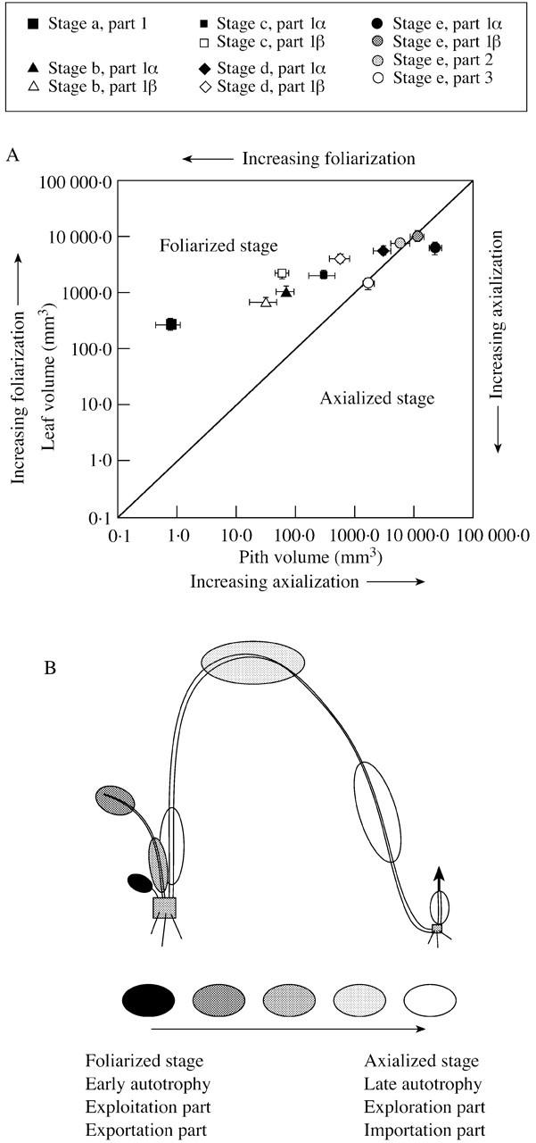
Fig. 5. A, Relationship between mean pith volume and mean leaf volume for metamers forming the different axis parts for individuals at different developmental stages. A 95% confidence interval is associated with each mean. The axes are graded on a logarithmic scale. B, Interpretation of Rubus alceifolius organization and functioning on the basis of the axialization and foliarization concepts. The shading (dark to pale) highlights changes for each developmental stage and along different axes with respect to level of axialization and foliarization, indicating the autotrophic and environment‐tapping or exploring potential of plants during their development. Grey rectangles indicate the presence of a rooted stock.
Using the terms proposed by Lauri (1988), and Lauri and Térouanne (1991, 1995), young plants of R. alceifolius can be described as beginning their growth with a very foliarized stage (the foliar component overrides the caulinary com ponent), then developing towards a more axialized stage (the caulinary component overrides the foliar component), which is expressed at stage e (Fig. 5A), and which corresponds to the acquisition of reproductive capacity.
Assessing maturity using axialization and foliarization concepts
At a given developmental stage, all axis parts were not in the same state. For stage a–d individuals, part 1α was generally less foliarized than part 1β. Similarly, for stage e, part 1 and 3 metamers were rather axialized, whereas those of part 2 were more foliarized. The latter part is the only one bearing fruiting twigs. This association between flowering and foliarized structures has also been described in Alstonia vieillardii (Apocynaceae) by Lauri (1991), who associated flower propensity with the foliarized metamer stage. In this species, flowering starts when the axes formed revert to an original foliarized stage. This reversion cannot solely justify the expression of sexuality as young axes are foliarized but sterile. Sexuality starts only when a certain dimensional value is exceeded (Lauri and Térouanne, 1991, 1995). This trend also applies to R. alceifolius, since the propensity to flower does not affect the axialized part (parts 1 and 3 at stage e) or the foliarized part formed at a young age. To flower, the meristems have to form metamers with a degree of foliarization close to the limit between the foliarized state and the axialized state (Fig. 5A), and with dimensions great enough to induce flowering and ensure fruit development, as was the case in part 2 for stage e individuals. In R. alceifolius, structural maturity (ripeness to flower; Klebs, 1918) can be characterized by a certain leaf quality, and a certain caulinary tissue organization of metamers, as noted in some other plants (Nicolini and Chanson, 1999).
Interpretation of Rubus alceifolius behaviour through axialization and foliarization concepts
The concepts of axialization and foliarization take us back to the interpretation of the balance between stems and leaves, and of the exchanges that occur between them. To what extent can the a posteriori description of R. alceifolius structure inform us about the functioning of the different plant parts, and to what extent can we qualify these respective functions? Previously published observations enable us to further interpret developmental patterns of R. alceifolius.
Studying apple trees, Hansen (1977) and Lakso (1984) showed that short twig leaves, representing foliarized structures (Lauri and Térouanne, 1991; Lauri and Kelner, 2001), start to export carbohydrates 10 d after the onset of growth, whereas rather axialized long shoots (Lauri and Térouanne, 1991; Lauri and Kelner, 2001) do not export carbohydrates until 3–4 weeks after budding. In agreement with Hansen’s (1977) results, Lakso (1984), Johnson and Lakso (1986), Lakso and Corelli‐Grappelli (1992) and Lauri and Kelner (2001) showed that, contrary to long shoots, the capacity of short shoots to precociously export carbohydrates to other parts of the plant could be linked, in part, to the development of a reduced quantity of caulinary tissues by these structures (Johnson and Lakso, 1986). Thereafter, less time is required to complete stem structure formation after lengthening of leafy shoots. Autotrophy and carbohydrate export phases are reached more rapidly in short shoots than in long shoots. Lauri and Kelner (2001) consider that early achievement of autotrophy is a major feature of structures with a high degree of foliarization.
The first developmental stage of R. alceifolius (stage a), corresponding to very reduced axis formation exhibiting high foliarization with reduced secondary growth, can be considered as a short shoot formation; such a structure is likely to reach the autotrophy phase rapidly with subsequent carbohydrate export towards other plant parts (stock, roots). This structure taps the environment more than it explores it.
The inverse pattern is noted in the first stages of shoot formation in stage e individuals. The production phase of axialized part 1α (very thick stem bearing reduced leaves, Fig. 1A) can be considered as long shoot formation—this part of the axis takes longer to reach the autotrophy phase with carbohydrate export towards other plant parts (stock, roots). This structure does not substantially tap the resources of the environment, but it explores it using its own reserves (stock, roots) to meet its needs for important secondary growth (Fig. 3D).
The autotrophic phase of a shoot at stage e is gradually reached after the complete lengthening of part 1α and 1β leaves, and especially of part 2 leaves. This highly foliarized part has a relatively thin axis (Table 1) with limited secondary growth (Fig. 2B), and bears large leaves of high specific weight (weight : area ratio, data not shown) that are well exposed in the canopy. This appears to be the most suitable part for tapping the environment. Its function is ultimately to ensure autotrophy of all shoots by exporting carbohydrates towards the basal parts (part 1) to enable their high secondary growth, towards the stock and roots, and also towards the shoot periphery where part 3 is constructed by the terminal meristem.
The axialized part 3, with a thin flagelliform stem bearing small leaves, can be considered as a structure that reaches the autotrophic phase later, i.e. a structure that explores the environment more than it taps it, probably utilizing carbohydrates from part 2 or other parts of the plant.
Shoots formed during the intermediate developmental stages (stages b–d) exhibit transient stages, as shown in Fig. 5B, which illustrates graphically these concepts with respect to R. alceifolius.
CONCLUSION
The results of this study highlight important aspects of the developmental strategy of this species, which goes through a series of stages from the seedling to the fruiting adult. The growth reconstitution method developed here, using morphological and architectural markers, allowed us to classify Rubus alceifolius as an organism midway between a bush and a liana able to bear fruit. Different stages were identified and characterized by morphological and architectural criteria, which could provide many useful markers to facilitate the identification of this species. The different developmental stages of this plant can be analysed using these markers. They could also be used to determine the growth patterns of this species in order to formulate eradication strategies for invasive plant control programmes.
ACKNOWLEDGEMENTS
This study was conducted under, and financed by, the Region Réunion project, REG/97/0307. The authors thank David Manley, Pascale Besse, Sarah Kirman, Laetitia Baret, Karine and Nicolas Cottis for the English correction and also Professor Donald R. Kaplan and Dr J. Clemens for insightful and constructive criticisms of a first version of the manuscript.
Fig. 4. A typical developmental sequence in Rubus alceifolius. Different parts (1: α and β, 2, 3, 4, 5) are specified for each developmental stage.
Supplementary Material
Received: 10 July 2002; Returned for revision: 2 September 2002; Accepted: 1 October 2002 Published electronically: 21 November 2002
References
- AllsoppA.1967. Heteroblastic development in vascular plants. Advances in Morphogenesis 6: 127–171. [DOI] [PubMed] [Google Scholar]
- AmorRL.1974. Ecology and control of blackberry (Rubus fruticosus L. agg.): II. Reproduction. Weed Research 14: 231–238. [Google Scholar]
- AmorRL, Richardson RG.1980. The biology of Australian weeds. 2. Rubus fruticosus L. Agg. Journal of the Australian Institute of Agricultural Science 46: 87–97. [Google Scholar]
- AmsellemL, Noyer JL, Le Bourgeois T, Hossaert‐McKey M.2000. Comparison of genetic diversity of the invasive weed Rubus alceifolius Poir. (Rosaceae) in its native range and in areas of introduction, using amplified fragment length polymorphism (AFLP) markers. Molecular Ecology 9: 433–455. [DOI] [PubMed] [Google Scholar]
- BarnolaP.1970. Recherche sur le déterminisme de la basitonie chez le framboisier (Rubus idaeus L.). Annales des Sciences Naturelles, Botanique 12: 129–152. [Google Scholar]
- BarnolaP.1971a Recherche sur le déterminisme du marcottage de l’extrémité apical des tiges de ronce (Rubus fructicosus L.). Revue générale de botanique 78: 185–199. [Google Scholar]
- BarnolaP.1971b Recherche sur le déterminisme de la basitonie chez la ronce (Rubus fruticosus). Beiträge zur Biologie der Pflanzen 47: 469–480. [Google Scholar]
- BarthélémyD, Edelin C, Hallé F.1989. Architectural concepts for tropical trees. In: Holm‐Nielsen LB, Baslev H, eds. Tropical forests: botanical dynamics, speciation and diversity London: Academic Press, 89–100. [Google Scholar]
- BlancP.1980. Observations sur les flagelles des Araceae. Andosonia 20: 325–338. [Google Scholar]
- CaballéG.1980. Caractéristiques de croissance et multiplication végétative en forêt dense du Gabon de la ‘liane à eau’ Tetracera alnifolia Willd. (Dilleniaceae). Andosonia 19: 467–475. [Google Scholar]
- CaballéG.1998. Le port autoportant des lianes tropicales: une synthèse des stratégies de croissance. Canadian Journal of Botany 76: 1703–1716. [Google Scholar]
- ChampagnatP.1947. Les principes généraux de la ramification des végétaux ligneux. Revue Horticole 2143: 335–341. [Google Scholar]
- CrabbéJ.1976. Surprises et promesses des recherches sur l’élaboration de la forme chez les végétaux ligneux. Avant propos: arbres et buissons. Le fruit Belge 374: 85–91. [Google Scholar]
- DayJS, Gould KS, Jameson PE.1997. Vegetative architecture of Elaeocarpus hookerianus Transition from juvenile to adult. Annals of Botany 79: 617–624. [Google Scholar]
- DoorenbosJ.1965. Shortening the breeding cycle of rhododendron. Euphytica 4: 141–146. [Google Scholar]
- EdelinC.1977. Images de l’architecture des conifères. PhD Thesis, University of Montpellier II, Montpellier, France. [Google Scholar]
- EdelinC.1984. L’architecture monopodiale: l’exemple de quelques arbres d’Asie tropicale. PhD Thesis, University of Montpellier II, Montpellier, France. [Google Scholar]
- GoebelK.1900. Organography of plants. Part I. General organography. Oxford: Clarendon Press (translated by IB Balfour). [Google Scholar]
- HalléF, Oldeman RAA.1970. Essai sur l’architecture et la dynamique de croissance des arbres tropicaux. Paris: Masson. [Google Scholar]
- HalléF, Oldeman RAA, Tomlinson PB.1978. Tropical trees and forests An architectural analysis. New York: Springer‐Verlag. [Google Scholar]
- HansenP.1977. Carbohydrate allocation. In: Landsberg JJ, Cutting CV, eds. Environmental effects on crop physiology London: Academic Press, 247–258. [Google Scholar]
- Heslop‐HarrisonY.1959. Natural and induced rooting of the stem apex in Rubus Annals of Botany 23: 307–318. [Google Scholar]
- HeuretP, Barthélémy D, Nicolini E, Atger C.2000. Analyse des composantes de la croissance en hauteur et de la formation du tronc chez le chêne sessile, Quercus petraea (Matt.) Liebl. (Fagaceae) en sylviculture dynamique. Canadian Journal of Botany 78: 361–373. [Google Scholar]
- HudsonJP.1959. Effects of environment on Rubus idaeus L. I. Morphology and development of the raspberry plant. Journal of Horticultural Science 34: 163–169. [Google Scholar]
- JohnsonRS, Lakso AN.1986. Carbon balance model of growing apple shoot: II. Simulated effects of light and temperature on long and short shoots. Journal of the American Society for Horticultural Science 111: 164–169. [Google Scholar]
- KaplanDR.2001. The science of plant morphology: definition, history, and role in modern biology. American Journal of Botany 88: 1711–1741. [PubMed] [Google Scholar]
- KlebsG.1918. Über die Blütenbildung von Sempervivum. Flora 111: 128–151. [Google Scholar]
- LaksoAN.1984. Leaf area development patterns in young pruned and unpruned apple trees. Journal of the American Society for Horticultural Science 109: 861–865. [Google Scholar]
- LaksoAN, Corelli‐Grappadelli L.1992. Implications of pruning and training practices to carbon partitioning and fruit development in apple. Acta Horticulturae 322: 231–239. [Google Scholar]
- LauriPE.1988. Le mouvement morphogénétique. Approche morphométrique et restitution graphique. L’esemple de quelques plantes tropicales. PhD Thesis, University of Montpellier II, Montpellier, France. [Google Scholar]
- LauriPE.1991. Données sur le contexte végétatif lié à la floraison chez le cerisier (Prunus avium). Canadian Journal of Botany 70: 1848–1859. [Google Scholar]
- LauriPE, Kelner JJ.2001. Shoot type demography and dry matter partitioning. A morphometric approach in apple (Malus × domestica). Canadian Journal of Botany 79: 1270–1273. [Google Scholar]
- LauriPE, Térouanne E.1991. Eléments pour une approche morphométrique de la croissance végétale et de la floraison: le cas d’espèces tropicales du modèle de Leeuwenberg. Canadian Journal of Botany 69: 2095–2112. [Google Scholar]
- LauriPE, Térouanne E.1995. Analyse de la croissance primaire de rameaux de pommier (Malus × domestica Borkh.) au cours d’une saison de végétation. Canadian Journal of Botany 73: 1471–1489. [Google Scholar]
- LeeDW, Richards JH.1991. Heteroblastic development in vines. In: Putz EF, Mooney HA, eds. The biology of vines. Cambridge: Cambridge University Press. [Google Scholar]
- MadisonM.1977. A revision of Monstera (Araceae). Contributions from the Gray Herbarium of Harvard University 207: 3–100. [Google Scholar]
- NicoliniE.1998. Architecture et gradients morphogénétiques chez de jeunes hêtres (Fagus sylvatica L.) en milieu forestier. Canadian Journal of Botany 76: 1232–1244. [Google Scholar]
- NicoliniE, Caraglio Y.1994. L’influence de divers caractères architecturaux sur l’apparition de la fourche chez Fagus sylvatica L., en fonction de l’absence ou de la présence d’un couvert. Canadian Journal of Botany 72: 1723–1734. [Google Scholar]
- NicoliniE, Chanson B.1999. La pousse courte, un indicateur du degré de maturation chez le hêtre (Fagus sylvatica L.). Canadian Journal of Botany 77: 1539–1550. [Google Scholar]
- NicoliniE, Barthélémy D, Heuret P.2000. Influence de la densité du couvert forestier sur le développement architectural de jeunes chênes sessiles, Quercus petraea (Matt.) Liebl. (Fagaceae), en régénération forestière. Canadian Journal of Botany 78: 1531–1544. [Google Scholar]
- NicoliniE, Chanson B, Bonne F.2001. Stem growth and epicormic branch formation in understorey beech trees (Fagus sylvatica L.). Annals of Botany 87: 737–750. [Google Scholar]
- PietersGA.1983. Growth of Populus euramericana Physiologia Plantarum 57: 455–462. [Google Scholar]
- PietersGA.1985. Effects of irradiation level on leave growth of sunflower. Physiologia Plantarum 65: 263–268. [Google Scholar]
- RichardsJH.1983. Heteroblastic development in the water hyacinth, Eichhornia crassipes Solms. Botanical Gazette 144: 247–259. [Google Scholar]
- SabatierS, Barthélémy D.1999. Growth dynamics and morphology of annual shoots, according to their architectural position, in young Cedrus atlantica (Endl.) Manetti ex Carriere (Pinaceae). Annals of Botany 84: 387–392. [Google Scholar]
- SabatierS, Barthélémy D.2001. Bud structure in relation to shoot morphology and position on the vegetative annual shoots of Juglans regia L. (Juglandaceae). Annals of Botany 87: 117–123. [DOI] [PMC free article] [PubMed] [Google Scholar]
- TomlinsonPB, Zimmermann MH.1966. Anatomy of the palm Rhapis excelsa III. Juvenile phase. Journal of the Arnold Arboretum 47: 301–312. [Google Scholar]
- TrollW.1939. Vergleichende Morphologie der höheren Pflanzen. Erster Band:Vegetationsorgane. Teil 2. Gebrüder Borntraeger, Berlin (Reprint, 1967, O. Koeltz, Koeningstein‐Taunus). [Google Scholar]
- TrollW, Rauh W.1950. Das Erstarkungswachstum krautiger Dikotylen, mit besonderer Berücksichtigung der primären Verdickungs vorgänge. Sitzungsberichte der Heidelberger Akademie der Wissen schaften. Mathematisch‐naturwissentschaftliche Klasse. Jahrgang 1950 1 Abhandlung, 1–86. [Google Scholar]
- WallerDM.1986. The dynamics of growth and form. In: Crawley MJ, ed. Plant ecology Oxford: Blackwell Scientific Publications, 291–320. [Google Scholar]
- WhiteJ.1979. The plant as a metapopulation. Annual Review of Ecology and Systematics 10: 109–145. [Google Scholar]
- WhiteJ.1984. Plant metamerism. In: Dirczo R, Sarukhan J, eds. Perspectives on plant ecology. Sunderland: Masson, Dirczo, Sarukhan Publishers. [Google Scholar]
Associated Data
This section collects any data citations, data availability statements, or supplementary materials included in this article.


