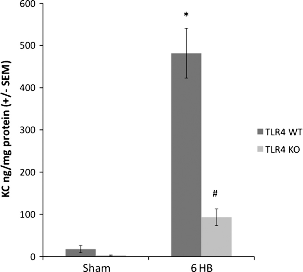
Fig. 5. Mouse lung keratinocyte-derived chemokine.
Lung KC levels measured by ELISA were significantly more increased 6 h after burn in TLR4 WT than TLR4 KO. Lung KC levels were minimal in both sham animal groups. Keratinocyte-derived chemokine levels were significantly increased in TLR4WT animals 6 h after burn (*P < 0.001 compared with both TLR4 WT sham and TLR4 KO sham). Although TLR4 KO animals did show a significant increase in KC levels over both sham groups 6 h after burn (#P < 0.01), the KO deletion led to a significantly lower production of KC compared with TLR4 WT 6 h after burn (* = P < 0.001). At least four animals per group were used.

