


Abstract
Objective
The purpose of this case report is to describe the use of chiropractic care for a patient with neurogenic heterotopic ossification of the anterior longitudinal ligament in the cervical spine and soft tissues of the right hip after a traumatic brain injury and right femur fracture.
Clinical Features
A 25-year-old military officer was referred to a hospital-based chiropractic clinic with complaints of pain and stiffness of the neck and back along with reduced respiratory excursions that began several months after a motor vehicle accident in which he had a traumatic brain injury. The patient had a fractured right femur from the accident, which had since been treated surgically, but had complications of heterotopic ossification in the soft tissues of the hip. His overall pain level was 3 of 10 on a verbal pain scale during use of oxycodone HCL/acetaminophen. Chest excursion was initially measured at .5 cm.
Intervention and Outcome
With the intent to restore respiratory chest motion and to reduce the patient's back and neck pain, the patient was placed on a program of chiropractic and myofascial manipulation, exercise therapy, and respiratory therapy. After a year of care, the patient rated overall pain at 3 of 10 verbal pain scale level but was no longer taking medications for pain and an increase in respiratory chest excursions measured at 3.5 cm.
Conclusion
This case demonstrated that chiropractic treatment provided benefit to a patient with heterotopic ossification concurrent with musculoskeletal pain.
Key indexing terms: Ossification, Heterotopic, Chiropractic, Military personnel, Brain injuries, Manipulation, Spinal
Introduction
Heterotopic ossification (HO) is the anomalous formation of mature bone in soft tissues.1 It should not be mistaken for soft tissue calcification (the accrual of calcium salts in soft tissues). Soft tissue calcification lacks the presence of osteoblasts and mature bone formation. Three types of HO have been identified and described: myositis ossificans progressiva (a rare metabolic disease), traumatic HO (resulting from trauma to the affected region), and neurogenic HO (NHO) (which we address in this case report). Little is known about the pathogenesis of NHO, but there appears to be a clinical relationship between traumatic brain injury (TBI) and predisposition to NHO.2 This is believed to be due to osteoinductive factors released from the site of the brain trauma. Heterotopic ossification also commonly occurs after spinal cord injuries and burns.3,4 One study revealed that taking serum from polytraumatized patients and introducing it to osteoblasts and fibroblasts in vitro resulted in enhanced osteogenesis.5
Heterotopic ossification may occur after trauma or surgery, but NHO is more prevalent in high-energy combat-related injuries of military service members.6–8 Neurogenic HO is a common complication of trauma to the brain or spinal cord, and although the epidemiology is not fully understood, it can clearly be surmised that high-velocity neurotrauma greatly increases the likelihood of NHO. In combat-related blast injuries to limbs, the prevalence of HO is 64.6%, and there is a strong contributory relationship with TBI and long bone fractures.8 The increase of combat veterans with NHO entering civilian health care markets, combined with the growing number of doctors of chiropractic working in military and Veterans Health Administration systems of health care, makes this topic increasingly relevant.
The combination of TBI and long bone fracture increases the likelihood of NHO formation.6–8 Heterotopic ossification associated with TBI typically occurs rapidly, usually 1 to 4 months after the injury.9–11 Prospective animal studies have shown a contributory relationship between TBI, long bone fractures, and NHO.12,13 However, it has not been demonstrated that spinal ligaments, as demonstrated in this case report, are particularly susceptible to NHO after a TBI.
The early stages of NHO are characterized by systemic inflammation. Measuring erythrocyte sedimentation rate (ESR) and C-reactive protein (CRP) appears to be useful in evaluating the inflammatory activity of NHO in patients with spinal cord injury.14 Elevated creatine phosphokinase and alkaline phosphatase levels also have predictive value in identifying NHO.15
Heterotopic ossification of the anterior longitudinal ligament (ALL) has been cited as a cause of dysphasia and hoarseness.16,17 Ossification of the posterior longitudinal ligament has been identified as a cause of cervical myelopathy.18
To date, there have been no reports of chiropractic management of a patient with NHO. Therefore, the purpose of this case report is to describe the use of chiropractic care for a patient with NHO of ALL in the cervical spine and soft tissues of the right hip after a TBI and right femur fracture.
Case Report
A 25-year-old male military officer sustained a traumatic head injury and a fractured right femur in a high speed motor vehicle accident while serving overseas. He was assessed with spinal, cranial, extremity (inclusive of all extremities), and chest radiographs, in addition to computed tomography (CT) of the head, cervical spine, both femurs, and pelvis. The cervical radiologic studies were negative for fracture, soft tissue injury, or pathology. The images did reveal a congenitally blocked vertebra at C2-3 (Fig 1). He was diagnosed with a right hip fracture and a TBI. After his condition was stabilized, he was medically air transported to a tertiary care medical center within the continental United States. His femur was treated surgically with an open reduction and internal fixation. Then, the patient began the process of rehabilitation of his femur and TBI.
Fig 1.
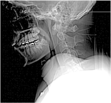
This CT scout image was taken 17 days after the accident occurred. Note that there is no HO of the ALL. It should be noted that this patient has congenital block vertebrae at C2-C3.
The patient's case was managed by his primary care manager along with an orthopedic surgeon and physical medicine and rehabilitation physician working as consulting specialists. A follow-up radiograph of the right femur revealed HO of the soft tissues of the hip (Fig 2). In addition to the right femur fracture and his diagnosis of TBI and hip HO, he began to develop neck pain and upper and middle back pain. He was referred to the physical therapy department where his neck pain and upper back pain was treated with hot packs and cervical traction. Cervical traction caused an increase in neck pain and produced head pain. Sixteen months after his initial injury, his orthopedic surgeon referred him to a hospital-based chiropractic clinic.
Fig. 2.
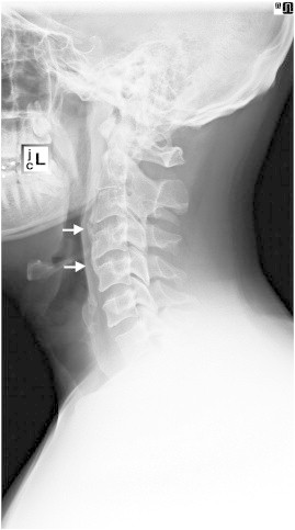
This lateral cervical radiograph was taken 16 months after the accident. Note that the ALL is ossified from C3 through C5. This is an atypical presentation of HO.
The initial examination at the chiropractic clinic revealed a young man with blunted affect. At the time of his initial examination, his primary complaint was neck and upper back pain. He also had complaints of bilateral hip pain, knee pain, and wrist and shoulder pain. His overall pain level was 3 of 10 on a verbal pain scale, but this was mitigated by the use of oxycodone HCL/acetaminophen, 5 to 325 mg, 1 to 2 tabs every 4 hours. His cervical range of motion was overtly limited in all planes of motion; the degrees of active range of motion were not recorded. His chest excursions were measured with a tape measure at the level of greatest chest circumference with the patient's arms at his sides. They were found limited to 0.5 cm change in chest circumference from maximal exhalation to maximal inhalation.19,20 Forestier's bowstring sign revealed restriction in thoracolumbar lateral flexion (less than 10° lateral flexion bilaterally).21,22 The patient actively contracted the ipsilateral paraspinal muscles in an attempt to laterally flex his thoracolumbar spine. This observation was present bilaterally. This was considered abnormal because gravity is typically sufficient for lateral bending in the absence of ankylosis, and contraction of the paraspinal muscles is not normally observed in lateral bending.
Palpation revealed the absence of perceptible passive joint play of the thoracic spine and the costovertebral joints inclusive. The cervical spinal segments also showed diffuse hypomobility. There was increased muscle tone of the levator scapula, the superior trapezius, and the scalene muscles bilaterally. Tender points were noted in the superior trapezius muscles bilaterally and in the suboccipital region bilaterally. Deep tendon reflexes were active, brisk, and normal at the Achilles, patella, brachioradialis, the biceps, and triceps bilaterally. Nerve tension and motor tests of the lower extremities were deferred because of orthopedic injuries affecting both hips. Cervical orthopedic tests were deferred because of the extremely restricted cervical range of motion.
Cervical spine radiographs were taken 16 months after the initial injury and spinal CT. These new images revealed ossification of the ALL from C3-5 and a congenitally blocked C2-3 (Fig 3). This was a relevant interval change because ALL ossification was not visible on the initial CT and radiographs taken 16 months prior. Chest radiographs were also taken and found to be unremarkable, showing no signs of injury or ossification.
Fig. 3.
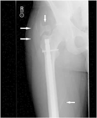
One year after injury and subsequent orthopedic surgical repair of a right femur fracture, HOs in the soft tissues of the right hip are visible (white arrows).
Laboratory tests were ordered at the time of his chiropractic evaluation, and these results are summarized in Table 1. He had elevated CRP, ESR, calcium, and alkaline phosphate. Antinuclear antibodies test and HLA-B27 were negative. The patient was diagnosed with HO and ankylosis of the ALL from C3-5 and decreased respiratory excursions of the chest wall.
Table 1.
The Early Stages of HO Are Characterized by Inflammation (Indicated in This Table With the Patient's Increased CRP and ESR Levels) and Elevated Alkaline Phosphatase
| Test | Results | Normal Range |
|---|---|---|
| CRP | 0.745 (H) mg/dL | 0.000-0.500 mg/dL |
| ESR | 19 (H) mm/h | 0-15 mm/h |
| Antinuclear antibodies test | Negative | Negative |
| HLA-B27 | Negative | Negative |
| Ca | 10.5 (H) mg/dL | 8.6-10.2 mg/dL |
| Alkaline phosphatase | 108 (H) U/L | 25-100 U/L |
CRP, C-reactive protein; ESR, erythrocyte sedimentation rate.
These laboratory studies were obtained 16 months after his injuries.
The chiropractic management plan included manual high-velocity low-amplitude articular manipulation of all the costovertebral joints in an attempt to increase joint mobility and costovertebral excursion. The costovertebral manipulations were performed with posterior to anterior thrusts upon the patient lying prone.
The patient was instructed in the use of an incentive spirometer and provided a unit for home use. He was instructed in techniques of sustained inspiratory exercises to maximize chest excursions, increase respiratory volume, and maintain rib motion. Using the incentive spirometer, his respiratory therapy exercises included performing 1 protracted inhalation, attempting to maintain inhalation as long as possible. He was instructed to obtain a package of balloons and told to inflate one with as few breaths as possible. His instructions were to perform this respiratory therapy routine, one prolonged inhalation and one balloon inflation, every hour for 10 hours per day. He was encouraged to integrate normal activities of daily living into his lifestyle and provided instructional support on activities of daily living. After his fourth chiropractic treatment, the patient was prescribed aqua therapy. He performed aqua therapy 3 times per week for 12 weeks. He was told to return weekly to the chiropractic clinic for a trial of treatment. He was evaluated and treated once per week for 13 weeks.
On the 13th chiropractic visit, the evaluation revealed that his inspiratory chest excursions had increased to 2.5 cm and his verbal pain scale level was 3 of 10 without the use of oxycodone HCL/acetaminophen. He continued with chiropractic care for once a week for 8 more weeks. It is noted that a chest CT was taken 16 months after initial injury, and there was no evidence of HO affecting the costovertebral joints. He was treated every 2 weeks for 26 more weeks. Nearly a year after initiating chiropractic care, his chest excursions had increased to 3.5 cm. The ethics board of the National Naval Medical Center, Bethesda/Walter Reed National Military Medical Center approved this case report for publication.
Discussion
This case identifies the need for practitioners to be alert for HO of soft tissues after traumatic brain injuries, burns, and polytrauma. Spinal ligaments may rapidly ossify after traumatic brain injuries. Heterotopic ossification is a complex malady that is best treated by an integrated team of health care providers working together and maintaining open communication. This case also shows that chiropractic care can be included in a multifaceted approach to managing patients with HO. However, it is important for all musculoskeletal practitioners to be aware of this condition and its impact on care.
The incidence of NHO in civilian patients with TBI is relatively infrequent at 20%, but when TBI and a femur fracture are concomitant, the rate of NHO exceeds 50%.30 The prevalence rates of NHO is remarkably common in high-energy combat injuries that result in TBI and long bone fracture or amputation.8,31 Doctors of chiropractic will likely encounter patients with NHO more frequently as war-wounded veterans seek care in military and Veterans Health Administration chiropractic treatment facilities, as well as when they leave military service and seek care with civilian providers. Doctors of chiropractic should be familiar with the clinical presentation of HO, its pathogenesis, treatment options, and contraindications. Every effort should be made to understand and define a patient's condition before initiating treatment, and in patients with NHO, a collaborative approach to care is preferred.
In the United States, between 1.5 and 2 million TBIs occur annually. The occurrence of HO adjacent to long bone fractures after a TBI is well documented in the literature, whereas ALL involvement is unusual.23 This case reveals a patient with HO near the site of a right femur fracture and of spinal ligaments after a TBI. The possibility of spinal ligament ossification has clinical implications for patients at risk for HO. The progressive nature of this condition may be an indication for sequential radiographic imaging over time for patients with HO risk factors and clinical indications such as pain and progressive loss of joint motion.
The conventionally recommended treatment of HO includes nonsteroidal anti-inflammatory drugs,24,25,26,27 passive range of motion therapy, and in some cases, surgical removal of the ossification with postsurgical radiation therapy to inhibit regrowth of the HO.27
Manual practitioners should be aware of the progressive nature of HO and the possibility that osteoneogenesis may occur in a relatively short time span after TBI, spinal cord injuries, polytrauma, or burns. On the basis of the positive outcome of this case, careful chiropractic comanagement of patients with NHO should be considered in multidisciplinary settings. Although this patient's ALL ossification was not reversed, his chest excursions increased to normal, and his pain was reduced while under chiropractic care. Because there are numerous variables affecting this case, it must be considered that his positive outcome may have been the result of one or more of these variables. His symptoms may have resolved as the natural course of this condition, the respiratory therapy exercises, or a delayed response to his previous physical therapy or pharmaceutical interventions.
There are certain contraindications to treatment of HO. High-velocity manipulations and mechanical traction should not be performed on ankylosed joints and only be performed with caution on patients with joints trending toward ankylosis. It is conceivable that high-velocity adjustments could cause a fracture of the HO or contribute to disc derangement.28,29
Limitations
This case report has limitations and unanswered questions. The cause of this patient's reduced respiratory chest excursions is unknown, so the mechanism or rationale for improvement is not clear. The treating author could have kept more scrupulous patient care notes and used the preferred multiple site method for measuring chest excursions, as well as additional measurements of spinal motion on the initial examination.19,20 It is possible that the patient improved due to natural history of the disorder and improved regardless of treatment. Because this is only one case, this case should not be used as an authoritative reference on treating patients with HO, but as a small point of observation in a much broader search for answers to the mysteries involving the etiology, pathogenesis, and treatment of HO. In addition to NHO causing ossification, there could be other explanations for the finding of ALL ossification. One such explanation could be localized trauma to the cervical spine. The authors hope that future research efforts would include studies addressing the safety, efficiency, and appropriateness of manual treatment options in the care of patients with HO.
Conclusion
This case demonstrated that chiropractic care aimed to address pain and loss of motion in a patient with NHO resulted in patient improvement and that manipulation may be utilized in the comanagement of this multifaceted malady.
Funding Sources and Conflicts of Interest
No funding sources or conflicts of interest were reported for this study.
Footnotes
Disclaimer: The views expressed in this article are those of the authors and do not necessarily reflect the official policy or position of the Department of the Navy, Department of Defense, the US Capitol, nor the US Government.
References
- 1.Toffoli A.M., Gautschi O.P., Frey S.P., Filgueira L., Zellweger R. From brain to bone: evidence for the release of osteogenic humoral factors after traumatic brain injury. Brain Inj. 2008;22(7–8):511–518. doi: 10.1080/02699050802158235. [DOI] [PubMed] [Google Scholar]
- 2.Garland D.E., Blum C.E., Waters R.L. Periarticular heterotopic ossification in head injured adults: incidence and location. J Bone Joint Surg Am. 1980;62-A:1143–1146. [PubMed] [Google Scholar]
- 3.Gautschi O.P., Toffoli A.M., Joesbury KA, Skirving AP, Filguera L, Zellweger R. Osteoinductive effect of cerebrospinal fluid from brain-injured patients. J Neurotrauma. 2007;24(1):154. doi: 10.1089/neu.2006.0166. [DOI] [PubMed] [Google Scholar]
- 4.Gautschi O.P., Cadosch D., Frey S.P., Skirving A.P., Filgueira L., Zellweger R. Serum-mediated osteogenic effect in traumatic brain-injured patients. ANZ J Surg. 2009;79(6):449–455. doi: 10.1111/j.1445-2197.2008.04803.x. [DOI] [PubMed] [Google Scholar]
- 5.Eid K., Labler L., Ertel W., Trentz O., Keel M. Systemic effects of severe trauma on the function and apoptosis of human skeletal cells. J Bone Joint Surg (Br) 2006;88-B:1394–1400. doi: 10.1302/0301-620X.88B10.17139. [DOI] [PubMed] [Google Scholar]
- 6.Forsberg J.A., Potter B.K. Heterotopic ossification in wartime wounds. J Surg Orthop Adv. 2010;19:54–61. [PubMed] [Google Scholar]
- 7.Potter B.K., Bums T.C., Lacap A.P., Granville R.R., Gajewski D.A. Heterotopic ossification following traumatic and combat-related amputations. Prevalence, risk factors, and preliminary results of excision. J Bone Joint Surg Am. 2007;89:476–486. doi: 10.2106/JBJS.F.00412. [DOI] [PubMed] [Google Scholar]
- 8.Forsberg J.A., Pepek J.M., Wagner S. Heterotopic ossification in high-energy wartime extremity injuries: prevalence and risk factors. J Bone Joint Surg Am. 2009;91(5):1084–1091. doi: 10.2106/JBJS.H.00792. [DOI] [PubMed] [Google Scholar]
- 9.Rossier A.B., Bussat P., Infante F. Current facts on para-osteoarthropathy (POA) Paraplegia. 1973;11:36–78. doi: 10.1038/sc.1973.5. [DOI] [PubMed] [Google Scholar]
- 10.Wharton G.W., Morgan T.H. Ankylosis in the paralyzed patient. J Bone Joint Surg Am. 1970;52:105–112. [PubMed] [Google Scholar]
- 11.Freed J.H., Hahn H., Menter R., Dillon T. The use of three-phase bone scan in the early diagnosis of heterotopic ossification (HO) and in the evaluation of Didronel therapy. Paraplegia. 1982;20:208–216. doi: 10.1038/sc.1982.39. [DOI] [PubMed] [Google Scholar]
- 12.Scherbel U., Riess P., Khunrana J. Expression of bone morphogenic proteins in rats with and without brain injury and a tibia fracture. Univ Penn Orthop J. 2001;14:85–91. [Google Scholar]
- 13.Boes M., Kain M., Sanjeev K. Osteogenic effects of traumatic brain injury on experimental fracture-healing. J Bone Joint Surg Am. 2006;88:738–743. doi: 10.2106/JBJS.D.02648. [DOI] [PubMed] [Google Scholar]
- 14.Estores I.M. C-reactive protein and erythrocyte sedimentation rate in patients with heterotopic ossification after spinal cord injury. J Spinal Cord Med. 2004;27(5):434–437. doi: 10.1080/10790268.2004.11752233. [DOI] [PubMed] [Google Scholar]
- 15.Singh R.S., Craig M.C., Katholi C.R. The predictive value of creatine phosphokinase and alkaline phosphatase in identification of heterotopic ossification in patients after spinal cord injury. Arch Phys Med Rehabil. 2003;84(11):1584–1588. doi: 10.1053/s0003-9993(03)00347-2. [DOI] [PubMed] [Google Scholar]
- 16.Epstein N.E., Hollingsworth R. Ossification of the cervical anterior longitudinal ligament contributing to dysphagia. J Neurosurg. 1999;90(2 Suppl.):261–263. doi: 10.3171/spi.1999.90.2.0261. [DOI] [PubMed] [Google Scholar]
- 17.Song J., Mizuno J., Nakagawa H. Clinical and radiological analysis of ossification of the anterior longitudinal ligament causing dysphagia and hoarseness. Neurosurgery. 2006;58(5):913–919. doi: 10.1227/01.NEU.0000209936.46946.99. [DOI] [PubMed] [Google Scholar]
- 18.Chen Y., Guo Y., Chen D. Diagnosis and surgery of ossification of posterior longitudinal ligament associated with dural ossification in the cervical spine. Eur Spine J. 2009;18(10):1541–1547. doi: 10.1007/s00586-009-1029-2. [DOI] [PMC free article] [PubMed] [Google Scholar]
- 19.Bockenhauer S.E., Chen H., Julliard K.N., Weedon J. Measuring thoracic excursion: reliability of the cloth tape measure technique. J Am Osteopath Assoc. 2007;107(5):191–196. [PubMed] [Google Scholar]
- 20.Mohan V., Dzulkifli N.H., Justine M., Haron R., H L.J., Rathinam C. Intrarater reliability of chest expansion using cloth tape measure technique. Bangladesh J Med Sci. 2012;11(04):307–311. [Google Scholar]
- 21.Forestier J., Jacqueline F., Rotesquerol J. Masson & Cie; Paris: 1951. La spondylarthrite ankylosante: clinique, radiologie anatomie pathologique, traitement; pp. 22–23. [Google Scholar]
- 22.Masi A.T., Sierakowski S., Kim J.M. Jacques Forestier's vanished bowstring sign in ankylosing spondylitis: a call to test its validity and possible relation to spinal myofascial hypertonicity. Clin Exp Rheumatol. 2005;23:760–766. [PubMed] [Google Scholar]
- 23.Czyrny J.J., Glasauer F.E. An unusual location for heterotopic ossification: lumbar anterior longitudinal ligament. J Spinal Cord Med. 1995;18(3):194–199. doi: 10.1080/10790268.1995.11719392. [DOI] [PubMed] [Google Scholar]
- 24.Banovac K., Williams J.M. Prevention of heterotopic ossification after spinal cord injury with indomethacin. Spinal Cord. 2001;39:370–374. doi: 10.1038/sj.sc.3101166. [DOI] [PubMed] [Google Scholar]
- 25.Banovac K., Williams J.M. Prevention of heterotopic ossification after spinal cord injury with COX-2 selective inhibitor (rofecoxib) Spinal Cord. 2004;42:707–710. doi: 10.1038/sj.sc.3101628. [DOI] [PubMed] [Google Scholar]
- 26.Right Fijn, Koorevaar R.T., Brouwers J.R. Prevention of heterotopic ossification after total hip replacement with NSAIDS. Pharm World Sci. 2003;25(4):138–145. doi: 10.1023/a:1024830213832. [DOI] [PubMed] [Google Scholar]
- 27.Herd R., Strassmann G., Schopohl B. Radiation therapy for the treatment of heterotopic ossification at the elbow. J Bone Joint Surg (Br) 2001;83-B:332–334. doi: 10.1302/0301-620x.83b3.11428. [DOI] [PubMed] [Google Scholar]
- 28.Mestan M.A., Bassano J.M. Fractured heterotopic bone in myositis ossificans traumatica. J Manipulative Physiol Ther. 2001;24(4):296–299. doi: 10.1067/mmt.2001.114360. [DOI] [PubMed] [Google Scholar]
- 29.Hsieh J.H., Wu C.T., Lee S.T. Cervical intradural disc herniation after spinal manipulation therapy in a patient with ossification of posterior longitudinal ligament: a case report and review of the literature. Spine. 2010;35(5):E149–E151. doi: 10.1097/BRS.0b013e3181bee8a7. [DOI] [PubMed] [Google Scholar]
- 30.Garland G.W. A clinical perspective on common forms of acquired heterotopic ossification. Clin Orthop. 1991;263:13–29. [PubMed] [Google Scholar]
- 31.Potter B.K., Forsberg J.A., Davis T.A. Heterotopic ossification following combat-related trauma. J Bone Joint Surg Am. 2010;92(Suppl. 2):74–89. doi: 10.2106/JBJS.J.00776. [DOI] [PubMed] [Google Scholar]