Abstract
Objective:
Chronic autoimmune thyroiditis (CAT) (chronic lymphocytic thyroiditis—Hashimoto's thyroiditis), which is the most common inflammatory disorder of the thyroid gland, causes hypothyroidism. Ultrasound elastography is a newly developed sonographic technique that provides an estimation of tissue elasticity by measuring the degree of tissue displacement under the application of an external force. In this study, our aim was to evaluate the accuracy of strain index ratio with real-time ultrasound elastography and to calculate the cut-off point for the diagnosis of CAT. Our aim was also to lead further studies on other pathological changes such as lymphoma, malign nodules etc. based on CAT by using this cut-off point. The gains from this study and further studies will assist clinical diagnoses and follow-up.
Methods:
Aplio™ 500 ultrasound machine (Toshiba Medical Systems Co. Ltd, Otawara, Japan) with linear 4.8–11.0 MHz transducers and elastography software was used. Routine B-mode (dimensions and parenchymal echogenicity) ultrasound evaluation was performed prior to the ultrasound elastography.
Results:
A total of 31 randomized patients (3 males, 28 females) with a mean age of 39.13 ± 10.16 years (range, 16–58 years) with CAT and 21 healthy controls (6 males, 15 females) with mean age of 34.67 ± 16.31 years (range, 14–81 years) were prospectively examined. The mean values of thyroid-stimulating hormone (TSH; normal TSH value is 0.27–4.20 IU ml−1) and anti-thyroid peroxidase (anti-TPO; normal anti-TPO value is 0–34 IU ml−1) were 3.40 ± 2.70 and 373.66 ± 148.94 IU ml−1, respectively. No correlation was detected between serum TSH and thyroid tissue strain index (Spearman r coefficient of TSH was −0.290). Positive-sided correlation was detected between anti-TPO values and thyroid tissue strain index ratio (Spearman r coefficient of anti-TPO was 0.682). The median strain index ratio of patients with CAT (1.39 ± 0.72) was significantly higher than the mean ratio of the controls (0.76 ± 0.55). The area under the receiver operating characteristic curve was 0.775 (95% confidence interval). The optimal cut-off value (in which the sum of sensitivity and specificity was highest) for the prediction of diffuse thyroid pathology was 0.677. For this cut-off ratio, thyroid stiffness had 96% sensitivity and 67% specificity. A total of 30 of 31 patients (96%) and a total of 7 of 21 healthy controls (33%) exceeded the cut-off points.
Conclusion:
The strain index ratio was higher in CAT than in normal thyroid parenchyma in real-time ultrasound elastography. Thus, it seems to be a useful method for the assessment of CAT with real-time ultrasound elastography, and further studies assessing the correlation of sonoelastography findings and histopathological subtypes of CAT would enrich the findings of the present study.
Advances in knowledge:
In our study, we detected the stiffness ratio of the thyroid tissue in patients with CAT. The cut-off value should be helpful for diagnosis or follow-up of the recently developed lesions such as lymphoma, malign nodule, etc. based on CAT. This study should also encourage new studies about CAT and ultrasound elastography.
Chronic autoimmune thyroiditis (CAT) (chronic lymphocytic thyroiditis—Hashimoto's thyroiditis), which is the most common inflammatory disorder of the thyroid gland, causes hypothyroidism.1 It occurs between 30 and 50 years of age, and up to 95% of the patients are females.2 It has a dominant inheritance, and thyroid lymphoma is a rare and serious complication of the disease.3
Ultrasound elastography, which is a newly developed sonographic technique, provides an estimation of tissue elasticity by measuring the degree of tissue displacement under the application of an external force.4 This ultrasound-based method can accurately evaluate the elasticity of superficial tissues such as the breast, prostate, scrotum, neck and thyroid.5–7 Several studies have used ultrasound elastography to differentiate benign and malignant thyroid nodules with colour scale scoring.8–11 In addition to visualization of elasticity on colour-coded elastographic images, the strain of tissues can be defined using numerical strain values and compared by cross-correlation of radiofrequency signals.12
Although there are many studies investigating stiffness of thyroid nodules, only one article has evaluated patients with subacute thyroiditis (SAT) in the literature.4 To the best of our knowledge, there is no report in the literature about the utility of real-time ultrasound elastography with the strain index ratio method for CAT. In this study, our aim was to evaluate the accuracy of strain index ratio with real-time ultrasound elastography and calculate the cut-off point for the diagnosis of CAT. Our aim was also to lead further studies on other pathological changes such as lymphoma, malign nodules etc. based on CAT by using this as a cut-off point. The gains from this study and further studies will assist clinical diagnosis and follow-up.
METHODS AND MATERIALS
A total of 31 randomized patients who were admitted to our endocrinology outpatient clinic for work-up of CAT were included in this work. The diagnosis of CAT was based on clinical and laboratory findings consistant with high titres of antithyroid antibodies [anti-thyroid peroxidase (TPO) and/or antiTg].13–16 Exclusion criteria were other causes of thyroiditis, Graves disease, nodular abnormalities, malignancies and patient non-compliance to ultrasound examination. All patients agreed to participate in our study, which was approved by the local ethics committee. The study also had no conflict of interest. A healthy control group Was selected from the volunteers working in the hospital.
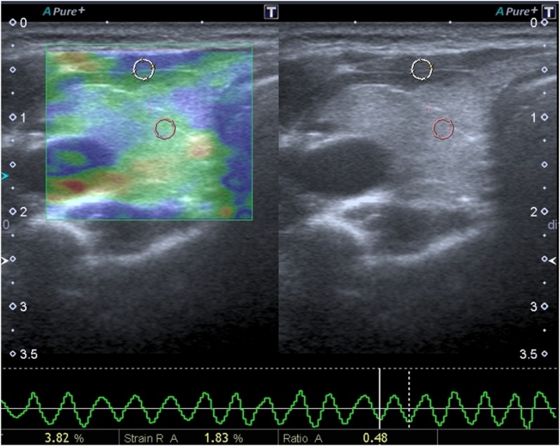
The demographic and laboratory data, including age, gender, titres of thyroid-stimulating hormone (TSH) and antithyroid antibodies (anti-TPO), were recorded. Then, all patients were examined using high-resolution B-mode greyscale ultrasound and sonoelastography by a radiologist with 7 years' experience in sonography and 2 years' experience in sonoelastography (MSM). All the ultrasound evaluations were performed in supine neutral position. The Aplio™ 500 ultrasound machine (Toshiba Medical Systems Co. Ltd, Otawara, Japan) with linear 4.8–11.0 MHz transducers and elastography software was used. Routine B-mode (dimensions and parenchymal echogenicity) ultrasound evaluation was performed prior to the ultrasound elastography. After the activation of the elastography system (Elasto-Q) of the machine, images were obtained by applying repetitive light compression on the skin above the targeted area by using a free-hand technique.17 The probe was positioned perpendicular to the skin when applying pressure, avoiding lateral movement. The acquired images were evaluated through cinememory, which permitted the retrospective assessment of the changes occurring at the target lesion during compression and decompression. The ideal pressure on the target lesion was confirmed based on the cinememory data. After seven to eight compression–relaxation cycles, the elastographic examinations were finalized. Strain value measurements were obtained from appropriate relaxation waves, which had a regular sign curve (sinusoidal shape) on the velocity profile (Figure 1). We used the strain index that represents the ratio of the strain of thyroid parenchyma to that of strap muscles. There were no space-occupying lesions or systemic and local muscle disease on the related strap muscles. This index increases when the parenchyma is harder (stiffer) than the strap muscles. After placing the region of interest (ROI) on the strap muscles, we placed the ROI of a target on the thyroid parenchyma. We measured strain index ratio by placing standard-sized (4 mm) ROIs determined by the system automatically on the thyroid parenchyma and strap muscles. We took care that the location of the ROIs should be placed at the same depth level as much as possible.18 Because changes in the distance of control ROI to the ultrasound probe significantly influences strain ratio values both in vitro and in vivo,19 three measurements were obtained for each lobe and for each patient. The mean of the total three measurements was used as representative of the strain index ratio. Elastography measurements were obtained when the sinusoidal wave was regular. These measurements were considered as valid.
Figure 1.
The elastography calculation of thyroid parenchyma. Left side of the windows is colour-coded elastography image and the right side is greyscale image. The circles indicate the regions of interest where we measured the stiffness ratios. One is on the strap muscle and one is on the normal thyroid parenchyma. At the bottom of the screen, normal sinusoidal wave can be seen. It indicates pressure of the probe is appropriate and regular. The numbers indicate the strain values, and “%” indicates the strain ratio.
Statistical analyses
IBM SPSS® v. 21 (IBM Corporation, Armonk, NY) was used for statistical analyses. Three measurements were obtained for each lobe and for each patient. The mean of the total three measurements was used for each lobe. The mean value of both right and left lobes was calculated and only one value obtained for each person as representative for the strain index ratio. Kolmogorov–Smirnov test was used to evaluate the distribution of the data. Spearman correlation coefficient was used to correlate serum TSH and anti-TPO values with the strain index ratio. Student's t-test was used to compare the ages of the groups, and Mann–Whitney U test was used to compare the strain index ratio of the groups. The receiver operating characteristic (ROC) curve was used to determine the cut-off point of strain index ratio of normal and CAT subjects. A p-value of <0.05 was considered statistically significant.
RESULTS
A total of 31 randomized patients (3 males, 28 females) with a mean age of 39.13 ± 10.16 years (range, 16–58 years) with CAT and 21 healthy controls (6 males, 15 females) with a mean age of 34.67 ± 16.31 years (range, 14–81 years) were prospectively examined. The mean values of TSH (normal TSH value is 0.27–4.20 IU ml−1) and anti-TPO (normal anti-TPO value is 0–34 IU ml−1) were 3.40 ± 2.70 and 373.66 ± 148.94 IU ml−1, respectively. No correlation was detected between serum TSH and thyroid tissue strain index ratio (Spearman r coefficient of TSH was −0.290). Positive-sided correlation was detected between anti-TPO values and thyroid tissue strain index ratio (Spearman r coefficient of anti-TPO was 0.682).
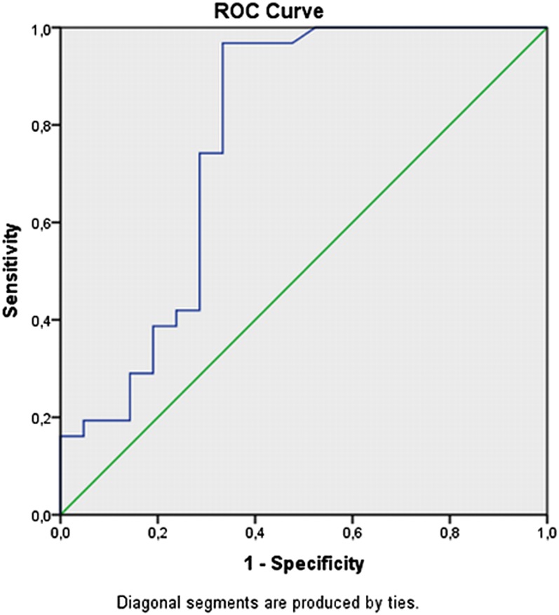
The median strain index ratio of patients with CAT (1.39 ± 0.72) was significantly higher than the mean ratio of the controls (0.76 ± 0.55). The area under the ROC curve (AUC) was 0.775 (95% confidence interval). The optimal cut-off value (in which the sum of sensitivity and specificity was the highest) for the prediction of diffuse thyroid pathology was 0.677. For this cut-off ratio, thyroid stiffness had 96% sensitivity; 67% specificity; +likelihood ratio (LR), 2.9; −LR, 0.05; positive-predictive value, 81%; and negative-predictive value, 83%. A total of 30 of 31 patients (97%) and a total of 7 of 21 healthy controls (33%) exceeded the cut-off points (Figure 2).
Figure 2.
The receiver operating characteristic (ROC) curve of the study. The area under curve was 0.775. The optimal cut-off value was 0.677.
DISCUSSION
We found significantly higher strain index ratio in CAT patients than those in the control group. This indicates that the stiffness of the thyroid parenchyma is higher than normal thyroid parenchyma.
It has been described that the typical pathological features of CAT consisted lymphoplasmacytic infiltration and lymphoid follicles with well-developed germinal centres. Although several subtypes of CAT were described, the most recognized subtype is the fibrous variant of the disease characterized by marked fibrous replacement of the thyroid parenchyma.20 In the literature, in a study evaluating the assessment of sonoelastography in SAT, Xie et al4 affirmed that the elasticity scores that the lesions had were higher than those of normal thyroid tissue, and they suggested that replacement of granulation tissue might have been the reason for these differences. Based on this information, the high elasticity score of CAT in our study might be owing to the replacement of hyperplastic fibrous tissue in thyroid parenchyma of the patients with CAT. In our study, we detected the stiffness ratio of the thyroid tissue in patients with CAT. The cut-off value should be helpful for diagnosis or follow-up of recently developed lesions such as lymphoma, malign nodules etc. based on CAT. This study should encourage new studies about CAT and ultrasound elastography.
Shear wave elastography, which is one of the techniques of sonoelastography, has been used to evaluate diffuse thyroid gland pathologies by an acoustic radiation force impulse imaging (ARFI).13,21 Ruchala et al21 used shear wave elastography for assessment of patients with SAT and CAT, and they found that the tissue stiffness of thyroid in CAT had been significantly higher than in healthy control subjects compatible with our results. Moreover, Sporea et al13 have reported significant differences between quantitative ARFI values of patients with CAT and healthy subjects. In comparison with our study, they yielded lower sensitivity (62.5% vs 96%) and higher specificity (79.5% vs 67%) for the prediction of CAT. However, we exposed approximately similar accuracy with an AUC of 0.775 as compared with ARFI imaging quantification (AUC, 0.804) for the prediction of the disease, and the lower specificity of 67% in our study may be as a result of wide range and overlap of strain index ratios between the two groups.13
We have also calculated the serum TSH and anti-TPO levels of patients with CAT. The value of these parameters was higher than normal intervals. No correlation was detected between serum TSH and thyroid tissue strain index ratio (Spearman r coefficient of TSH was −0.290). Positive-sided correlation was detected between anti-TPO values and thyroid tissue strain index ratio (Spearman r coefficient of anti-TPO was 0.682). Our results were consistent with the results of Sporea et al13 revealing no correlation between TSH and thyroid tissue stiffness.
Ultrasound elastography is a standardized technique and presents quantitative values. This is not dependent on the operator, which makes this technique reproducible.
There are some limitations of our study. The first limitation of our study was the small sample size and female predilection. CAT occurs mostly in the female population, and our results were consistent with the percentages found in the literature. The cause of the small sample size is the location where the study was performed. The city was small and the number of patients was not as many as we expected, so we could not reach many more patients during this period, and we ended the study with 31 patients. Secondly, we did not perform pathological evaluation. However, it has been well described that the characterization of the disease is based on high titres of circulating antibodies to thyroid peroxidase and thyroglobulin.15 Thirdly, we could not compare our threshold value with the studies in literature. We used real-time sonoelastography method, which was different from the study of Spoera et al.13 They performed their study with the ARFI method, and they reported the threshold value obtained with ARFI.
The weakness of the present study is a lack of inter–intraobserver data. Big sample size and inter–intraobserver studies could improve the technique.
In conclusion, the strain index ratio is higher in CAT than in normal thyroid parenchyma in real-time ultrasound elastography. Thus, it seems to be a useful method for the assessment of CAT with real-time ultrasound elastography, and further studies assessing the correlation of sonoelastography findings and histopathological subtypes of CAT would enrich the findings of the present study.
REFERENCES
- 1.Hamburger JI. The various presentations of thyroiditis. Diagnostic considerations. Ann Intern Med 1986; 104: 219–24. [DOI] [PubMed] [Google Scholar]
- 2.Sakiyama R. Thyroiditis: a clinical review. Am Fam Physician 1993; 48: 615–21. [PubMed] [Google Scholar]
- 3.Dayan CM, Daniels GH. Chronic autoimmune thyroiditis. N Engl J Med 1996; 335: 99–107. [DOI] [PubMed] [Google Scholar]
- 4.Xie P, Xiao Y, Liu F. Real-time ultrasound elastography in the diagnosis and differential diagnosis of subacute thyroiditis. J Clin Ultrasound 2011; 39: 435–40. doi: 10.1002/jcu.20850 [DOI] [PubMed] [Google Scholar]
- 5.Koizumi Y, Hirooka M, Kisaka Y, Konishi I, Abe M, Murakami H, et al. Liver fibrosis in patients with chronic hepatitis C: noninvasive diagnosis by means of real-time tissue elastography—establishment of the method for measurement. Radiology 2011; 258: 610–17. doi: 10.1148/radiol.10100319 [DOI] [PubMed] [Google Scholar]
- 6.Wojcinski S, Cassel M, Farrokh A, Soliman AA, Hille U, Schmidt W, et al. Variations in the elasticity of breast tissue during the menstrual cycle determined by real-time sonoelastography. J Ultrasound Med 2012; 31: 63–72. [DOI] [PubMed] [Google Scholar]
- 7.Fleury Ede F, Fleury JC, Piato S, Roveda D, Jr. New elastographic classification of breast lesions during and after compression. Diagn Interv Radiol 2009; 15: 96–103. [PubMed] [Google Scholar]
- 8.Cakir B, Aydin C, Korukluoğlu B, Ozdemir D, Sisman IC, Tüzün D, et al. Diagnostic value of elastosonographically determined strain index in the differential diagnosis of benign and malignant thyroid nodules. Endocrine 2011; 39: 89–98. doi: 10.1007/s12020-010-9416-3 [DOI] [PubMed] [Google Scholar]
- 9.Vorländer C, Wolff J, Saalabian S, Lienenlüke RH, Wahl RA. Real-time ultrasound elastography—a noninvasive diagnostic procedure for evaluating dominant thyroid nodules. Langenbecks Arch Surg 2010; 395: 865–71. doi: 10.1007/s00423-010-0685-3 [DOI] [PubMed] [Google Scholar]
- 10.Hong Y, Liu X, Li Z, Zhang X, Chen M, Luo Z. Real-time ultrasound elastography in the differential diagnosis of benign and malignant thyroid nodules. J Ultrasound Med 2009; 28: 861–7. [DOI] [PubMed] [Google Scholar]
- 11.Rubaltelli L, Corradin S, Dorigo A, Stabilito M, Tregnaghi A, Borsato S, et al. Differential diagnosis of benign and malignant thyroid nodules at elastosonography. Ultraschall Med 2009; 30: 175–9. doi: 10.1055/s-2008-1027442 [DOI] [PubMed] [Google Scholar]
- 12.Onur MR, Poyraz AK, Ucak EE, Bozgeyik Z, Özercan IH, Ogur E. Semiquantitative strain elastography of liver masses. J Ultrasound Med 2012; 31: 1061–7. [DOI] [PubMed] [Google Scholar]
- 13.Sporea I, Sirli R, Bota S, Vlad M, Popescu A, Zosin I. ARFI elastography for the evaluation of diffuse thyroid gland pathology: preliminary results. World J Radiol 2012; 4: 174–8. doi: 10.4329/wjr.v4.i4.174 [DOI] [PMC free article] [PubMed] [Google Scholar]
- 14.Caturegli P, De Remigis A, Rose NR. Hashimoto thyroiditis: clinical and diagnostic criteria. Autoimmun Rev 2014; 13: 391–7. doi: 10.1016/j.autrev.2014.01.007 [DOI] [PubMed] [Google Scholar]
- 15.Farwell AP, Braverman LE. Inflammatory thyroid disorders. Otolaryngol Clin North Am 1996; 29: 541–56. [PubMed] [Google Scholar]
- 16.Basaria S, Cooper DS. Amiodarone and the thyroid. Am J Med 2005; 118: 706–14. [DOI] [PubMed] [Google Scholar]
- 17.Orman G, Özben S, Hüseyinoğlu N, Duymuş M, Orman KG. Ultrasound elastographic evaluation in the diagnosis of carpal tunnel syndrome: initial findings. Ultrasound Med Biol 2013; 39: 1184–9. doi: 10.1016/j.ultrasmedbio.2013.02.016 [DOI] [PubMed] [Google Scholar]
- 18.Ciledag N, Arda K, Aribas BK, Aktas E, Köse SK. The utility of ultrasound elastography and MicroPure imaging in the differentiation of benign and malignant thyroid nodules. AJR Am J Roentgenol 2012; 198: W244–9. doi: 10.2214/AJR.11.6763 [DOI] [PubMed] [Google Scholar]
- 19.Havre RF, Waage JR, Gilja OH, Odegaard S, Nesje LB. Real-time elastography: strain ratio measurements are influenced by the position of the reference area. Ultraschall Med Jun 2011. Epub ahead of print. [DOI] [PubMed] [Google Scholar]
- 20.Katz SM, Vickery AL, Jr. The fibrous variant of Hashimoto's thyroiditis. Hum Pathol 1974; 5: 161–70. [DOI] [PubMed] [Google Scholar]
- 21.Ruchala M, Szczepanek-Parulska E, Zybek A, Moczko J, Czarnywojtek A, Kaminski G, et al. The role of sonoelastography in acute, subacute and chronic thyroiditis: a novel application of the method. Eur J Endocrinol 2012; 166: 425–32. doi: 10.1530/EJE-11-0736 [DOI] [PubMed] [Google Scholar]


