Abstract
A 73-year-old man with gallstone disease was admitted with right upper quadrant abdominal pain. He was treated for cholecystitis with intravenous antibiotics. Two days later, he reported of new onset left iliac fossa pain, with tenderness and guarding. An abdominal X-ray demonstrated small bowel obstruction, a CT scan demonstrated an impacted gallstone within the proximal ileum. He was treated for a gallstone ileum and underwent an uncomplicated laparotomy, small bowel enterotomy and removal of a faceted gallstone. Three months later, the patient re-presented with generalised abdominal pain, guarding and rebound tenderness. Small bowel obstruction was again demonstrated with an impacted gallstone within the distal ileum seen on CT scan. A second laparotomy revealed two further faceted gallstones, which were removed through an enterotomy. The densely adherent gallbladder to the duodenum precluded a surgical repair of the cholecystoduodenal fistula. He made an uneventful recovery and was subsequently discharged home.
Background
Gallstone ileus accounts for 25% of non-strangulated small-bowel obstruction in those above 65 years of age. With an increasing elderly population, this condition may become more prevalent, and hence an increased awareness is important. Furthermore, recurrent gallstone ileus is less commonly described, and its management can be challenging.
Case presentation
About 1–3% of mechanical small bowel obstruction cases in the general population can be attributed to gallstone ileus. Among the elderly, the incidence rises and accounts for up to 25% of non-strangulated small-bowel obstructions.1
We present a case of recurrent gallstone ileus, which is less commonly described, and propose a treatment strategy for the condition.
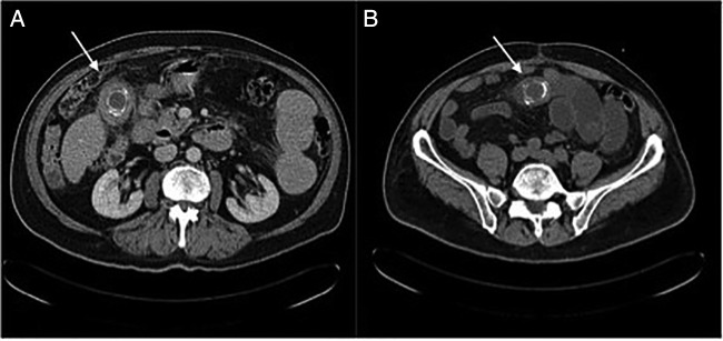
A 73-year-old man known to have gallstone disease was admitted with sudden onset, colicky and sharp right upper quadrant abdominal pain, which radiated to his back. Clinical examination revealed a non-distended soft abdomen with mild tenderness in the patient's right upper quadrant. Except for an elevated white cell count level of 15.4×10⁹/L and a C reactive protein of 40 mg/L, the rest of his blood investigations were within normal limits. A diagnosis of cholecystitis was made and he was treated conservatively with intravenous antibiotics. However, 2 days later, he reported of new onset left iliac fossa pain, with tenderness and guarding. An abdominal X-ray demonstrated features of small bowel obstruction. A subsequent CT scan (figure 1A) demonstrated an impacted gallstone within the proximal ileum. The clinical and radiological picture was therefore consistent with a gallstone ileus.
Figure 1.

(A) CT scan of the abdomen demonstrating an impacted gallstone (white arrow) within the proximal ileum during the first presentation. (B) CT scan of the pelvis demonstrating an impacted recurrent gallstone within the distal ileum (white arrow).
The patient underwent an uncomplicated laparotomy, small bowel enterotomy and removal of a faceted gallstone (figure 2A). He made an uneventful recovery and was subsequently discharged home.
Figure 2.

The faceted gallstones extracted following laparotomy and enterotomy. (A) At the initial laparotomy, a solitary gallstone was removed through an enterotomy. Subsequently, a further two gallstones were removed at the second laparotomy. (B) When combined together, the gallstones appear to adopt the shape of a gallbladder.
Three months later, the patient represented with generalised abdominal pain with associated guarding and rebound tenderness. Small bowel obstruction was again demonstrated on abdominal X-ray. A repeat CT scan this time demonstrated an impacted gallstone within the distal ileum (figure 1B). He underwent a second laparotomy during which two further faceted gallstones were removed through an enterotomy (figure 2A, B). The gallbladder was densely adhered to the duodenum and thus a surgical repair of the cholecystoduodenal fistula was not considered. He made an uneventful recovery and was subsequently discharged home.
Discussion
Recurrent gallstone ileus is an uncommon condition, with an incidence of between 2% and 5%, with a significant mortality rate of 12–20%. Approximately 57% of recurrences occur within 6 months of initial surgery.1
Gallstone ileus occurs as a consequence of acute or chronic gallstone disease. An inflamed gallbladder erodes through the mucosal wall of a segment of the gastrointestinal tract (GI tract) creating a cholecystoenteric fistula, the commonest of which are cholecystoduodenal fistulas, followed by cholecystocolonic fistulas.2
Recurrent gallstone ileuses are normally caused by stones already present at first presentation, either within the gallbladder, which makes a subsequent escape through the fistula, or within the bowel and was overlooked at initial surgery.
Gallstones escape through the fistula, and their passage down the GI tract and subsequent impaction is responsible for the patient's symptoms and clinical presentation.
Although enteric gallstones will pass spontaneously in 80% of the time,1 those with a diameter greater than 2.5 cm invariably become impacted.3 The terminal ileum is where impaction commonly occurs, followed by the jejunum and stomach.1 If a stone impacts in the duodenal bulb, this can present as gastric outlet obstruction (Bouveret's syndrome).4
Gallstone ileus has a typical radiological appearance known as the Rigler's triad, first described in 1941,3 which comprises of small bowel obstruction, air in the biliary tree and ectopic gallstone. The diagnosis of gallstone ileus is indicated if at least two of these signs are present, and about 50% of cases have demonstrated these.5 6 CT scans, however, are more readily utilised now as diagnostic tools and have an increased sensitivity and specificity in the diagnosis of gallstone ileus.7
How we manage patients with gallstone ileus remains controversial.2 Options include the one-stage procedure, which involves enterolithotomy with cholecystectomy and repair of the biliary fistula, the two-stage procedure, which involves an emergency enterolithotomy followed by a cholecystectomy 4–6 weeks later after the inflammation has settled,1 or an enterolithotomy alone.
Those who prefer the one-stage procedure believe that this approach prevents future complications that arise from the gallbladder that is left behind. This may include recurrent gallstone ileus, cholecystitis, cholangitis and an increased incidence of gallbladder carcinoma.1 2 8 Clavien et al8 found no complications following the one-stage procedure and yet noted a 56% biliary disorder rate after enterolithotomy alone, without undertaking cholecystectomy and closure of the fistula. The one-stage procedure, however, carries the risk of a biliary or enteric leak following closure of the fistula. It is also a more technically demanding procedure and requires a lengthier operative time.9 This may not be ideal as these patients are normally elderly with other comorbidities and may also be deemed high risk for general anaesthesia.10
The necessity to repair the biliary fistula in the same setting is a question asked by those who favour performing enterolithotomy alone. They argue that this would prolong the operative time unnecessarily, in a group of patients who are already vulnerable. Furthermore, as spontaneous closure of the biliary fistula can occur, particularly when the cystic duct is patent, the need of a fistula repair is debatable.6
The morbidity and mortality rates between the two procedures also differ. Some studies have shown no difference,8 9 while others have. Reisner and Cohen1 found a mortality rate of 16.9% in patients undergoing a one-stage procedure. In contrast, a mortality rate of 11.7% was found in 80% of patients who underwent simple enterolithotomy.
Rodriguez-Sanjuan et al11 demonstrated postoperative morbidity for one -stage procedure to be 67% versus 50% for enterolithotomy alone, while others have reported a 61.1% for one-stage versus 23.7% for the two-stage surgery.12 Thus advocates of simple enterotomy procedure believe it is a safer operation, technically easier and with shorter operating times.1 9 11 12
We believe that the choice of surgical treatment is ultimately influenced by the general health and medical comorbidities of the patient, coupled with the experience of the surgeon involved. The one-stage procedure is usually reserved for patients who are in general good health with a mild degree of cholecystitis. For patients who are haemodynamically unstable and have other comorbidities, thus necessitating shorter operative time, simple enterolithotomy is favoured. This is also true in those patients who have extensively inflamed gallbladders where a cholecystectomy would prove technically difficult.
The role of laparoscopic surgery is adopting an important role in the diagnosis and treatment of gallstone ileus.13 This will be particularly useful in treating this group of patients who are normally elderly with various comorbidities.
We therefore think it is helpful to have a management plan for these patients, with additional steps to reduce the chances of a recurrent gallstones ileus. Patients who clinically are suspected to have gallstone ileus should undertake a CT scan to confirm or exclude the presence of gallstones. This should also help determine the level of GI obstruction.
Multiple gallstones are found in 3–15% of patients with gallstone ileus14 and thus it has been advocated that thorough palpation of the entire bowel should be undertaken at the initial laparotomy to exclude additional stones or stone remnants. If the stone is faceted, it is likely that further stones may be present.15 16
Our patient recovered well following his second laparotomy and enterolithotomy procedure. Appropriate surgical treatment following careful patient selection is key to ensure a successful outcome from this challenging condition.
Learning points.
Consider a diagnosis of gallstone ileus in the elderly who present with non-strangulated small bowel obstruction.
A thorough palpation of the entire bowel must be undertaken at the first laparotomy to avoid the possibility of further undetected gallstones, which may lead to a recurrent gallstone ileus in the future.
The patient's overall condition and comorbidites must be considered when deciding the final operative management.
Footnotes
Competing interests: None.
Patient consent: Obtained.
Provenance and peer review: Not commissioned; externally peer reviewed.
References
- 1.Reisner RM, Cohen JR. Gallstone ileus: a review of 1,001 reported cases. Am Surg 1994;60:441–6. [PubMed] [Google Scholar]
- 2.Kirchmayr W, Muhlmann G, Zitt M et al. Gallstone ileus: rare and still controversial. ANZ J Surg 2005;75:234–8. [DOI] [PubMed] [Google Scholar]
- 3.Rigler LG, Borman CN, Noble JF. Gallstone obstruction: pathogenesis and roentgen manifestations. JAMA 1941;117:1753–9. [Google Scholar]
- 4.Masson JW, Fraser A, Wolf B et al. Bouveret's syndrome: gallstone ileus causing gastric outlet obstruction. Gastrointest Endosc 1998;47:104–5. [DOI] [PubMed] [Google Scholar]
- 5.Heuman R, Sjodahl R, Wetterfors J. Gallstone ileus: an analysis of 20 patients. World J Surg 1980;4:595–8. [DOI] [PubMed] [Google Scholar]
- 6.Deitz DM, Standage BA, Pinson CW et al. Improving outcome in gallstone ileus. Am J Surg 1986;151:572–6. [DOI] [PubMed] [Google Scholar]
- 7.Swift SE, Spencer JA. Gallstone ileus: CT findings. Clin Radiol 1998; 53:451–4. [DOI] [PubMed] [Google Scholar]
- 8.Clavien PA, Richon J, Burgan S et al. Gallstone ileus. Br J Surg 1990; 77:737–42. [DOI] [PubMed] [Google Scholar]
- 9.Tan YM, Wong WK, Ooi LL. A comparison of two surgical strategies for the emergency treatment of gallstone ileus. Singapore Med J 2004;45:69–72. [PubMed] [Google Scholar]
- 10.Ayantunde AA, Agrawal A. Gallstone ileus: diagnosis and management. World J Surg 2007;31:1292–7. [DOI] [PubMed] [Google Scholar]
- 11.Rodriguez-Sanjuan J, Casado F, Fernandez MJ et al. Cholecystectomy and fistula closure versus enterolithotomy alone in gallstone ileus. Br J Surg 1997; 84:634–7. [PubMed] [Google Scholar]
- 12.Doko M, Zovak M, Kopljar M et al. Comparison of surgical treatments of gallstone ileus: preliminary report. World J Surg 2003;27:400–4. [DOI] [PubMed] [Google Scholar]
- 13.Behrens C, Amson B. Laparoscopic management of multiple gallstone ileus. Surg Laparosc Endosc Percutan Tech 2010;20:e64–5. [DOI] [PubMed] [Google Scholar]
- 14.Van Hillo M, van der Vliet JA, Wiggers T et al. Gallstone obstruction of the intestine: an analysis of ten patients and a reviewof the literature. Surgery 1987;101:273–6. [PubMed] [Google Scholar]
- 15.Davies JB, Sedman PC, Benson EA. Gallstone ileus-beware the silent second stone. Postgrad Med J 1996;72:300–1. [DOI] [PMC free article] [PubMed] [Google Scholar]
- 16.Keogh C, Brown JA, Torreggiani WC et al. Recurrent gallstone ileus. Can Assoc Radiol J 2003;54:90–2. [PubMed] [Google Scholar]


