

Abstract
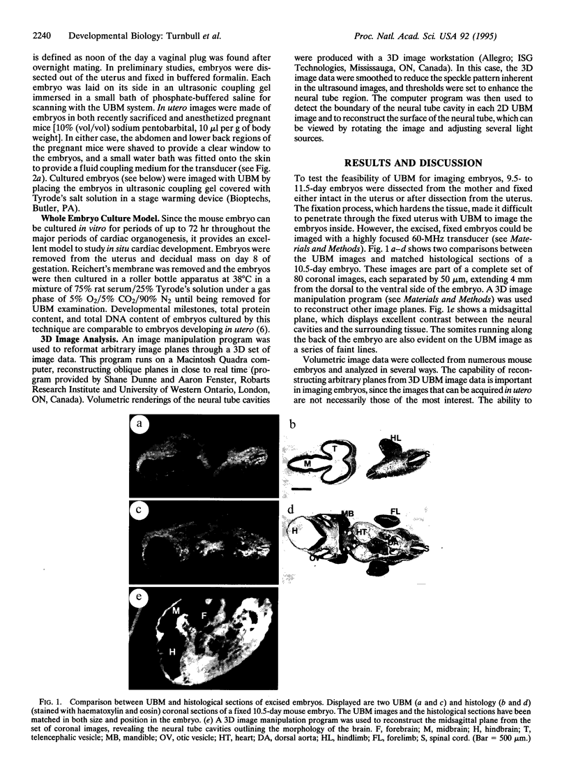
The history of developmental and genetic analysis in the mouse has made it the model of choice for studying mammalian embryogenesis. Presently lacking is a simple technique for efficiently analyzing early mouse mutant phenotypes in utero. We demonstrate application of a real-time imaging method called ultrasound backscatter microscopy for visualizing mouse early embryonic neural tubes and hearts. This method was used to study live embryos in utero between 9.5 and 11.5 days of embryogenesis, with a spatial resolution close to 50 microns. Ultrasound backscatter microscope images of cultured embryos made it possible to visualize the heart chambers. This noninvasive imaging method was also used for analyzing a neural tube defect. The midhindbrain deletion associated with a null mutation of the Wnt-1 protooncogene was easily recognizable on ultrasound backscatter microscope images of 10.5- and 11.5-day embryos. Computer-generated volumetric renderings of the neural tube cavities were made from three-dimensional image data. This allowed a much clearer definition of the Wnt-1 mutant phenotype. These imaging techniques should be of considerable use in studying mouse development in utero.
Full text
PDF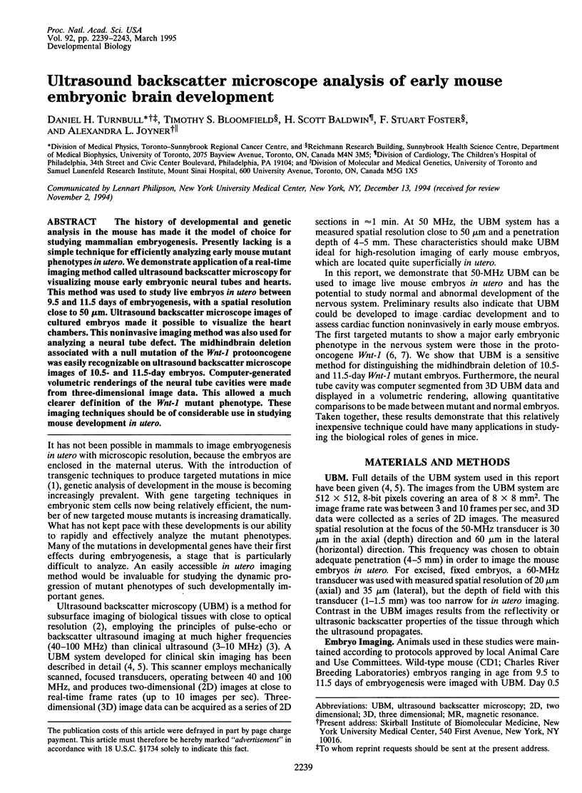

Images in this article
Selected References
These references are in PubMed. This may not be the complete list of references from this article.
- Capecchi M. R. Altering the genome by homologous recombination. Science. 1989 Jun 16;244(4910):1288–1292. doi: 10.1126/science.2660260. [DOI] [PubMed] [Google Scholar]
- Jacobs R. E., Fraser S. E. Magnetic resonance microscopy of embryonic cell lineages and movements. Science. 1994 Feb 4;263(5147):681–684. doi: 10.1126/science.7508143. [DOI] [PubMed] [Google Scholar]
- Joyner A. L., Guillemot F. Gene targeting and development of the nervous system. Curr Opin Neurobiol. 1994 Feb;4(1):37–42. doi: 10.1016/0959-4388(94)90029-9. [DOI] [PubMed] [Google Scholar]
- McMahon A. P., Bradley A. The Wnt-1 (int-1) proto-oncogene is required for development of a large region of the mouse brain. Cell. 1990 Sep 21;62(6):1073–1085. doi: 10.1016/0092-8674(90)90385-r. [DOI] [PubMed] [Google Scholar]
- Sherar M. D., Noss M. B., Foster F. S. Ultrasound backscatter microscopy images the internal structure of living tumour spheroids. Nature. 1987 Dec 3;330(6147):493–495. doi: 10.1038/330493a0. [DOI] [PubMed] [Google Scholar]
- Smith B. R., Johnson G. A., Groman E. V., Linney E. Magnetic resonance microscopy of mouse embryos. Proc Natl Acad Sci U S A. 1994 Apr 26;91(9):3530–3533. doi: 10.1073/pnas.91.9.3530. [DOI] [PMC free article] [PubMed] [Google Scholar]
- Thomas K. R., Capecchi M. R. Targeted disruption of the murine int-1 proto-oncogene resulting in severe abnormalities in midbrain and cerebellar development. Nature. 1990 Aug 30;346(6287):847–850. doi: 10.1038/346847a0. [DOI] [PubMed] [Google Scholar]
- Turnbull D. H., Starkoski B. G., Harasiewicz K. A., Semple J. L., From L., Gupta A. K., Sauder D. N., Foster F. S. A 40-100 MHz B-scan ultrasound backscatter microscope for skin imaging. Ultrasound Med Biol. 1995;21(1):79–88. doi: 10.1016/0301-5629(94)00083-2. [DOI] [PubMed] [Google Scholar]
- Wurst W., Auerbach A. B., Joyner A. L. Multiple developmental defects in Engrailed-1 mutant mice: an early mid-hindbrain deletion and patterning defects in forelimbs and sternum. Development. 1994 Jul;120(7):2065–2075. doi: 10.1242/dev.120.7.2065. [DOI] [PubMed] [Google Scholar]