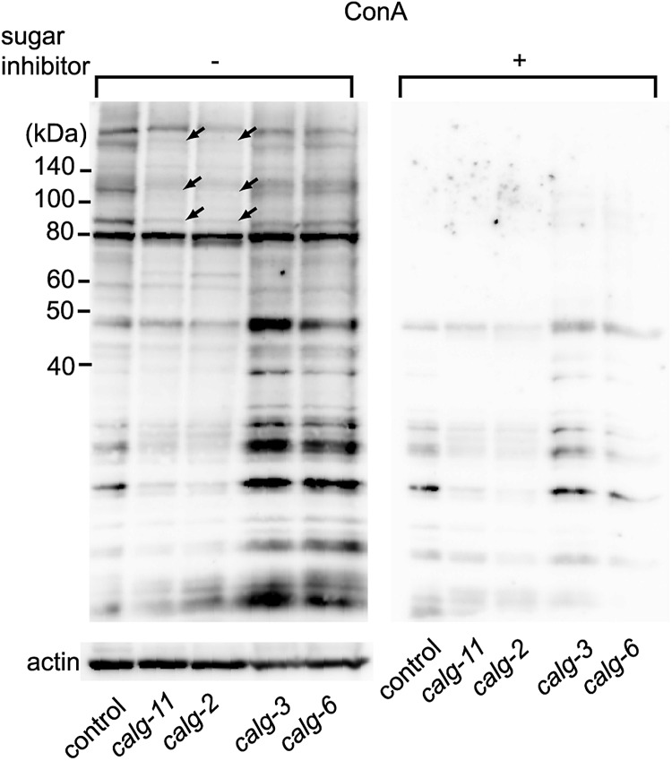
Fig. 7.
Knockdown of calg-11 or calg-2 shows reduction of ConA staining. ConA staining was decreased in the RNAi-mediated inhibition of cytoplasmic alg genes (calg-2 and calg-11) compared with the control. The arrows indicate the bands showing strong reduction. This reduction was not detected under the inhibition of ER-lumen alg genes (calg-3 and calg-6). The blotted membrane of the same samples shown at the right panel was incubated with sugar inhibitor plus HRP-conjugated ConA (Plus sign), and the left membrane was incubated with HRP-conjugated ConA in the absence of the sugar inhibitor (minus sign), followed by ECL detection. The bottom panel shows the anti-actin staining of the same lanes of the same samples as a control.

