Abstract
Cylindrospermopsin (CYN) is a cytotoxic alkaloid produced by cyanobacteria. The distribution of this toxin is expanding around the world and the number of cyanobacteria species producing this toxin is also increasing. CYN was detected for the first time in Turkey during the summer months of 2013. The responsible species were identified as Dolichospermum (Anabaena) mendotae and Chrysosporum (Aphanizomenon) ovalisporum. The D. mendotae increased in May, however, C. ovalisporum formed a prolonged bloom in August. CYN concentrations were measured by LC-MS/MS and ranged from 0.12 µg·mg−1to 4.92 µg·mg−1 as dry weight, respectively. Both species were the only cyanobacteria actively growing and CYN production was attributed solely to these species. Despite CYN production by C. ovalisporum being a well-known phenomenon, to our knowledge, this is the first report of CYN found in D. mendotae bloom.
Keywords: LC-MS/MS, cyanobacteria, Lake Iznik, cylindrospermopsin, Turkey
1. Introduction
Toxins produced by cyanobacteria are increasingly documented around the world and problems related to toxins in water bodies have received more attention in recent years. As a result, awareness of risk posed by cyanotoxins increased substantially and regulatory approaches for cyanotoxin risk management have been put in place. Apart from a few countries (Australia, New Zealand and Brazil), microcystin is considered as a toxin for regulation or guidance value. However, Cylindrospermopsin (CYN) is increasingly encountered worldwide, including all continents with the exception of Antarctica [1].
Cylindrospermopsin was originally identified and implicated as the causative agent of the Palm Island mystery disease in 1979 [2] and its structure elucidated by Ohtani et al. [3]. It is a biologically active alkaloid with hepatotoxic, cytotoxic and neurotoxic effects [4]. Recent studies showed that CYN has also genotoxic effects on hepatocytic and enterocytic model cell lines of the human [5] and even lymphocyte cells [6].
Although it is named after Cylindrospermopsis raciborski which originates from tropical lakes of Australia and Africa, there are other cyanobacterial species (Anabaena bergii, Anabaena lapponica, Aphanizomenon gracile, Aphanizomenon ovalisporum, A. flos-aquae, Lyngbya wollei, Raphidiopsis curvata, R. mediterranea and Umezakia natans), known to produce cylindrospermopsin [7]. C. raciborskii is considered an invasive species because of its dispersal capacity around the world, especially into temperate climates, and successful populations have been established in various areas in recent years. On the other hand, the blooms of C. raciborskii generally appear to be non-toxic in Europe [8]. Another important cylindrospermopsin producer is Chrysosporum (Aphanizomenon) ovalisporum. This species was first described by Forti [9] from Lake Küçükçekmece, Turkey. However, it has not been recorded in this lake after that. In contrast to C. raciborskii, all populations of C. ovalisporum produce cylindrospermopsin. Toxic populations forming blooms have been found in Israel [10], Greece [11], Spain [12], Italy [7], and also in Australia [13]. CYN production is also attributed to other Aphanizomenon and Anabaena species with some uncertainty. Brient et al. [14] detected CYN in four waterbodies dominated by either Aphanizomenon flos-aquae or Anabaena planktonica. Cylindrospermopsin was also found in two German lakes without clear evidence of a known producer [15].
It is important to detect the responsible species for cyanotoxin production, especially for risk management. This study describes the presence of CYN in Dolichospermum (Anabaena) mendotae bloom for the first time in the world and contributes to the increasing prevalence and distribution of CYN, since it is the first report of CYN from Turkey.
2. Results and Discussion
Lake Iznik is a deep, alkaline lake with a high conductivity. Recorded water temperatures were greater than 20 °C when the cyanobacteria bloom samples were collected and reaching 27.4 °C in August. Water transparency was high in May and a dramatic decrease was observed in August reflected in the chlorophyll-a values (Table 1). Nutrient values refer to meso-eutrophic character with a high chlorophyll-a, TP values and low transparency. SRP was higher in late spring; however, it decreased to under detection limits by the end of August when the C. ovalisporum bloom was over.
Table 1.
Some physicochemical characteristics of Lake Iznik taken on sampling days.
| Parameters | Units | Date | ||
|---|---|---|---|---|
| 22 May 2013 | 22 August 2013 | 28 August 2013 | ||
| Water Temperature | °C | 20.6 | 27.0 | 27.4 |
| pH | - | 8.9 | 8.6 | 9.2 |
| Conductivity | µS·cm−1 | 900 | 1037 | 1056 |
| Secchi depth | m | 7.9 | 1.4 | 1.4 |
| Nitrate + Nitrite | µg·L−1 | 125.6 | 391.4 | 69.6 |
| Soluble Reactive Phosphorus | µg·L−1 | 11.3 | 2.5 | <2 |
| Total Phosphorus | µg·L−1 | 24.8 | 12.7 | 13.4 |
| Total Nitrogen | µg·L−1 | 1889 | 1562 | 1253 |
| Chlorophyll-a | µg·L−1 | 4.2 | 20.9 | 23.1 |
The genus Dolichospermum is distinguished from Anabaena with some characteristics including the obligatory presence of gas vesicles, intercalary and frequently solitary heterocysts, undifferentiated apical cells and solitary or small clusters of filaments [16].
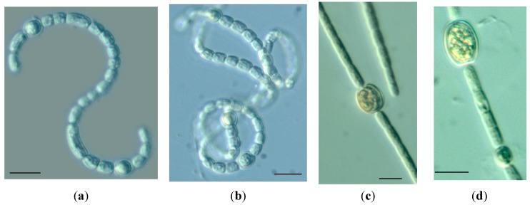
The trichomes of Dolichospermum from Lake Iznik are free floating, loosely coiled, solitary or sometimes in small clusters. Vegetative cells are cylindrical and there is no difference in the apical cell. Heterocysts are oval, slightly wider than vegetative cells. They are found solitary and intercalary in the trichome. Akinets are intercalary, solitary and elongated with rounded ends (Figure 1). The morphometric characteristics of the vegetative cells, heterocysts and akinetes of Dolichospermum are given in Table 2.
Figure 1.
Trichomes of Dolichospermum mendotae (a,b) and Chrysosporum ovalisporum (c,d) showing vegetative cells, heterocysts and akinets. Scale bars indicate 10 µm.
Table 2.
The size and the length/width ratio of vegetative cells, akinetes and heterocysts of D. mendotae and C. ovalisporum.
| Species | Morphological characters | Form | Mean ± SD | Min | Max | n |
|---|---|---|---|---|---|---|
| D. mendotae | ||||||
| Cell | ||||||
| length | 4.8 ± 0.7 | 3.4 | 5.8 | 50 | ||
| width | 4.2 ± 0.3 | 3.5 | 5 | |||
| l:w | 1.1 | 0.98 | 1.2 | |||
| Heterocyst | ||||||
| length | 6.5 ± 0.8 | 4.7 | 8.4 | 50 | ||
| width | 5.6 ± 0.5 | 4.1 | 6.6 | |||
| l:w | 1.2 | 1.15 | 1.3 | |||
| Akinete | ||||||
| length | 8.96 ± 1.8 | 7.2 | 11.3 | 5 | ||
| width | 4.8 ± 0.7 | 4.1 | 5.7 | |||
| l:w | 1.9 | 1.8 | 2 | |||
| C. ovalisporum | ||||||
| Cell | ||||||
| length | 6.1 ± 1.8 | 2.9 | 13.4 | 50 | ||
| width | 3.4 ± 0.4 | 2.7 | 4.4 | |||
| l:w | 1.8 | 1.1 | 3 | |||
| Heterocyst | ||||||
| length | 6.2 ± 0.7 | 4.9 | 7.9 | 50 | ||
| width | 4.3 ± 0.7 | 2.7 | 6.5 | |||
| l:w | 1.4 | 1.8 | 1.2 | |||
| Akinete | ||||||
| length | 12.1 ± 2 | 7.9 | 15.5 | 15 | ||
| width | 9.6 ± 1.8 | 6.9 | 14.5 | |||
| l:w | 1.3 | 1.1 | 1.07 |
The 16S rRNA gene regions were also amplified from Dolichospermum bloom sample from Lake Iznik and BLAST search was done to confirm the species. 16S rRNA gene sequence showed 99% identity to previously published 16S rRNA gene sequence of D. mendotae strain CHAB 4408 and CHAB 3512.
The species was identified as Dolichospermum mendotae. The dimensions of vegetative cells, akinetes and heterocysts are smaller in comparison to the literature. On the other hand, there is a direct link between the cell dimensions and nutrient availability. The phosphorus concentrations have a clear effect on cell dimensions. Zapomelova et al. [17] found that the cell length and length/width ratio decreased at higher phosphorus concentrations. Morphologically, the dimensions of the cells and the length/width ratio are important characteristics to identify the species. This situation was discussed on the identification of D. mendotae and D. sigmoideum, since the only morphological difference between them is the width and the length/width ratio of vegetative cells and cell wall constriction [18]. Therefore, these are considered as a joint morphological group. On the other hand, Zapomelova et al. [17] indicated that the cell dimensions of these two species are only slightly different and spanned in the range of both species in culture conditions. Therefore, we decided to define the species as D. mendotae instead of using the term of D. mendotae/sigmoideum complex, since it possesses a priority over D. sigmoideum according to Botanical code [17]. Moreover, 16S rRNA results confirmed the species as D. mendotae.
D. mendotae is a part of phytoplankton community of meso to eutrophic lakes [19]. The distribution area is very wide from the Southern to Northern hemisphere and has been detected in more than ten countries around the world (Table 3). Although it was recorded in different continents, generally these records are based on strains in culture collections or floristic studies in the water bodies and there is very little information about its biomass in environmental samples. The blooms are reported from Lednice ponds, Czech Republic [20], Lake Karhijarvi in Finland [21], and in Lake Stechlin [22].
Table 3.
The examples of geographical distribution and toxicity of D. mendotae and C. ovalisporum.
| Species | Status | Geographical Area | Toxicity | Reference |
|---|---|---|---|---|
| D. mendotae | Environmental | Finland | NA | [21] |
| Bangladesh | NA | [23] | ||
| Brazil | NA | [24] | ||
| Hungary | NA | [22] | ||
| Germany | NA | [25] | ||
| Poland | NA | [26] | ||
| Czech Republic | NA | [20] | ||
| Greece | NA | [27] | ||
| Strain | Finland | Ana | [28] | |
| Japan | NA | [29] | ||
| Denmark | NT | [30] | ||
| Spain | NT* | [31] | ||
| C. ovalisporum | Environmental | Israel | CYN | [10] |
| Greece | MCY | [11] | ||
| Spain | CYN | [12] | ||
| Italy | CYN | [7] | ||
| Strain | Israel | CYN | [10] | |
| Australia | CYN | [13] | ||
| USA | CYN | [32] | ||
| Spain | CYN | [31] |
NA: Not Analyzed; NT: No Anatoxin, Microcystin; NT*: No Anatoxin, Microcystin, Cylindrospermopsin and PSP; Ana: Anatoxin-a; CYN: Cylindrospermopsin; MCY: Microcystin.
Chrysosporum (Aphanizomenon) ovalisporum was identified in accordance with morphological features [19]. C. ovalisporum has straight, solitary, free-floating filaments. Vegetative cells are barrel shaped and are longer than wide (Table 2). Apical cell is rarely hyaline. Heterocysts are elipsoidal and placed in the middle of trichome and connected to the neighboring cell with a small bridge. Akinetes are oval to nearly spherical and distant from each other (Figure 1).
According to 16S rRNA analysis, the similarity between published sequences and the environmental sample in this study was 100% for C. ovalisporum. The identification of C. ovalisporum was therefore confirmed by both morphologic and molecular methods.
C. ovalisporum, now considered to be an invasive species [8] in Europe, was first detected in Lake Küçükçekmece located in the Istanbul metropolitan area in 1910 by Forti and dominated the phytoplankton community in Lake Kinneret in 1994. During the last decades, its distribution area expanded from Greece to the Iberian Peninsula (Table 3). Up to now, there has been no bloom record in Turkish freshwaters; however, this could be a result of insufficient monitoring efforts. This species is not restricted by special environmental conditions. It can be found in both deep, stratified waterbodies and shallow ponds [8]. The bloom in Lake Iznik was observed at the beginning of August when the water temperature was over 25 °C and conductivity was 1034 µS·cm−1. Temperature is an important driving factor for C. ovalisporum growth as well as dispersion. The bloom in different reservoirs took place at a temperature over 25 °C [11,12,33].
A total of three bloom samples were analyzed for cylindrospermopsin (Table 4). CYN was analyzed from one sample taken in May 2013 which contained D. mendotae and 2 samples collected in August 2013 when C. ovalisporum formed a bloom. D. mendotae was collected on a GF/C filters (Whatman, Maidstone, UK), whereas filaments of C. ovalisporum were collected with a plankton net and then separated from other species using its buoyancy and subsequently freeze-dried. Both species are the only cyanobacteria found in the bloom samples. A 100 µm length of C. ovalisporum filament was regarded as one unit, however, cell counts was done for D. mendotae,since it has tangled filaments. D. mendotae formed stripes on the surface in the morning of a calm day at Lake Iznik. On the other hand, C. ovalisporum reached higher numbers producing a heavy bloom as also indicated in biomass results. Moreover, it lasted for a longer period, nearly a month.
Table 4.
The abundance and biomass of D. mendotae and C. ovalisporum and cylindrospermopsin (CYN) concentrations.
| Species and toxin | Units | Date | |||
|---|---|---|---|---|---|
| 22 May 2013 | 22 August 2013 | 28 August 2013 | |||
| D. mendotae | Abundance | Cell·L−1 | 4.9 × 10⁷ | - | - |
| Biomass | µg·L−1 | 3471 | - | - | |
| C. ovalisporum | Abundance | Fil·L−1 | - | 2.3 × 10⁷ | 2.0 × 10⁷ |
| Biomass | µg·L−1 | - | 20,960 | 18,413 | |
| Cylindrospermopsin | µg·L−1 | 0.12 | 3.91 | 4.92 | |
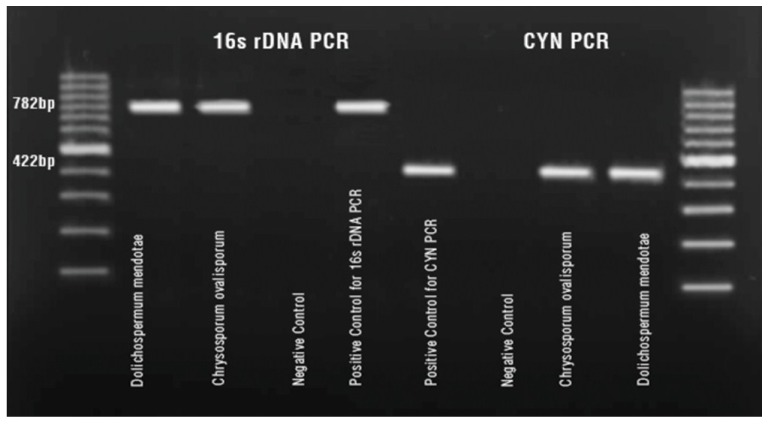
D. mendotae and C. ovalisporum bloom samples were investigated for the presence of polyketide synthase (cyrC and aoaC) genes of the CYN cluster. A PCR product at 422 bp was obtained in bothbloom samples showing the potential of CYN production of these two species (Figure 2) [34].
Figure 2.
PCR assay for 16SrRNA and polyketide synthase genes of CYN in bloom samples.
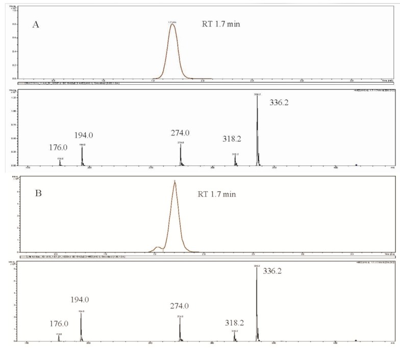
CYN production was also detected using LC-MS/MS based on extracted ion chromatograms and MS/MS spectra of CYN standards and samples. The retention time of samples matched that of the standard, 1.7 min (Figure 3).
Figure 3.
Extracted Ion Chromatogram (EIC) of m/z 416 > 194 (Upper panel) and MS/MS spectrum of m/z 416 (Lower panel) of CYN standart (A), C. ovalisporum (B), D. mendotae (C).
This is the first report of CYN production of D. mendotae bloom. It is confirmed both the presence of polyketide synthase genes and LC-MS/MS. The only toxin reported to be produced by D. mendotae in the literature was anatoxin-a (ATX-a) [29]. The Ana producing strain was isolated from Lake Säyhteenjärvi located in the Southern part of Finland. Cires et al. [32] screened the cyanotoxin production of Nostocalean strains including D. mendotae from Spain and could not detect any MC, ATX, CYN and PSP toxin production or responsible genes in their strain. Similarly, MC and ATX production were not detected in D. mendotae strain 57 isolated from Lake Velje Sø, Denmark [31]. The lack of information of toxin production by this species might be due to the bloom formation patterns. This species generally produce blooms at the beginning of the growth season, in May, as it is observed in Lake Iznik and the bloom formation is generally very weak. The stripes can be found on the surface of water when it is very calm and disappear easily with a light wind. This makes it difficult to detect the bloom.
C. ovalisporum became the main CYN producer in Southern Europe. Our results showed that the C. ovalisporum bloom in Lake Iznik had higher CYN concentration compared to the reported concentrations from other countries [7,12]. Moreover, the detected concentrations fall within the upper range of maximum CYN concentrations found in isolated strains of different CYN producers [32]. On the other hand, the concentration of CYN detected in the D. mendotae bloom is markedly lower (0.12 mg·g−1) in comparison to C. ovalisporum. As only intracellular CYN was detected in D. mendotae sample, the result could be an underestimate. The bloom period coincided with the growth season of Cladocera species in Lake Iznik and their gut contents were full of cyanobacteria. Therefore, it is necessary to confirm the extra- and intracellular CYN production with isolated strains of D. mendotae. Moreover, the effect of grazers on the changes of intra- and extracellular CYN concentrations also needs to be clarified.
3. Experimental Section
3.1. Study Site
Lake Iznik is a natural lake located in the western part of Turkey. It is the fifth biggest lake of Turkey with a surface area of 308 km2 and maximum depth of 65 m. Partially treated sewage and non-point pollution of fertilizers and pesticides from surrounding agricultural areas reach the lake [35]. Lake water is also used for irrigation and is a popular place for recreation during summer months.
3.2. Sample Collection
Samples were collected from surface waters when there was a surface bloom of Dolichospermum (Anabaena) mendotae in May 2013 and another bloom of Chrysosporum (Aphanizomenon) ovalisporum in August 2013. The bloom of C. ovalisporum persisted nearly one month and samples were collected twice with one week intervals.
Samples for microscopic observation were immediately fixed with 1% Lugol’s solution. Water samples for nutrient and chlorophyll-a analysis were stored in cold and dark conditions and transported to the laboratory. Temperature, conductivity, dissolved oxygen and pH was measured by portable multiparameter (Sonde Model 6600, YSI Incorporated, Yellow Springs, OH, USA) and Secchii disc measurements were done with a 20 cm black and white device.
Cyanobacterial species were enumerated according to Utermöhl [36] and 100 µm length of C. ovalisporum filament was regarded as one unit. However, since D. mendotae had tangled filaments, the measurement of filament lengths was not possible; therefore, cell counts were performed. After the measurement of the dimensions biovolumes were calculated using the formulae of Hillebrand et al. [37]. Chlorophyll-a measurements were performed according to Nusch [38]. Nutrient analysis was done according to APHA, AWWA and WEF [39].
3.3. Molecular Analysis
Total genomic DNA extraction from lyophilized samples was performed using XS extraction buffer containing 1% potassium-methylxanthogenate; 800 mM ammonium acetate; 20 mM EDTA; 1% SDS; 100 mM Tris-HCI (pH 7.4) [40].
16s rDNA amplification was performed using forward primer (27F; 5' AGA GTT TGA TCC TGG CTC AG 3') and reverse primer (809R; 5' GCT TCG GCA CGG CTC GGG TCG ATA 3'). Thermal cycling was performed at 92 °C for 2 min followed by 35 cycles of 94 °C for 10 s, 60 °C for 20 s and 72 °C for 1 min and a final extension step at 72 °C for 5 min [41].
Amplified PCR products of 16S rDNA were visualized on 1.5% agarose gels stained with ethidium bromide and photographed under UV transillumination. Published sequences were obtained from NCBI databases (http://www.ncbi.nlm.nih.gov). Genbank accession numbers of the sequences obtained in this study are KM360484 for D. mendotae and KM360485 for C. ovalisporum.
CYN polyketide synthase (aoaC and cyrC) genes of CYN gene cluster were amplified using forward primer (K18; 5' CCTCGCACATAGCCATTTGC 3') and reverse primer (M4; 5' GAAGCTCTGGAATCCGGTAA 3'). Thermal cycling conditions for the PCR were 94 °C for 10 min; 94 °C for 30 s, 45 °C for 30 s, and 72 °C for 30 cycles; 72 °C for 7 min [34]. PCR products were visualized on agarose gel and DNA molecular weight marker was used to indicate the size of the amplification products. Cylindrospermopsis raciborskii AWT 205 was used as a positive control.
3.4. CYN Extraction
Water sample was filtered through a GF/C filter in May 2013 when D. mendotae increased in high numbers and C. ovalisporum was separated from water by using its buoyancy in August 2013. Samples were lyophilized and extracted with 100% methanol containing 0.1% TFA and the extracts dried with nitrogen. The dry extracts were dissolved in 300 μL of water and filtered through GHP Acrodisc 13 mm syringe filters (Pall Life Sciences, Ann Arbor, MI, USA) with 0.2 µm GHP membrane. Then samples were analyzed for cylindrospermopsin using LC-MS/MS as described below.
3.5. CYN Analysis Using the LC-MS/MS
The LC-MS/MS experiments were carried out on an Agilent Technologies (Waldbronn, Germany) 1200 Rapid Resolution (RR) LC coupled to a Bruker Daltonics HCT Ultra Ion trap MS (Bremen, Germany) with electrospray (ESI) source. The 1200 RR LC system included a binary pump, vacuum degasser, SL autosampler, and thermostated column compartment set at 40 °C. Separation of the toxins was achieved on a Supelco (Bellefonte, PA, USA) Ascentis C18 column, (50 mm × 3 mm I.D. with 3 µm particles) protected by a 4 × 2 mm C8 guard column. The mobile phase consisted of solvents A, 99% water–1% acetonitrile–0.1% formic acid; B, acetonitrile–0.1% formic acid with the following linear gradient program: 0 min 0% B, 2.5 min 0% B, 2.6 min 50% B, 4 min 50% B, 4.1 min 0% B; stop time 10 min. The flow-rate was 0.5 mL·min−1 and the injection volume was 5 µL.
The ion trap was operated utilizing positive electrospray ion mode. The ion source parameters were set as follows: evaporator temperature 350 °C, nebulizer pressure 40 psi and dry gas flow 10.0 L·min−1. The capillary voltage was set at 4.0 kV. The MS scan range was m/z 395 to 440 and MS/MS fragmentation of the target mass m/z 416 was employed to get MS/MS spectra. The ICC target was set to 200,000 with a maximum accumulation time of 100 ms.
CYN in the samples was identified by comparing the retention time (1.7 min) and MS/MS fragmentation with those of the pure CYN standard (12.5 µM CRM-CYN, NRC-IMB, Halifax, NS, Canada). 1:10, 1:50, 1:100, 1:200 and 1:500 aqueous dilutions of the standard were analyzed for quantification purposes.
4. Conclusions
In conclusion, reports on the CYN distribution around the world have been expanding due to an increasing awareness of cyanotoxin problems. CYN became the second most studied freshwater toxin after microcystin and pose more hazards to water users since the liberation of CYN to surrounding water is much higher compared to other toxins. Therefore, the identification of CYN producers is of great importance and the determination of CYN in the bloom of D. mendotae was revealed for the first time with this study. Moreover, this is the first report of CYN in Turkish freshwaters. In terms of water management, there are very limited national regulations or guideline values considering CYN in contrast to microcystin [42]. On the other hand, the production of CYN by two different cyanobacteria species growing in the same water body in different time of growth season increase the risk to exposure to the toxin. Therefore, to identify the producers and also to detect the CYN concentrations from different regions will be helpful to manage and prevent potential health hazards to people.
Acknowledgments
This study was financially supported by the TUBITAK Project (102Y209). The authors would like to acknowledge the European Cooperation in Science and Technology, COST Action ES 1105 “CYANOCOST” for adding value to this study through networking and knowledge sharing with European researchers. Also, we would like to thank Eliska (Zapomelova) Kozlíková for her help in the identification of cyanobacteria species and James Metcalf and anonymous reviewers for their valuable comments to improve the manuscript.
Author Contributions
Reyhan Akcaalan and Meric Albay designed the project. Latife Köker and Ayça Oğuz performed the analysis. Lisa Spoof and Jussi Meriluoto performed the CYN measurements and Reyhan Akcaalan and Meric Albay wrote the manuscript. Lisa Spoof and Jussi Meriluoto performed the final edition of the manuscript.
Conflicts of Interest
The authors declare no conflict of interest.
References
- 1.Kinnear S. Cylindrospermopsin: A decade of progress on bioaccumulation research. Mar. Drugs. 2010;8:542–564. doi: 10.3390/md8030542. [DOI] [PMC free article] [PubMed] [Google Scholar]
- 2.Griffiths D.J., Saker M.L. The Palm Island mystery disease 20 years on: A review of research on the cyanotoxin cylindrospermopsin. Environ. Toxicol. 2003;18:78–93. doi: 10.1002/tox.10103. [DOI] [PubMed] [Google Scholar]
- 3.Ohtani I., Moore R.E., Runnegar M.T. Cylindrospermopsin: A potent hepatotoxin from the blue-green alga Cylindrospermopsis raciborskii. J. Am. Chem. Soc. 1992;114:7941–7942. doi: 10.1021/ja00046a067. [DOI] [Google Scholar]
- 4.Gutiérrez-Praena D., Jos A., Pichardo S., Cameán A.M. Oxidative stress responses in tilapia (Oreochromis niloticus) exposed to a single dose of pure cylindrospermopsin under laboratory conditions: Influence of exposure route and time of sacrifice. Aquat. Toxicol. 2011;105:100–106. doi: 10.1016/j.aquatox.2011.05.015. [DOI] [PubMed] [Google Scholar]
- 5.Bazin E., Mourot A., Humpage A.R., Fessard V. Genotoxicity of a freshwater cyanotoxin, cylindrospermopsin, in two human cell lines: Caco-2 and HepaRG. Environ. Mol. Mutagen. 2010;51:251–259. doi: 10.1002/em.20539. [DOI] [PubMed] [Google Scholar]
- 6.Žegura B., Gajski G., Štraser A., Garaj-Vrhovac V. Cylindrospermopsin induced DNA damage and alteration in the expression of genes involved in the response to DNA damage, apoptosis and oxidative stress. Toxicon. 2011;58:471–479. doi: 10.1016/j.toxicon.2011.08.005. [DOI] [PubMed] [Google Scholar]
- 7.Messineo V., Melchiorre S., di Corcia A., Gallo P., Bruno M. Seasonal succession of Cylindrospermopsis raciborskii and Aphanizomenon ovalisporum blooms with cylindrospermopsin occurrence in the volcanic Lake Albano, Central Italy. Environ. Toxicol. 2010;25:18–27. doi: 10.1002/tox.20469. [DOI] [PubMed] [Google Scholar]
- 8.Sukenik A., Hadas O., Kaplan A., Quesada A. Invasion of Nostocales (cyanobacteria) to subtropical and temperate freshwater lakes—Physiological, regional, and global driving forces. Front. Microbiol. 2012;3 doi: 10.3389/fmicb.2012.00086. [DOI] [PMC free article] [PubMed] [Google Scholar]
- 9.Forti A. Diagnoses Myxophycearum Novarum, Series IV. Volume 12. Atti e Memorie dell’Accademia di Agricoltura, Scienze e Lettere, Arti e Commercia di Verona; Verona, Italy: 1911. pp. 3–5. [Google Scholar]
- 10.Banker R., Carmeli S., Hadas O., Teltsch B., Porat R., Sukenik A. Identification of cylindrospermopsin in Aphanizomenon ovalisporum (cyanophyceae) isolated from Lake Kinneret, Israel. J. Phycol. 1997;33:613–616. doi: 10.1111/j.0022-3646.1997.00613.x. [DOI] [Google Scholar]
- 11.Gkelis S., Moustaka-Gouni M., Sivonen K., Lanaras T. First report of the cyanobacterium Aphanizomenon ovalisporum Forti in two Greek lakes and cyanotoxin occurrence. J. Plankton Res. 2005;27:1295–1300. doi: 10.1093/plankt/fbi085. [DOI] [Google Scholar]
- 12.Quesada A., Moreno E., Carrasco D., Paniagua T., Wormer L., Hoyos C.D., Sukenik A. Toxicity of Aphanizomenon ovalisporum (Cyanobacteria) in a Spanish water reservoir. Eur. J. Phycol. 2006;41:39–45. doi: 10.1080/09670260500480926. [DOI] [Google Scholar]
- 13.Shaw G.R., Sukenik A., Livne A., Chiswell R.K., Smith M.J., Seawright A.A., Norris R.L., Eaglesham G.K., Moore M.R. Blooms of the cylindrospermopsin containing cyanobacterium, Aphanizomenon ovalisporum (Forti), in newly constructed lakes, Queensland, Australia. Environ. Toxicol. 1999;14:167–177. doi: 10.1002/(SICI)1522-7278(199902)14:1<167::AID-TOX22>3.0.CO;2-O. [DOI] [Google Scholar]
- 14.Brient L., Lengronne M., Bormans M., Fastner J. First occurrence of cylindrospermopsin in freshwater in France. Environ. Toxicol. 2009;24:415–420. doi: 10.1002/tox.20439. [DOI] [PubMed] [Google Scholar]
- 15.Fastner J., Rücker J., Stueken A., Preussel K., Nixdorf B., Chorus I., Koehler A., Wiedner C. Occurrence of the cyanobacterial toxin cylindrospermopsin in northeast Germany. Environ. Toxicol. 2007;22:26–32. doi: 10.1002/tox.20230. [DOI] [PubMed] [Google Scholar]
- 16.Wacklin P., Hoffmann L., Komárek J. Nomenclatural validation of the genetically revised cyanobacterial genus Dolichospermum (Ralfs ex Bornet et Flahault) comb. nova. Fottea. 2009;9:59–64. [Google Scholar]
- 17.Zapomělová E., Řeháková K., Jezberová J., Komárková J. (Polyphasic characterization of eight planktonic Anabaena strains (Cyanobacteria) with reference to the variability of 61 Anabaena populations observed in the field. Hydrobiologia. 2010;639:99–113. doi: 10.1007/s10750-009-0028-y. [DOI] [Google Scholar]
- 18.Zapomelova E., Rehakova K., Znachor P., Komarkova J. Morphological diversity of coiled planktonic types of the genus Anabaena (cyanobacteria) in natural populations: Taxonomic consequences. Cryptogam. Algol. 2007;28:353–371. [Google Scholar]
- 19.Komárek J. Cyanoprokaryota: 3rd Part: Heterocytous Genera. 1st ed. Springer-Verlag; Berlin/Heidelberg, Germany: 2013. p. 1130. [Google Scholar]
- 20.Kopp R., Skácelová O., Heteša J., Marvan P., Bešta T., Zapomělová E., Straková L., Bohunıcká M. A Hundred Years of Phycological Research in the Lednice Pond: The Impact of Environmental Conditions on the Development of Cyanobacteria and Algae. Moravian Museum; Brno, Czech Republic: 2012. [Google Scholar]
- 21.Nõges P., Viirret M. Environmental conditions and the development of Planktonema lauterbornii Schmidle in phytoplankton of Karhijärvi, a lake in SW Finland. Boreal Environ. Res. 2001;6:181–190. [Google Scholar]
- 22.Padisák J., Hajnal E., Krienitz L., Lakner J., Üveges V. Rarity, ecological memory, rate of floral change in phytoplankton—And the mystery of the Red Cock. Hydrobiologia. 2010;653:45–64. doi: 10.1007/s10750-010-0344-2. [DOI] [Google Scholar]
- 23.Affan A., Jewel A.S., Haque M., Khan S., Lee J.B. Seasonal cycle of phytoplankton in aquaculture ponds in Bangladesh. Algae. 2005;20:43–52. doi: 10.4490/ALGAE.2005.20.1.043. [DOI] [Google Scholar]
- 24.Werner V.R., Laughinghouse H.D., IV Bloom-forming and other planktonic Anabaena (Cyanobacteria) morphospecies with twisted trichomes from Rio Grande do Sul State, Brazil. Nova Hedwig. 2009;89:17–47. doi: 10.1127/0029-5035/2009/0089-0017. [DOI] [Google Scholar]
- 25.Täuscher L. Checklisten und Gefährdungsgrade der Algen des Landes Brandenburg. Verh. Bot. Ver. Berl. Brandenbg. 2011;144:177–192. [Google Scholar]
- 26.Paturej E., Gutkowska A., Mierzejewska J. A review of biological research in the Vistula Lagoon. Oceanol. Hydrobiol. Stud. 2012;41:81–88. doi: 10.2478/s13545-012-0042-y. [DOI] [Google Scholar]
- 27.Katsiapi M., Moustaka-Gouni M., Vardaka E., Kormas K.A. Different phytoplankton descriptors show asynchronous changes in a shallow urban lake (L. Kastoria, Greece) after sewage diversion. Fundam. Appl. Limnol. Arch. Hydrobiol. 2013;182:219–230. doi: 10.1127/1863-9135/2013/0362. [DOI] [Google Scholar]
- 28.Rantala J., Sivonen K., Luukkainen R., Niemelä S.I. Anatoxin-a concentration in Anabaena and Aphanizomenon under different environmental conditions and comparison of growth by toxic and non-toxic Anabaena-strains—A laboratory study. J. Appl. Phycol. 1993;5:581–591. doi: 10.1007/BF02184637. [DOI] [Google Scholar]
- 29.Li R., Watanabe M., Watanabe M.M. Taxonomic studies of planktic species of Anabaena based on morphological characteristics in cultured strains. Hydrobiologia. 2000;438:117–138. doi: 10.1023/A:1004170230774. [DOI] [Google Scholar]
- 30.Gugger M., Lyra C., Henriksen P., Couté A., Humbert J.F., Sivonen K. Phylogenetic comparison of the cyanobacterial genera Anabaena and Aphanizomenon. Int. J. Syst. Evol. Microbiol. 2002;52:1867–1880. doi: 10.1099/ijs.0.02270-0. [DOI] [PubMed] [Google Scholar]
- 31.Cirés S., Wörmer L., Ballot A., Agha R., Wiedner C., Velázquez D., Casero M.C., Quesada A. Phylogeography of cylindrospermopsin and paralytic shellfish toxin-producing Nostocales cyanobacteria from Mediterranean Europe (Spain) Appl. Environ. Microbiol. 2014;80:1359–1370. doi: 10.1128/AEM.03002-13. [DOI] [PMC free article] [PubMed] [Google Scholar]
- 32.Yılmaz M., Phlips E.J., Szabo N.J., Badylak S. A comparative study of Florida strains of Cylindrospermopsis and Aphanizomenon for cylindrospermopsin production. Toxicon. 2008;51:130–139. doi: 10.1016/j.toxicon.2007.08.013. [DOI] [PubMed] [Google Scholar]
- 33.Hadas O., Pinkas R., Malinsky-Rushansky N., Shalev-Alon G., Delphine E., Berner T., Sukenik A., Kaplan A. Physiological variables determined under laboratory conditions may explain the bloom of Aphanizomenon ovalisporum in Lake Kinneret. Eur. J. Phycol. 2012;37:259–267. doi: 10.1017/S0967026202003645. [DOI] [Google Scholar]
- 34.Fergusson K.M., Saint C.P. Multiplex PCR assay for Cylindrospermopsis raciborskii and cylindrospermopsin-producing cyanobacteria. Environ. Toxicol. 2003;18:120–125. doi: 10.1002/tox.10108. [DOI] [PubMed] [Google Scholar]
- 35.Albay M., Aykulu G. Invertebrate grazer-epiphytic algae interactions on submerged macrophytes in a mesotrophic Turkish lake. TrJFAS. 2002;19:247–258. [Google Scholar]
- 36.Utermöhl H. Zur vervollkommnung der quantitativen phytoplankton-methodik. Mitt. Int. Ver. Theor. Angew. Limnol. 1958;9:1–38. [Google Scholar]
- 37.Hillebrand H., Durselen C.D., Kirschtel D., Pollinger U., Zohary T. Biovolume calculation for pelagic and benthic microalgae. J. Phycol. 1999;35:403–424. doi: 10.1046/j.1529-8817.1999.3520403.x. [DOI] [Google Scholar]
- 38.Nusch E. Comparison of different methods for chlorophyll and phaeopigment determination. Arch. Hydrobiol. Beih. 1980;14:14–36. [Google Scholar]
- 39.American Public Health Association (APHA) American Water Works Association (AWWA) Water Environment Federation (WEF) Standart Methods for the Examination of Water and Wastewater. 17th ed. American Public Health Association; Washington, DC, USA: 1989. [Google Scholar]
- 40.Tillett D., Neilan B.A. Xanthogenate nucleic acid isolation from cultured and environmental cyanobacteria. J. Phycol. 2000;36:251–258. doi: 10.1046/j.1529-8817.2000.99079.x. [DOI] [Google Scholar]
- 41.Saker M.L., Jungblut A.D., Neilan B., Rawn D.F.K., Vasconcelos V.M. Detection of microcystin synthetase genes in health food supplements containing the freshwater cyanobacterium Aphanizomenon flos-aquae. Toxicon. 2005;46:555–562. doi: 10.1016/j.toxicon.2005.06.021. [DOI] [PubMed] [Google Scholar]
- 42.Chorus I. Current Approaches to Cyanotoxin Risk Assessment, Risk Management and Regulations in Different Countries. Federal Environment Agency (Umweltbundesamt); Dessau-Rosslau, Germany: 2012. [Google Scholar]


