Abstract
Transfection of DNA has been invaluable for biological sciences and with recent advances to organotypic brain slice preparations, the effect of various heterologous genes could thus be investigated easily while maintaining many aspects of in vivo biology. There has been increasing interest to transfect terminally differentiated neurons for which conventional transfection methods have been fraught with difficulties such as low yields and significant losses in viability. Biolistic transfection can circumvent many of these difficulties yet only recently has this technique been modified so that it is amenable for use in mammalian tissues.
New modifications to the accelerator chamber have enhanced the gene gun's firing accuracy and increased its depths of penetration while also allowing the use of lower gas pressure (50 psi) without loss of transfection efficiency as well as permitting a focused regioselective spread of the particles to within 3 mm. In addition, this technique is straight forward and faster to perform than tedious microinjections. Both transient and stable expression are possible with nanoparticle bombardment where episomal expression can be detected within 24 hr and the cell survival was shown to be better than, or at least equal to, conventional methods. This technique has however one crucial advantage: it permits the transfection to be localized within a single restrained radius thus enabling the user to anatomically isolate the heterologous gene's effects. Here we present an in-depth protocol to prepare viable adult organotypic slices and submit them to regioselective transfection using an improved gene gun.
Keywords: Neuroscience, Issue 92, Biolistics, gene gun, organotypic brain slices, Diolistic, gene delivery, staining
Introduction
Originally the biolistic technique, a turn-of-phrase for “biological ballistics”, was established for particle-mediated gene transfer into plant cells1. This physical method of cell transformation accelerates micro- or nanoparticles at high velocity to overcome the physical barriers of the impermeable cell membranes in order to deliver cargos such as DNA or dyes. Because it does not depend on specific ligand-receptors and/or the biochemical properties at the cell surface membranes, particle-mediated gene transfer can be readily applied to a variety of biological systems such as organotypic brain slices.
Using organotypic slices have advantages over other in vitro platforms since they maintain many anatomical and biochemical properties that are pertinent to in vivo biology2-4. The slices mostly conserve the local architectural characteristics from where they have originated and preserve neurochemical activity and connectivity of the synapses. The use of brain slices for basic research, and in pharmaceutical endeavors, has concomitantly increased with the number of possible biotechnological manipulations to measure and monitor the neurobiological behaviors of the brain in an in vivo like context3,5-7. The major advantages for using organotypic slice-based assay systems is that it provides easy experimental control and allows precise manipulations of extracellular environments.
Fruitfully, organotypic slice culture systems have been established from a variety of brain regions such as, but not restricted to, the cortex, spinal cord, and cerebellum8-10. Furthermore, a number of cocultures have been demonstrated, which allow the assessment of intercellular communication across distal brain regions as well as between neurons and pathological cells11,12. Many protocols have already been established to successfully culture organotypic slices and can maintain long-term viability and many recent studies now utilize the membrane interface methods and various modifications to it13. This principle maintains the organotypic slices at the interface between the medium and the incubator's humidified atmosphere by placing the slices on a porous membrane filter. The medium can thus provide sufficient nutrients to feed the organotypic slices via capillary motion. Typically slices have been prepared from early postnatal animals (3 - 9 days old; P3 - 9). However, brain tissues from these slices display a high level of cellular plasticity and have an inherent resistance to mechanical stresses, which is helpful to obtain viable cultures, yet mature synapses and neuroanatomical circuitry have not fully developed in vivo until 2 to 3 weeks of age14. For example, previous observations had shown that hippocampal slices obtained from P0 - 1 neonates, although highly viable following preparation, gradually lost some morphological characteristics. Essentially, they were shown to be unsuitable for long-term cultures suggesting immature cells were more likely to de-differentiate compared to organotypic cultures from older animals15,16. For this reason our method has been optimized for adult organotypic brain slices at which maturation and architectural development have reached their terminal stages13,17-21. Nevertheless, this method is also suitable for neonate and juvenile organotypic slices.
Once the viable organotypic slices have been produced the entire plate containing the slices can be brought to the biolistic mount and submitted to regioselective delivery and transfection. Proper mounting of the gene gun (as described in Figure 1), oriented 90° at a distance of 10 mm directly over the slices (from the aperture to the tissue), permits the rapid biolistic delivery of the 40 nm gold particle coated cargos. These cargos such as dyes and fluorescent DNA vectors, as well as any gene of interest, are readily delivered into the viable slices with ease. This method thus describes the protocol necessary to deliver, in a regioselective manner, cargo coated gold nanoparticles into viable organotypic brain slices. The gene gun barrel and optimal mounting can be seen in Figure 1. For further information regarding the improved barrel and nanoparticle ballistics, please see O'Brien, et al.17.
Subsequent refinements are also indicated for the user to optimize conditions such as the proper amount of gold delivered per target area, defined as Microcarrier Loading Quantity (MLQ), and to determine the amount of DNA loaded per mg of gold, defined as DNA Loading Ratio (DLR). Prior to precipitating DNA onto gold particles and loading them into the Tefzel tubing that comprises the actual gene gun 'cartridges', it is necessary to calculate the amount of DNA and gold required for each transfection which can differ slightly among tissues and conditions (ratio should be maintained between 1 mg gold and 1 µg DNA to 1:5). It is vital to prepare the proper proportion of MLQ to DLR as otherwise DNA coated gold particles could adhere together forming larger than expected agglomeration, which could reduce the overall homogeneity of the biolistic spread as well as increase tissue damage and cytotoxicity.
This method shows the recent improvements in biolistic delivery, which have been tested and determined to be amenable for neonatal, juvenile and adult organotypic brain slices. Furthermore, by exploiting the gene gun barrel's reduced biolistic spread, the user is now able to selectively transfect a punctual region in the brain. Following the appropriate incubation time, the expression of the fluorescent proteins, which reaches its maximum rates between 24 and 48 hr, can be visualized by epifluorescence and confocal microscopy. These fluorophores permit the morphological analysis and localization of individual cells within biologically relevant structures. However, the regioselective modulation of the organotypic slices by other heterologous genes can also be monitored by any other tests amenable to these preparations. Possible working strategies for the localized gene delivery are presented in Figure 2.
Protocol
The use of animals and animal tissues should strictly adhere to ethical committee approval under local rules and regulation. All tissues obtained during this study adhered to the MRC-LMB's animal experimentation guidelines.
1. Preparation of Materials & Culture Media
Prepare all solutions using Millipore water; use only analytical grade reagents. Prepare and store all reagents at RT (unless indicated otherwise).
Make 500 ml of HEPES-buffered saline (10 mM HEPES, 120 mM NaCl pH 7.2). Filter through a 0.2 µm sterile filter unit. Store at 4 °C.
Make 500 ml of Phosphate-buffered saline (PBS; 10 mM Na2HPO4, 2 mM KH2PO4, 137 mM NaCl, 2.7 mM KCl, pH 7.4). Sterilize by autoclaving.
Prepare neuronal medium by supplementing DMEM (Dulbecco’s modified eagle’s medium) with 25 mM HEPES, 10% Fetal calf serum, 30 mM glucose, 1:100 N2 supplement, penicillin-streptomycin 1,100 U/ml; pH 7.2. Filter with a 0.2 µm sterile filter unit. Store at 4 °C.
Make polyvinylpyrrolidone (PVP) stock solution with 20 mg PVP in 1 ml 100% Ethanol. Aliquot this into 1 ml amounts, freeze and store at -20 °C. Working solution is 0.05 mg/ml, therefore add 10 µl of PVP stock solution to every 4 ml of 100% ethanol.
Prepare a 0.05 M spermidine stock solution in Millipore water; adjust the pH to 7.2.
Make a 2% agarose solution in DMEM (2 g electrophoresis grade agarose in 100 ml DMEM in a sterilized screw cap bottle). Microwave this until the agarose has dissolved. Store in the 4 °C fridge for several weeks, if necessary.
2. Preparation of Organotypic Slices
Euthanize C57 Black 6 mice of the desired age by CO2 asphyxiation followed by decapitation. Remove the brain using lateral skull cuts starting at the foramen magnum and ending at the olfactory lobes. Gently lift the skull from the rear to expose the brain.
Cut between the olfactory lobes and the frontal cortex, and rostral to the cerebellum. Gently lift the rostral part of the brain and cut the optic nerves. Finally, remove the brain and place it in a petri dish filled with ice cold HEPES-buffered saline on ice.
Using a dissecting microscope, peel away the pia using fine forceps; carefully remove blood vessels and meninges around the brain.
Place the fresh brain into a small recipient mold and cover it with the agarose solution cooled to slightly above RT. Then rapidly cool the mold to 4 °C by placing it on ice. NOTE: Process the slices as rapidly as possible to avoid loss of viability.
When the agarose has set (5 - 10 min), remove the agarose-embedded brain from the mold (trim if necessary), and then glue it to the vibroslicer platform. Place this into a vibroslicer chamber containing sterile ice cold PBS. Disinfect the vibroslicer chamber before being used with 70% ethanol to minimize subsequent contaminations.
Cut the brain using a custom built vibroslicer or commercial equivalent. The model concept was to design a tissue slicer that could generate large amplitude and high frequency movements horizontal to the edge of the blade while minimizing vertical vibrations.
Use the oscillation frequency of 90 Hz, at an amplitude of 1.5 - 2.0 mm, and the cutting blade positioned at an angle of 15° to the horizontal plane; set the mounting block holding the agarose-embedded brain to move at 1.7 mm/min towards the blade. Collect sections (150 µm) into a chamber containing ice cold culture media (DMEM Pen/Strep + 10% FCS). View cutting and manipulation of the tissue slices as a supplementary video in Arsenault and O'Brien13.
Gently place the slices into cell culture inserts (0.4 µm, 30 mm diameter) in a 6 multi-well tray with the culture media on the outside of the insert, and incubate in a humidified incubator at 37 °C with 5% CO2. NOTE: For proper tissue viability, be extremely gentle while transferring and manipulating the slices. Also, too much fluid will prevent the organotypic slices from adhering to the membrane of the filter insert. If this happens, remove the excess fluid or plate again.
Although organotypic slices can be cultured for a few weeks, proceed to transfection no later than 4 days after plating for best transfection yields. NOTE: To avoid glial overgrowth in long term culture conditions, use AraC or serum free medium2.
3. Preparation of DNA-coated Micro & Nano-projectiles
- Prepare nano-projectiles using 40 nm diameter gold particles. Briefly, add 50 µl of 0.05 M spermidine and 10 µl DNA at 1 mg/ml (pEYFP-N1) to 10 mg of gold particles.
- Add calcium and spermidine in the solution mixture during DNA-gold micro particles preparation in order to aid in the binding of DNA molecules to the gold nanoparticles. NOTE: The amount of DNA used per mg of gold carriers (DLR), should range between 1 and 5 µg DNA per 1 mg of gold. Furthermore, the amount of gold carrier particles that will be shot per cartridge (MLQ), should itself range from 0.1 to 0.5 mg of gold. The DLR and MLQ should be tried and optimized for each user depending on the particular system under investigation as well as the type of particle and gene gun used.
Mix using a vortex while slowly adding 50 µl of 1 M CaCl2 in fractions of 10 - 15 µl. Following 5 min of occasional vortexing, centrifuge at 1,000 x g for 30 sec then remove the supernatant. Resuspend the gold particle pellet in 3.5 ml of 0.075 M PVP.
Draw this suspension into tubing (2.36 mm internal diameter) using a syringe. Place the tubing in the tubing preparation station. Allow the gold particles to settle and remove the supernatant by aspiration with a syringe. Rotate the tubing to evenly spread the gold particles, which were subsequently dried under a constant nitrogen flow at 5 L per min.
To create DNA-bullets, cut the tubing using a tubing cutter into 1 cm lengths. Either insert immediately into the gene gun cartridge or keep desiccated (4 °C) until required.
4. Biolistic Transfection on Organotypic Slices
Perform the following steps within a sterile laminar flow hood.
Insert a 9 V battery and an empty cartridge holder into the Helios gene gun.
Attach the gene gun to the helium tank with the helium hose. Fire 2 - 3 blanks (empty slots) at 50 psi to pressurize the helium hose and the reservoirs in the gun.
Load the cartridges containing the DNA-coated gold into the cartridge holders and load the holder into the gene gun.
Unscrew the gene gun spacer and attach the modified gene gun barrel to the gene gun.
Using sterile forceps, place a filter insert containing the organotypic slice into a sterile plastic dish.
Remove media from organotypic slices.
Set the gas pressure at 50 psi. NOTE: Eye protection is essential and ear protection advisable.
Place the gene gun at the appropriate distances by using the mounting measured to 10 mm. Carefully aim the centre of the barrel over the desired region for genetic delivery.
After firing, replace the inserts back into their dish with fresh media and return them to normal growth conditions, i.e., 37 °C with 5% CO2 incubator.
Once finished, close the helium tank, release the pressure from the gene gun and detach the gene gun from the helium tank.
Remove the cartridge holder and discard the cartridges and clean the gene gun .
Check the tissue for morphology and for successful penetration by visualizing the slices using an inverted microscope. If using microparticles, evenly disperse the gold; there should be no dense areas of gold particles seen on the slice. Nanoparticles due to their small size can't be seen as single units by conventional microscopy techniques.
After the appropriate time interval (usually 1 - 2 days), fix the slices in 4% paraformaldehyde (PFA) for microscopy observation. NOTE: Fixing in PFA is not always desired. For live cell imaging avoid this step.
5. Fixing and Visualization of Brain Slices
After biolistic transfection, wash slices twice in PBS for 2 min.
Fix slices by incubating in freshly made, ice cold 4% PFA in PBS for 20 min.
Wash slices twice in PBS for 2 min.
Gently cut round the membrane supports using a scalpel, without disturbing the slices mounted on them. Use forceps to place each membrane support/slice on a microscope slide. Rest the organotypic slices on top of the membrane on the glass slide.
Add one drop of mounting media on top of the slice and add a coverslip gently without any force directly in contact with the organotypic slice.
Secure the coverslip with a thin layer of nail varnish.
View the slices on an appropriate microscope.
6. Confocal Applications
Visualize the slices by using an upright scanning laser confocal microscope with a 60X/1.4 numerical aperture (NA) oil immersion objective lens. NOTE: The most problematic feature of the confocal microscope is the pinhole size (set at 1 AU) which is capable of isolating and collecting a plane of focus, thus eliminating the out of focus 'haze' normally seen with a fluorescent sample. Fine details are often obscured by this haze and cannot be detected in a nonconfocal microscope.
- Set the Argon laser to an excitation wavelength of 488 nm and optimize for collection of emitted light in the 500 to 520 nm band. Typically, collect images at 1,024 x 1,024 pixels and with stacks of z-sections at 0.5 μm intervals to encompass the full width of each nerve cell that was analyzed.
- To observe the morphology of the nucleus, counter stain these cells with DAPI (excitation wavelength set at 405 nm and emission light collected between 420 - 4 40 nm). Finally, for examining red fluorescence, set the laser at 561 nm for excitation and 565 - 620 nm for emission.
Typically, acquire images by using a 4X scan zoom that covered an area of 57.6 μm2; image sizes were in the region of 1,024 x 1,024 pixels. Record stacks of z-sections in 0.5 μm intervals and projections of 5 - 8 frames combined.
Representative Results
Organotypic slice viability can be monitored with lactate dehydrogenase activity, propidium iodide labeling, and dUTP staining as was previously reported13,22. Evidently, the viability and integrity of the slices are important for the long-term sustained expression of the delivered genetic cargo. Following biolistic delivery into the organotypic slices, the expression of the fluorescent heterologous gene was monitored by confocal microscopy. The tightness of the biolistic spread using the modified barrel can be seen in Figure 3A. Figure 3B shows a representative image of the transfected hippocampus region using DNA coated gold nanoparticles. Figure 4 shows representative pictures of transfected adult mouse organotypic slices. As can be seen in Figure 4A, a Purkinje neuron with a remarkable dendritic harbor found at the interface of the Purkinje layer and the molecular layer of the cerebellum shows pronounced labeling following transient biolistic transfection with pEGFP-N1. Figure 4B shows a higher magnification of these dendrites where spines can be observed. Figure 4C shows a pyramidal cell found in the CA1 region of the hippocampus following biolistic delivery of pDSredFP coated gold nanoparticles.
Dyes instead of fluorescent encoded DNA cargos can also be used in a similar manner to visualize morphological characteristics in organotypic slices. DiO, which labels the plasma membranes, can more rapidly delineate individual cell morphologies or can be used in conjunction with a DNA coated biolistic delivered to the same region to confirm the regioselection. As can be seen in Figure 5A a neuron stained with DiO in the hippocampus also shows long dendrites with numerous spines. An arrow indicates the axon which is identified by the presence of synaptic boutons in comparison to the dentritic spines on the other neurites. Figure 5B shows another example of the CA1, which highlights numerous hippocampal projections with a DAPI counter stain to label the nucleus. Figure 5C shows a higher magnification of these types of parallel dendrites with numerous spines.
Evidently, these images illustrate morphological characteristics following cellular labeling. Nothing precludes the same methodology from being applied to deliver any number of exogenous genes coated onto the gold nanoparticles. To permit a visual confirmation of successful transfection, a single plasmid can be constructed to co-express both the gene of interest and a fluorescent reporter protein on the same vector.
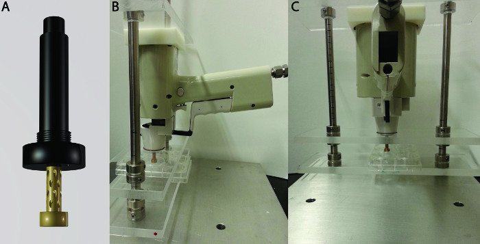 Figure 1. Gene gun barrel and mounting. (A) The improved barrel developed by Dr. O'Brien at the MRC-LMB that has a restricted biolistic spread that minimizes tissue damage by lowering the required pressure for particle acceleration. (B) The side view of an optimized gene gun mounting developed by Dr. Henderson and Dr. Andras Nagy at the University of Toronto. The mounting holds the gene gun at precisely 90˚ perpendicular to the organotypic slices while permitting the user to accurately control the distance of biolistic delivery. For optimal biolistic conditions the barrel aperture should be placed directly over the region of interest at a 10 mm distance from the organotypic slices. (C) Back view of the gene gun mounting. The region of interest to be transfected needs to be placed directly underneath the barrel aperture. Targeting can be confirmed with dye (diolistic) or fluorescent protein tranfection (biolistic). Please click here to view a larger version of this figure.
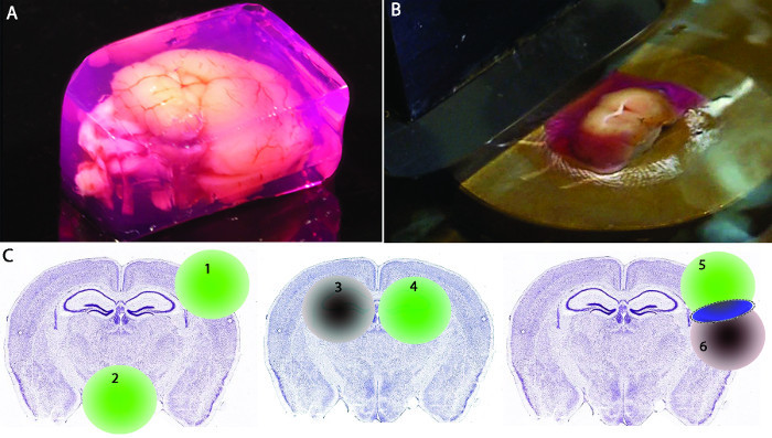 Figure 2. Organotypic slice preparation and regioselective targeting. (A) A DMEM embedded adult mouse brain ready for slicing. (B) The mould was glued onto the vibroslicer mounting with the blade ready to cut a slice. (C) Regioselective heterologous gene expression strategies for biolistic delivery into coronal organotypic brain slices. Images prepared from pictures obtained from the Allen Brain Atlas anatomical reference atlas23. Left panel shows two different brain regions targeted as an example. To restrict the gene expression (1) to the cortical area of the left hemisphere. To restrict the heterologous gene expression (2) to the hypothalamus region of the brain. Middle panel shows that the expression of two heterologous genes (3 and 4) can be isolated and compares the hippocampus of two hemispheres in the same organotypic slice, essentially eliminating any bias from using different sections and/or granting the possibility to address distal crosstalk from the effects of those genes. Right panel shows the possible overlap of transgenic delivery to express the first gene (5) in a precise cortical region while expressing another gene (6) in a disparate yet overlapping cortical area. This permits to analyze the effect of genetic modulation of each heterologous gene alone and also in combination within the same slice. Please click here to view a larger version of this figure.
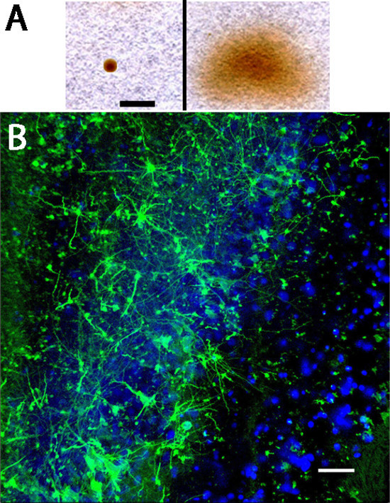 Figure 3. Biolistic scatter pattern seen on filter paper and low magnification image. (A) The improved barrel was fired onto filter paper to determine the spread of gold particles. Left side shows the distribution of the improved barrel while the right panel shows the normal barrel. Black bar: 1 cm. Image taken from O'Brien, et al.17. (B) Low magnification image showing the stochastic transfection patter within the 3 mm biolistic spread within the hippocampal region of 6 week old mice. White bar: 100 µm. Please click here to view a larger version of this figure.
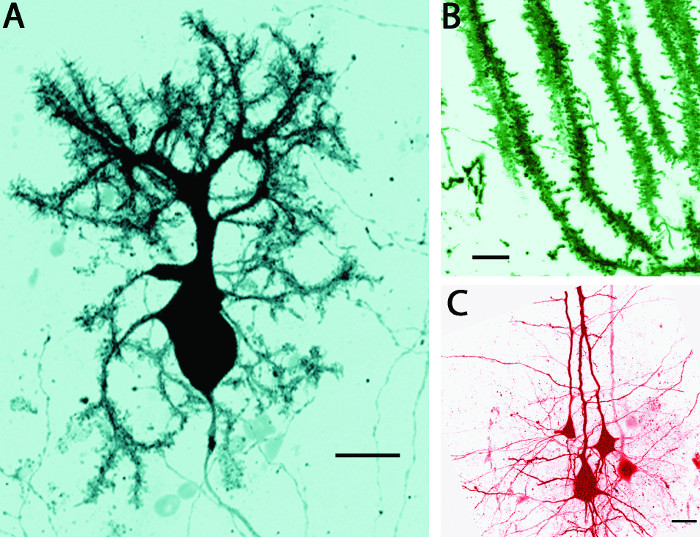 Figure 4. Biolistic delivery of fluorescent protein encoded DNA into organotypic slices of 6 week old mice. (A) Inverted confocal images of a fixed organotypic slice of the cerebellum of a pEGFP-N1 coated gold particles transfected Purkinje cell. Image was taken at 40X magnification. Black bar: 30 µm. (B) Shows a 60x magnification of another Purkinje cell's dentritic harbor to highlight the spines. Black bar: 5 µm. (C) Shows a confocal image of fixed slices taken from the hippocampal region at 60x magnification of four DSredFP transfected pyramidal cells. Black bar: 10 µm. Please click here to view a larger version of this figure.
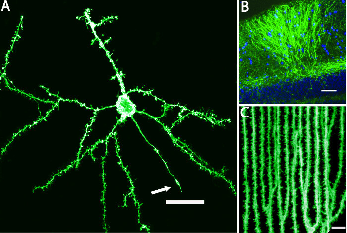 Figure 5. Diolistic delivery of membrane labeling fluorescent dyes into organotypic slices. (A) shows the live imaging of a hippocampal neuron labeled with DiO under 20X magnification. The dentritic spines can clearly be observed as well as the axon as identified by the absence of dentritic spines and the presence of synaptic boutons indicated by a white arrow. White bar: 30 µm. (B) Pronounced staining of the hippocampal region labeled with DiO and counterstained with DAPI. White bar: 30 µm. (C) Higher magnification (60x) of the dentritic spines found in the CA1 region of the hippocampus. White bar: 5 µm. Please click here to view a larger version of this figure.
Discussion
The protocol describes approaches to deliver dyes and genetic materials into adult organotypic slices by using an enhanced gene gun. Essentially, the types of dyes and the variety of possible genes make this method multifaceted and amenable to answer a wide range of biological questions. The regioselective delivery method used, in this case to specific brain region, opens new avenues for experimentation as heterologous gene modulation of localized areas within a biologically relevant architecture hasn't been easily feasible before. We have previously determined that this method can be used on adult organotypic slices, where mature synapses are fully formed, as it showed improved cell survival and heterologous gene expression for many weeks13. With the advent of new and improved culture conditions for mammalian brains as well as other tissues, the use of regioselective genetic modulations will become increasingly useful for a wide variety of biochemical analyses.
As we shall highlight here and previously mentioned by others24, many different factors could readily affect the accuracy and reliability of biolistic delivery. Firstly, the preparation of the gold particle linked to dyes or DNA must be adequately balanced to prevent aggregation and to facilitate the cartridge cargo expulsion. Calcium and spermidine in the DNA-gold micro particles solution can aid the binding of DNA molecules to the gold nanoparticles. The ability for the nanoparticles to penetrate the cells under reduced pressure was previously determined17. Secondly, the gene gun must be mounted as steady as possible with the correct angle and distance. A reliable pressure gauge is also necessary for reproducibility, to maintain tissue integrity, and to properly accelerate the gold particles to their intended targets. Indeed, the distance, orientation, and stability of the equipment are also a crucial factors for the targeting and precision of regioselective delivery. As these brain structures are very small, minor variations in the firing angle could easily render the entire procedure less reliable. And finally, the preparation and maintenance of viable organotypic slices have been fraught with difficulties for many decades yet abundant and reliable protocols for different animals, brain regions, ages and cocultures are currently available2,4,25,26. These procedures should be user-optimized before submitting the tissues to biolistic procedures. This will more readily permit the user to unbiasedly ascertain the impact of the biolistic particles and the effect of the heterologous gene on their own cultures. To this end, the use of fluorescent genes such as EYFP and EGFP can both permit a novel user to test the targeting precision and also follow the cellular viability through heterologous gene expression over a sustained period of time13. Many of the critical steps for reliable and reproducible use of this protocol are highlighted in the following three paragraphs.
For efficient transfection, the DNA concentration must be appropriate. Concentrations that are too low would hinder the transfection yield while concentrations that are too high can cause agglomeration of the nanoparticles. Aggregates of gold can dramatically reduce transfection efficiency, cause uneven distributions, and can lead to increased cytotoxicity and oxidative stress. Problems with coating efficiency are likely due to the expiration of the spermidine solution (should be replaced every 2 to 3 months). For an adequate biolistic spread, the labeling must be even. A non-homogeneous labeling of the gold nanoparticle/DNA suspension may be due to the PVP solution. It should also be replaced every 2 to 3 months. Coating of the gold nanoparticles can be seen on agarose gel electrophoresis as a higher MW band27.
Each biological system under investigation as well as each different instrument used might require small changes in gas pressure to reach optimized levels of transfection and biolistic spread. The critical parameter is the pressure of the helium pulse required to strip the micro- or nano-carriers from the plastic cartridge and propel them into the tissue. A high gas pressure will affect cell survival, because it will create shock waves across the target and disrupt or detach the tissue from the matrix17. Aiming the gene gun barrel and having the apparatus exactly perpendicular to the tissue is important for accurate biolistic delivery. The selected area should be placed in the direct center of the barrel at precisely 10 mm from the outer aperture. This results in a 3 mm diameter of gold particle delivery around the epicenter. The gene gun barrel should be cleaned with 70% ethanol after each use. To protect the tissue from large particle aggregates, the barrel also contains a nylon mesh that should be replaced regularly to maintain optimum performance. In order to replace the mesh unscrew the cap then insert a new nylon mesh between the cap and the O-ring.
Fluorescently labeled cells should be visible at least 1 - 2 days after transfection around the epicenter. Absence or misalignment of labeling can result from inadequate preparation and instability of the gene gun mounting. The observations of cells using confocal microscopy depends on a number of factors: the wavelength of the excitation/emission light, pinhole size, NA of the objective lens, refractive index of components in the light path, depth of the tissue, and the alignment of the instrument. These factors should be optimized for each system under investigation.
Recent advances in biolistics were exploited for the success of this method. The gene gun barrel used in this study was modified in order to reduce the pressure necessary as well as funnel the biolistic scatter pattern into a more constrained and specific area17,19. Other barrel modifications have tried to restrict the biolistic spread and reduce the outflow pressure28 yet this custom designed gene gun barrel designed by Dr. O'Brien is a single component fitted to the standard Helios Gene gun. Subsequently, sub-micrometer gold particles were used to reduce the invasiveness of micrometer particles that we had previously determined caused cellular damage and ultimately, loss of sustained heterologous gene expression. These changes to the biolistic procedure improved the overall feasibility and reproducibility of the method13. Other improvements on slice preparation such as in-depth troubleshooting of the slicing procedure, using a DMEM-Agarose matrix to preserve the brain, and numerous other useful advances in organotypic brain slice preparation previously reported13 enabled us to explore the use of adult slices that have developed mature synaptic networks.
In our study we mainly used dyes and fluorescent heterologous genes to visualize cell morphology as a readout on transfection efficiency and an ability to image the morphological characteristics of neurons in mouse brain tissues. The signal to noise ratio of the stochastic delivery within a defined radius enabled us to completely isolate a single cell's dentritic harbor within its native context. As numerous efforts are used to investigate these morphological characteristics, mapping 'connectomics' or to label single cells for various analyses, this method could easily be combined with existing protocols such as electrophysiology and neurite measurements29,30. Evidently, combinations of fluorescent heterologous genes could enable a much more robust delineation of single cells as the stochastic distribution of fluorescent proteins, and their combination would determine the color of the individual cells31,32. The use of cell specific promoters such as synapsin and glial fibrillary acid protein upstream of the heterologous gene exon can also restrict the expression to subpopulations33. Indeed, the unending variety of genetic material could also be delivered in a regioselective manner by gold nanoparticles using this same procedure. The total transfected regions could thus be analyzed with traditional methods, such as dissection and submitting that region to western immunoblotting to analyze the localized effect of the heterologous gene.
Improvements in organotypic slice procedures will also permit wider use of brain tissues for long-term experimentation as well as permit the use of other tissues and cocultures and would facilitate transfection procedures. Lipid and polymer transfection have been previously reported to affect membrane homeostasis, internalization, protein permeability34 and frequently show very low efficiency to transfect terminally differentiated neurons. Cell specific cargo targeting is also amenable only to cells that express the cell surface receptors needed for internalization, and although this method can be highly selective, it can lack targeted regioselectivity35. Viral vectors otherwise are often difficult and costly to produce, require stringent regulations and have on occasion demonstrated safety concerns. Biolistic delivery thus has an unparalleled ability to regioselectively transfect neurons and other cell types within viable tissues. As gold is non toxic, further advances in the use of biolistics could pave the way for biomedical uses such as transdermal pharmaceutical delivery, noninvasive in vivo gene delivery, as well as localized nonviral gene therapies.
Disclosures
Dr. John A. O'Brien holds patents for the improved gene gun barrel. US patent number 10/380,452 and European patent Number 01974517.3.
Acknowledgments
The authors would like to thank the MRC-LMB workshop for the production of the enhanced gene gun barrel. We would also like to thank Dr. David R. Hampson at the University of Toronto for the use of equipment and resources.
References
- Klein RM, Wolf ED, Wu R, Sanford JC. High-velocity microprojectiles for delivering nucleic acids into living cells. Biotechnology. 1987;24:384–386. [PubMed] [Google Scholar]
- Kim H, Kim E, Park M, Lee E, Namkoong K. Organotypic hippocampal slice culture from the adult mouse brain: a versatile tool for translational neuropsychopharmacology. Progress in Neuro-Psychopharmacolog., & Biological Psychiatry. 2013;41:36–43. doi: 10.1016/j.pnpbp.2012.11.004. [DOI] [PubMed] [Google Scholar]
- Morin-Brureau M, De Bock F, Lerner-Natoli M. Organotypic brain slices: a model to study the neurovascular unit micro-environment in epilepsies. Fluids and Barriers of the CNS. 2013;10:11. doi: 10.1186/2045-8118-10-11. [DOI] [PMC free article] [PubMed] [Google Scholar]
- Millet LJ, Gillette MU. Over a century of neuron culture: from the hanging drop to microfluidic devices. The Yale Journal of Biology and Medicine. 2012;85:501–521. [PMC free article] [PubMed] [Google Scholar]
- Cho S, et al. Spatiotemporal evidence of apoptosis-mediated ischemic injury in organotypic hippocampal slice cultures. Neurochemistry International. 2004;45:117–127. doi: 10.1016/j.neuint.2003.11.012. [DOI] [PubMed] [Google Scholar]
- Daviaud N, et al. Modeling nigrostriatal degeneration in organotypic cultures, a new ex vivo model of Parkinson's disease. Neuroscience. 2013;256:10–22. doi: 10.1016/j.neuroscience.2013.10.021. [DOI] [PMC free article] [PubMed] [Google Scholar]
- Drexler B, Hentschke H, Antkowiak B, Grasshoff C. Organotypic cultures as tools for testing neuroactive drugs - link between in-vitro and in-vivo experiments. Current Medicinal Chemistry. 2010;17:4538–4550. doi: 10.2174/092986710794183042. [DOI] [PubMed] [Google Scholar]
- Oishi Y, Baratta J, Robertson RT, Steward O. Assessment of factors regulating axon growth between the cortex and spinal cord in organotypic co-cultures: effects of age and neurotrophic factors. J Neurotrauma. 2004;21:339–356. doi: 10.1089/089771504322972121. [DOI] [PubMed] [Google Scholar]
- Baratta J, Marienhagen JW, Ha D, Yu J, Robertson RT. Cholinergic innervation of cerebral cortex in organotypic slice cultures: sustained basal forebrain and transient striatal cholinergic projections. Neuroscience. 1996;72:1117–1132. doi: 10.1016/0306-4522(95)00603-6. [DOI] [PubMed] [Google Scholar]
- Barateiro A, Domingues HS, Fernandes A, Relvas JB, Brites D. Rat Cerebellar Slice Cultures Exposed to Bilirubin Evidence Reactive Gliosis, Excitotoxicity and Impaired Myelinogenesis that Is Prevented by AMPA and TNF-alpha Inhibitors. Molecular Neurobiology. 2013;49(1):424–439. doi: 10.1007/s12035-013-8530-7. [DOI] [PubMed] [Google Scholar]
- Franke H, Schelhorn N, Illes P. Dopaminergic neurons develop axonal projections to their target areas in organotypic co-cultures of the ventral mesencephalon and the striatum/prefrontal cortex. Neurochemistry International. 2003;42:431–439. doi: 10.1016/s0197-0186(02)00134-1. [DOI] [PubMed] [Google Scholar]
- Mingorance A, et al. Regulation of Nogo and Nogo receptor during the development of the entorhino-hippocampal pathway and after adult hippocampal lesions. Molecular and Cellular Neurosciences. 2004;26:34–49. doi: 10.1016/j.mcn.2004.01.001. [DOI] [PubMed] [Google Scholar]
- Arsenault J, O'Brien JA. Optimized heterologous transfection of viable adult organotypic brain slices using an enhanced gene gun. BMC Research Notes. 2013;6:544. doi: 10.1186/1756-0500-6-544. [DOI] [PMC free article] [PubMed] [Google Scholar]
- De Simoni A, Griesinger CB, Edwards FA. Development of rat CA1 neurones in acute versus organotypic slices: role of experience in synaptic morphology and activity. J Physiol. 2003;550:135–147. doi: 10.1113/jphysiol.2003.039099. [DOI] [PMC free article] [PubMed] [Google Scholar]
- Laywell ED, et al. Neuron-to-astrocyte transition: phenotypic fluidity and the formation of hybrid asterons in differentiating neurospheres. J Comp Neurol. 2005;493:321–333. doi: 10.1002/cne.20722. [DOI] [PMC free article] [PubMed] [Google Scholar]
- Walder S, Zhang F, Ferretti P. Up-regulation of neural stem cell markers suggests the occurrence of dedifferentiation in regenerating spinal cord. Dev Genes Evol. 2003;213:625–630. doi: 10.1007/s00427-003-0364-2. [DOI] [PubMed] [Google Scholar]
- Brien JA, Holt M, Whiteside G, Lummis SC, Hastings MH. Modifications to the hand-held Gene Gun: improvements for in vitro biolistic transfection of organotypic neuronal tissue. J Neurosci Methods. 2001;112:57–64. doi: 10.1016/s0165-0270(01)00457-5. [DOI] [PubMed] [Google Scholar]
- Brien J, Unwin N. Organization of spines on the dendrites of Purkinje cells. Proceedings of the National Academy of Sciences of the United States of America. 2006;103:1575–1580. doi: 10.1073/pnas.0507884103. [DOI] [PMC free article] [PubMed] [Google Scholar]
- Brien JA, Lummis SC. Nano-biolistics: a method of biolistic transfection of cells and tissues using a gene gun with novel nanometer-sized projectiles. BMC Biotechnology. 2011;11(66):1472–6750. doi: 10.1186/1472-6750-11-66. [DOI] [PMC free article] [PubMed] [Google Scholar]
- Brien JA, Lummis SC. Diolistic labeling of neuronal cultures and intact tissue using a hand-held gene gun. Nature Protocols. 2006;1:1517–1521. doi: 10.1038/nprot.2006.258. [DOI] [PMC free article] [PubMed] [Google Scholar]
- Brien JA, Lummis SC. Diolistics: incorporating fluorescent dyes into biological samples using a gene gun. Trends in Biotechnology. 2007;25:530–534. doi: 10.1016/j.tibtech.2007.07.014. [DOI] [PMC free article] [PubMed] [Google Scholar]
- Han X, et al. Validation of an LDH assay for assessing nanoparticle toxicity. Toxicology. 2011;287:99–104. doi: 10.1016/j.tox.2011.06.011. [DOI] [PMC free article] [PubMed] [Google Scholar]
- Lein ES, et al. Genome-wide atlas of gene expression in the adult mouse brain. Nature. 2007;445:168–176. doi: 10.1038/nature05453. [DOI] [PubMed] [Google Scholar]
- Woods G, Zito K. Preparation of gene gun bullets and biolistic transfection of neurons in slice culture. Journal of Visualized Experiments. 2008;12 doi: 10.3791/675. [DOI] [PMC free article] [PubMed] [Google Scholar]
- Su T, Paradiso B, Long YS, Liao WP, Simonato M. Evaluation of cell damage in organotypic hippocampal slice culture from adult mouse: a potential model system to study neuroprotection. Brain Research. 2011;1385:68–76. doi: 10.1016/j.brainres.2011.01.115. [DOI] [PubMed] [Google Scholar]
- Mewes A, Franke H, Singer D. Organotypic brain slice cultures of adult transgenic P301S mice--a model for tauopathy studies. PloS One. 2012;7:45017. doi: 10.1371/journal.pone.0045017. [DOI] [PMC free article] [PubMed] [Google Scholar]
- Ito T, Ibe K, Uchino T, Ohshima H, Otsuka M. Preparation of DNA/Gold Nanoparticle Encapsulated in Calcium Phosphate. Journal of Drug Delivery. 2011;2011:647631. doi: 10.1155/2011/647631. [DOI] [PMC free article] [PubMed] [Google Scholar]
- Rinberg D, Simonnet C, Groisman A. Pneumatic capillary gun for ballistic delivery of microparticles. Appl Phys Lett. 2005;87:014103. [Google Scholar]
- Meijering E. Neuron tracing in perspective. Cytometry. Part A : the Journal of the International Society for Analytical Cytology. 2010;77:693–704. doi: 10.1002/cyto.a.20895. [DOI] [PubMed] [Google Scholar]
- Sasaki T, Matsuki N, Ikegaya Y. Targeted axon-attached recording with fluorescent patch-clamp pipettes in brain slices. Nature Protocols. 2012;7:1228–1234. doi: 10.1038/nprot.2012.061. [DOI] [PubMed] [Google Scholar]
- Gan WB, Grutzendler J, Wong WT, Wong RO, Lichtman JW. Multicolor 'DiOlistic' labeling of the nervous system using lipophilic dye combinations. Neuron. 2000;27:219–225. doi: 10.1016/s0896-6273(00)00031-3. [DOI] [PubMed] [Google Scholar]
- Livet J, et al. Transgenic strategies for combinatorial expression of fluorescent proteins in the nervous system. Nature. 2007;450:56–62. doi: 10.1038/nature06293. [DOI] [PubMed] [Google Scholar]
- Gholizadeh S, Tharmalingam S, Macaldaz ME, Hampson DR. Transduction of the central nervous system after intracerebroventricular injection of adeno-associated viral vectors in neonatal and juvenile mice. Human Gene Therapy Methods. 2013;24:205–213. doi: 10.1089/hgtb.2013.076. [DOI] [PMC free article] [PubMed] [Google Scholar]
- Arsenault J, et al. Botulinum protease-cleaved SNARE fragments induce cytotoxicity in neuroblastoma cells. Journal of Neurochemistry. 2014;129:781–791. doi: 10.1111/jnc.12645. [DOI] [PMC free article] [PubMed] [Google Scholar]
- Arsenault J, et al. Stapling of the botulinum type A protease to growth factors and neuropeptides allows selective targeting of neuroendocrine cells. Journal of Neurochemistry. 2013;126:223–233. doi: 10.1111/jnc.12284. [DOI] [PMC free article] [PubMed] [Google Scholar]


