SUMMARY
Introduction
Quadriceps heads are important in biomechanical stabilization and in the pathogenesis osteoarthritis of the knee. This is the first study to explore the relative distribution of quadriceps head anatomical cross-sectional areas (ACSA) and volumes, and their response to pain and to training intervention.
Methods
The relative proportions of quadriceps heads were determined in 48 Osteoarthritis Initiative participants with unilateral pain (65% women; age 45–78y). Quadriceps head volumes were also measured in 35 untrained women (45–55y) before and after 12 week training intervention. Cross-sectional areas of the vastus medialis (VM), inter-medius (VIM), and lateralis (VL), and of the rectus femoris (RF) were determined from axial T1-weighted MR images.
Results
The proportion of the VM on the total quadriceps ACSA increased from proximal to distal. The difference in quadriceps ACSA of painful (vs. pain-free) limbs was −5.4% for the VM (p<0.001), −6.8% for the VL (p<0.01), −2.8% for the VIM (p=0.06), and +3.4% for the RF (p=0.67) but the VM/VL ratio was not significantly altered. The muscle volume increase during training intervention was +4.2% (p<0.05) for VM, +1.3% for VL, +2.0% for VIM (p<0.05) and +1.6% for RF.
Conclusion
The proportion of quadriceps head relative to total muscle ACSA and volume depends on the anatomical level studied. The results suggest that there may be a differential response of the quadriceps heads to pain-induced atrophy and to training-related hypertrophy. Studies in larger samples are needed to ascertain whether the observed differences in response to pain and training are statistically and clinically significant.
Keywords: quadriceps heads, training, pain, MRI
INTRODUCTION
Loss of quadriceps muscle strength is known to have adverse effects on knee joint biomechanics, and to increase joint loading (Andriacchi et al., 2009; Hortobágyi et al., 2004; Jefferson et al., 1990; Lattanzio et al., 1997; Radin et al., 1991; Segal and Glass, 2011; Skinner et al., 1986; Winby et al., 2009). Further, reduction in quadriceps strength was found to contribute to functional disability and knee pain (McAlindon et al., 1993; O’Reilly et al., 1998; Sattler et al., 2012). Although it is still controversial whether muscle strengthening exercise can effectively reduce the incidence and progression of knee symptoms (Segal and Glass, 2011; Segal et al., 2012, 2009a, 2009b) and structural (particularly radiographic) incidence and progression of knee osteoarthritis (Amin et al., 2009; Brandt et al., 1999; Ding et al., 2008; Eckstein et al., 2013; Mikesky et al., 2006; Roos et al., 2011; Ruhdorfer et al., 2013; Segal and Glass, 2011; Segal et al., 2010b, 2009a, 2012a, 2010a; Sharma et al., 2003; Thorstensson et al., 2004), muscle strengthening exercise is currently recommended by the Osteoarthritis Research Society International (OARSI) therapeutic guidelines for knee osteoarthritis (Bennell et al., 2009; Roos et al., 2011; Zhang et al., 2008).
Anatomically, the quadriceps consists of three heads (vastus medialis [VM], intermedius [VIM], and lateralis [VL]) originating from the femur and inserting via the quadriceps tendon, patellar bone, and patellar tendon at the tibial tuberosity. The fourth head, the rectus femoris (RF), in contrast, originates from the inferior anterior iliac spine and, therefore, not only extends the knee, but also flexes the hip. The VM is known to play an important role in biomechanical stabilization of the femoropatellar joint by pulling the patella medially, keeping contact pressure in the lateral patella facet within limits, and preventing the patella from lateral (sub)luxation relative to the trochlear groove (Elias et al., 2009). Yet, several studies reported that VM strength variation had only a limited effect on patello-femoral biomechanics (Lee et al., 2002; Lorenz et al., 2012). However, the VM and VL have also been proposed to have antagonistic functions with regard to rotating the femur relative to the tibia; and have been suggested to maintain rotational stability of the knee and to be important in prevention of knee injuries (Schmitt and Mittelmeier, 1978). The quadriceps heads (specifically the VM) hence are suspected to play a role in pain and degenerative changes in the femoropatellar (Berry et al., 2008a, 2008b) and femorotibial joints (Fink et al., 2007; Hinman et al., 2002; Pan et al., 2011).
Recent cross sectional studies in participants with “pre-clinical” osteoarthritis suggested that larger VM (versus VL) ACSAs were associated with more cartilage defects in the patella (Berry et al., 2008a; Pan et al., 2011) and with inferior compositional cartilage properties (i.e. magnetic resonance imaging [MRI] transverse relaxation times; T2) in the femorotibial joint (Pan et al., 2011); these studies therefore indicated that a high VL/VM ratio was beneficial to cartilage health. A recent longitudinal study (Wang et al., 2012), in contrast, reported that the baseline VM ACSA was inversely associated (and hence protective) of current knee pain and of longitudinal medial tibial cartilage volume loss. Further, the longitudinal increase in VM ACSA from baseline to year 2 follow-up was found to be associated with a concurrent reduction in knee pain, with reduced medial tibial cartilage loss from 2 to 4.5 years follow-up, and with reduced risk of knee replacement over 4 years (Wang et al., 2012). The authors suggested that optimizing VM ACSA was important in reducing progression of knee osteoarthritis and knee replacement (Wang et al., 2012).
We previously reported that, in patients with the same grade of bilateral radiographic knee osteoarthritis, quadriceps ACSAs and isometric strength were significantly smaller in limbs with frequent knee pain, relative to a contralateral reference knee without pain (Sattler et al., 2012). We further reported that a supervised 12 week training intervention in untrained perimenopausal women (Ring-Dimitriou et al., 2009) involved a statistically significant increase in quadriceps ACSAs and volume (Hudelmaier et al., 2010). However, it is currently unknown whether, and if yes to what extent, there is a differential response of the quadriceps heads. In the current study, we therefore aimed to explore the relative distribution of the heads (i.e. the VL, VIM, VM, RF) to total quadriceps anatomical cross-sectional area (ACSA) and volume, and their individual response to pain and to training intervention.
MATERIAL AND METHODS
Subjects
We examined data from two cohorts that were previously described in detail, one from the US-based Osteoarthritis Initiative (Eckstein et al., 2014, 2012) that suffered from unilateral frequent knee pain (Sattler et al., 2012), and the other from a 12 week training intervention study performed in Salzburg, Austria (Hudelmaier et al., 2010; Ring-Dimitriou et al., 2009).
The unilateral knee pain sample comprised 48 participants who fulfilled the following criteria: frequent pain (i.e. pain, aching or stiffness in or around the knee for at least one month during the past 12 months) in one knee, no pain at all during the past 12 months in the contralateral knee, the same grade of radiographic knee osteoarthritis on fixed flexion radiographs (i.e. Kellgren Lawrence grade [KLG] 2 or 3 in both knees), and availability of thigh muscle MRIs and muscle strength measurements from the OAI data base (Sattler et al., 2012). This sample comprised 17 men and 31 women, aged 45–78 years (mean±standard deviation [SD] 63±9.3 years), and a body mass index (BMI) ranging from 21–44 (29.9±4.8). Twenty-one participants displayed bilateral KLG2, and 27 bilateral cKLG3 (Sattler et al., 2012). This intra-individual, between-knee comparison revealed that frequent knee pain was associated with an approximately 5% reduction in quadriceps ACSA relative to the painless limb; this “response” was similar between men and women, and similar between bilateral KLG2 or KLG3 cases (Sattler et al., 2012). The side differences in quadriceps ACSAs were also associated with side differences in extensor strength, but no reduction in hamstring ACSAs or flexor muscle strength were observed in painful limbs (Sattler et al., 2012).
The training intervention study was performed in Salzburg, Austria (Ring-Dimitriou et al., 2009) and recruited 41 untrained women with a physical activity level of less than one hour a week, no history of organized sports participation, and at the end of their menopause. The participants were assigned to three different training intervention groups: Strength training (ST; n=16), endurance training (ET; n=19) and autogenic training (controls; n=6). The women selected were 45–55 years old (50.8±3.2years) with a BMI of 26.5±5.2kg/m2 (Ring-Dimitriou et al., 2009). The participants had supervised training sessions 3 times per week for 60 min over twelve weeks (Ring-Dimitriou et al., 2009). The ST intervention significantly improved muscle quality, and both the ST and ET intervention improved cardiorespiratory fitness (i.e. VO(2)peak) (Ring-Dimitriou et al., 2009). Acquiring thigh MRIs before and after the training intervention, a 3.1% increase in total quadriceps volume was recorded after 12 weeks of ST, and a 3.7% increase after ET. There was no relevant or statistically significant change observed with AT (Hudelmaier et al., 2010).
Magnetic resonance imaging (MRI) and muscle segmentation
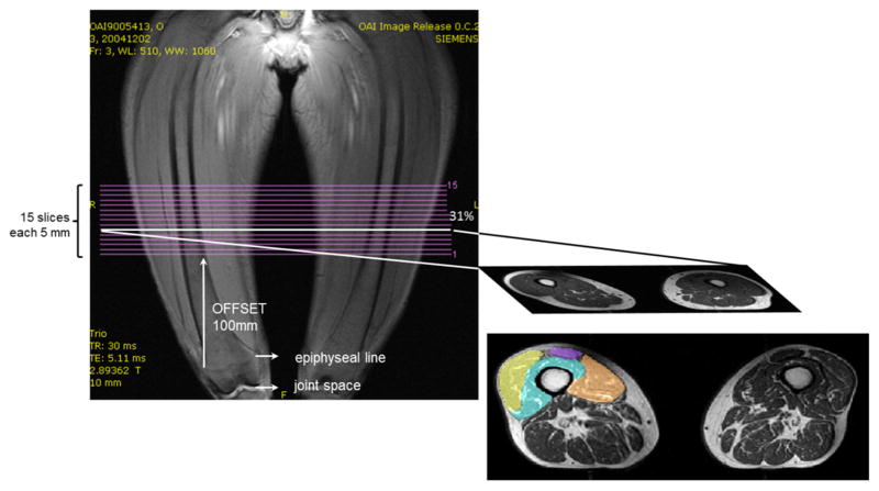
The analysis of the unilateral knee pain sample relied on the public-use baseline MRI data (0.E.1) from the Osteoarthritis Initiative (Eckstein et al., 2014, 2012). Fifteen axial contiguous 0.5cm slices (0.98mm x 0.98mm in-plane resolution) of both thighs were acquired using a T1-weighted spin echo sequence (TR 500ms, TE 10ms; Fig. 1) and a 3 Tesla Magnetom Trio scanner (Siemens Healthcare Erlangen, Germany). The image acquisition started 10cm proximal to the distal femoral epiphysis and extended 7.5cm proximally (Fig. 1). Details regarding the MRI techniques and protocols are available online (www.oai.ucsf.edu/datarelease/operationsmanuals.asp) and have been described previously (Eckstein et al., 2014, 2012; Sattler et al., 2012). It is important to note that, due to the fixed (10 cm) distance between the distal femoral epiphysis and the most distal MRI slice acquired, the position of the images relative to the femur and thigh muscles was variable, depending on individual femur length and body height. This variability prevented us from performing a volumetric assessment of the quadriceps (heads) in this part of the study, given lack of comparability between participants. Because the quadriceps heads can be better distinguished distally than proximally (see below), we selected the most distal slice that could be identified to be in an anatomically consistent location across all study participants (given their variability in body height – Fig. 1); this was based on a previously established relationship between femoral length, location of the distal femoral epiphysis, and body height (Dannhauer et al., 2010). These relationships were determined in another subsample of Osteoarthritis Initiative participants who had long limb radiographs available (Dannhauer et al., 2010). Applying these relationships, we selected variable slice numbers from the acquisition protocol amongst the participants, with the target of selecting an image located at 31% of the total femoral length from distal to proximal. Manual segmentation of the ACSAs of the four quadriceps heads was performed in the selected image by M.S. (Fig. 1). Please note that this slice differs in location from the 35% slice from which results were reported previously (Sattler et al., 2012). Segmentation of the VM was repeated once, one year later by the same observer (M.S.) without reference to the previous segmentations, to evaluate the test-retest reproducibility under (non-paired) re-segmentation conditions.
Fig. 1.
OAI thigh image acquisition and study analysis protocol. The image on the right shows the segmented quadriceps on the left [right thigh] (brown = vastus medialis: yellow = vastus lateralis; turquoise = vastus intermedius; purple = rectus femoris: and an unsegmented image on the right image side [left thigh]. The images are from a 57 year old women, with a body height of 167.5 cm and a body weight of 49.3 kg (BMI 17.6).
For the training intervention study, axial contiguous 1cm slices (0.78mm x 0.78mm in-plane resolution) of both thighs were acquired, again using a T1-weighted spin echo sequence (TR 1541ms, TE=15ms), and a 1.5 Tesla Phillips NT Intera scanner (Phillips Medical Systems, Best, Netherlands). The region of interest started distally at the level of the quadriceps tendon and extended proximally to the level of the femoral neck. The imaging parameters and the region of interest were kept identical between baseline and follow-up acquisitions. Because the quadriceps heads were difficult to delineate in the proximal part of the muscle, segmentation of the VM, VL, VIM and RF (of the dominant leg) was confined to the distal third of the original volume of interest.
For both parts of the study, quadriceps head ACSAs and volumes were computed by numerical integration of the segmented voxels.
Statistical analysis
To determine the test-retest reproducibility of VM ACSAs, the mean and standard deviations of the two independent measurements made one year apart were computed across both knees of all 48 participants, and the root-mean square coefficient of variation (RMS CV%) was used to express the precision error (Gluer et al., 1995). Descriptive statistics were provided by calculating the mean values and SDs of the quadriceps ACSAs and volumes in painful and in painless limbs, and in limbs before and after training intervention. The relative distribution of quadriceps ACSAs and volumes heads was reported in percent (%) of the total quadriceps ACSA or volume within the selected region of interest. Paired t-tests were used to compare quadriceps head ACSAs between painful and contralateral painless limbs (in the same person), and before and after training intervention. No adjustment for multiple statistical testing was performed given the exploratory nature of the study and the expected co-linearity of the changes seen within subcomponents of the quadriceps. Further, the VL/VM ratio was compared between painful and painless contra-lateral knees, and before and after the training interventions using the paired t-test, to determine whether the response (i.e. the percent difference due to pain or training intervention) differed between VL and VM. An unpaired t-test was used to explore whether the magnitude of the training effects differed between endurance and strength training intervention.
RESULTS
Test-retest reproducibility of vastus medialis (VM) ACSA measurement
The RMS CV% for segmentation and ACSA analysis by the same observer one year apart without referring to the previous segmentations, was 4.3% in painful knees and 4.7% in painless knees. The mean difference between painful vs. painless limbs was −5.7± 12% in the first analysis, and −5.4± 11% in the second analysis (one year later).
Quadriceps head ACSAs in painful vs. painless limbs
The absolute values measured for the quadriceps heads in painful vs. painless knees are shown in Table 1. In painless knees, the proportion taken by the quadriceps heads (at 31% of femoral length from distal to proximal) was 37±5.0% (mean±SD) for the VM, 29±3.8% for the VL, 30±3.7% for the VIM, and 4.5±1.8% for the RF. This distribution was similar in the painful knees (data not shown). Computing pairwise differences (in %), the mean±SD the quadriceps was by −5.2±8.1% lower (p=0.00004) in painful compared with contralateral painless limbs: The differences were −5.4±11% for the VM (p=0.0005), −6.8±16% for the VL (p=0.0018), −2.8±15% for the VIM (p=0.06), and +3.4±29.1 for the RF (p=0.67). As these values were computed from individual pairs, they diverged from the difference between group means in Table 1; the latter amounted to −6.3% for the VM, −7.8% for the VL, −4.2% for the VIM, and −1.5% for the RF. The VL/VM ratio did not differ significantly (p=0.86) between painful (0.8±0.2) vs. painless knees (0.8±0.2)
Table 1.
Anatomical cross-sectional areas (ACSAs) of the quadriceps (quad), vastus medialis (VM), vastus lateralis (VL), vastus intermedius (VIM) and rectus femoris (RF) in painful vs. contralateral painless limbs
| Painless | Painful | Difference (pairs) | ||||
|---|---|---|---|---|---|---|
| Mean | SD | Mean | SD | Mean | SD | |
| Quad cm2 | 49.7 | 12.8 | 46.7 | 11.3 | −2.9 | 4.4 |
| VM cm2 | 18.3 | 5.6 | 17.1 | 5.1 | −1.1 | 2.1 |
| VL cm2 | 14.2 | 4.0 | 13.1 | 3.7 | −1.1 | 2.3 |
| VIM cm2 | 14.9 | 4.1 | 14.3 | 3.7 | −0.6 | 2.2 |
| RF cm2 | 2.2 | 1.1 | 2.2 | 1.1 | −0.03 | 0.5 |
SD = standard deviation
Quadriceps ACSAs before and after training intervention
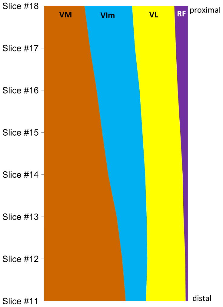
The total volume of the quadriceps in the region of interest (distal third of the thigh between the femoral neck and the quadriceps tendon) was 354±50cm3; the proportion taken by the VM was 36±3.1%, that by the VL 29±3.5%, that by the VIM 30±2.3%, and that by the RF 4.8±1.2%. In the most proximal slice of the region of interest, the relative proportions of ACSAs were 25±3.0% for the VM, 36±3.2% for the VL, 31±3.0% for the VIM, and 8.1±1.7% for the RF. This distribution was different at the most distal slice in the region of interest, where it amounted to 49±5.6% for the VM, 19±4.9% for the VL, 30±4.0% for the VIM, and 1.8±0.5% for the RF. A representative example of the change in the relative contribution of the quadriceps heads to the total quadriceps ACSA from proximal to distal is shown in Figure 2.
Fig. 2.
Representative example of the change in the relative contribution of the quadriceps heads to the total quadriceps ACSA from proximal to distal. The results are from a 50 year old women, with a body height of 170.0 cm and a body weight of 57.0 kg (BMI 19.7).
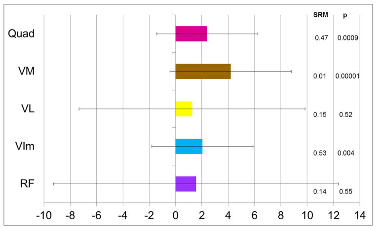
The gain in quadriceps volume by 12 week training intervention (both training groups taken together) was +2.4%±3.8% (p=0.0009) in the distal third of the region of interest (quadriceps tendon to femoral neck), in which the different quadriceps heads could be well differentiated (Fig. 3). The volume gain was 4.2±4.6% (p=0.000007) for the VM, 1.3±8.6% (p=0.15) for the VL, 2.0±3.9% for the VIM (p=0.004), and 1.6±10.8% for the RF (p=0.55). The paired t-test of the VL/VM ratio before and after training intervention did not confirm a significant difference in response (% increase) (p=0.44). Results for the mean volume change, including those for ST and ET intervention groups are shown in Table 2. ET appeared to be slightly more effective than ST in causing an increase in VM volume, but there was no statistically significant difference in the response between both groups (p=0.32).
Fig. 3.
Bar graphs showing the mean difference in muscle volume between baseline and follow-up (i.e. before and after 12 week training intervention) in percent (%), i.e. the intra-individual changes, in the 35 participants: Quad. = quadriceps femoris in the distal third of the region of interest, where the quadriceps heads could be well differentiated; VM = vastus medialis; VL = vastus lateralis; VIm = vastus intermedius; RF = rectus femoris. The error bars show the standard deviation.
Table 2.
Change (% increase) in muscle volume of the quadriceps (quad), vastus medialis (VM), vastus lateralis (VL), vastus intermedius (VIM) and rectus femoris (RF) during 12 week training intervention
| Endurance | Strength | |||||
|---|---|---|---|---|---|---|
| Mean | SD | p | Mean | SD | p | |
| cm3 | ||||||
| Quad | 2.7 | 4.1 | 0.0066 | 2.1 | 3.6 | 0.068 |
| VM | 4.9 | 4.6 | 0.0002 | 3.3 | 4.7 | 0.013 |
| VL | 1.9 | 9.8 | 0.43 | 0.5 | 7.1 | 0.96 |
| Vim | 1.8 | 3.6 | 0.039 | 2.4 | 4.2 | 0.057 |
| RF | 0.9 | 12 | 0.79 | 2.4 | 10 | 0.56 |
SD = standard deviation; p = results of a paired t-test
DISCUSSION
To our knowledge, this is the first study to explore the relative distribution of the heads to total quadriceps anatomical cross-sectional area (ACSA) and volume, and their response to pain and to training intervention. In its distal third of the quadriceps muscle, the VM took the largest (36%) and the RF the smallest proportion (4.8%) of the total quadriceps volume. The relative contribution of the VM to the total quadriceps increased from proximal to distal, whereas that of the VL, VIM and RF decreased. Painful limbs displayed significantly smaller quadriceps ACSAs than contralateral painless limbs, with the percent difference appearing largest for the VL and smallest for the RF. Training intervention (3x1h/week) over 12 weeks lead to a significant increase in quadriceps volume, with the effect appearing largest for the VM and smallest for the VL. However, statistical testing was unable to confirm that the response to pain or exercise differed significantly in magnitude between the VM and VL.
A limitation of the study is the small sample size; it may well be that statistically significant differences in the response of the quadriceps heads to pain or training may have been confirmed if larger number of participants had been studied. Yet, a strength of our approach was to compare painful vs. painless limbs in the same participants (i.e. a between-knee, intra-individual design (Neogi et al., 2009; Sattler et al., 2012), a design that eliminates person-specific confounders, such as variability in age, sex, body mass index, body height, training status, and other variables that are potentially associated with muscle ACSAs. Additionally, painful and painless knees had the same radiographic disease stage, so that the analysis was specific to knee pain, and not to knee osteoarthritis in general; these specific inclusion criteria were met by only 48 of 4796 participants of the Osteoarthritis Initiative (Sattler et al., 2012). Another limitation of the study is that no volumetric muscle analysis could be performed using the Osteoarthritis Initiative data, given the fixed 10cm distance of the 15 slices relative to the distal femoral metaphysis. This fixed distance of the volume of interest was associated with an inconsistent anatomical location across participants with variable body height and femoral length. Other investigators have performed volumetric measurement of the muscles in the above 15 slices and made cross-sectional and longitudinal comparisons between participants (Beattie et al., 2012; Pan et al., 2011). However, our volumetric analysis in the exercise intervention cohort showed that, as for the thigh muscle groups in general (Cotofana et al., 2010), the relative ACSA taken by the quadriceps heads varies substantially with anatomical location. In a subsample of Osteoarthritis Initiative participants, Pan et al. (Pan et al., 2011) reported a significantly greater VL/VM ratio in women than in men (p<0.0001). This finding, however, is not surprising, given that women have a shorter body height and shorter femora, and that the image slab (of the 15 slices) was therefore located more proximal relative to the quadriceps muscle (where VL is larger and VM is smaller) in women than in men. The reported difference in VL/VM ratio (Pan et al., 2011) is hence likely not specific to sex, but due to an inconsistent anatomical location of the images analysed. Not accounting for these differences can lead to systematic bias, as shown above, but also adds random error in between-subject comparisons. In a previous study (Sattler et al., 2012) and in the current investigation, we made an attempt to overcome this limitation by selecting a variable slice number amongst the 15 for analysis, which was estimated to be located at 31% length of the femur, based on available data for body height (Dannhauer et al., 2010). This approach was developed in another sample of Osteoarthritis Initiative participants who had long limb radiographs, and it was shown that the variability in anatomical location can be reduced substantially, if a specific slice is selected based on individual body height (Dannhauer et al., 2010). In addition, a benefit of the between-knee, intra-individual design chosen here is, that, because both thighs were acquired in the same MR images, comparisons between contra-lateral limbs are always made at the same anatomical level.
In the current study, the difference in total quadriceps ACSAs between painful vs. painless limbs was very similar to that reported in our previous analysis (Sattler et al., 2012), despite the analysis being performed one year later and despite the use of a different slice location (31% rather than 35% from distal). The previous study used the more proximal slice because the hamstrings and adductors were also measured and because these have a larger relative proportion in more proximal locations (Cotofana et al., 2010). In the current study, the only quadriceps head that appeared to somewhat differ in response to pain was the RF. However, the RF had by far the smallest ACSA amongst the quadriceps heads and was hence subject to greater measurement variability; its side differences were hence not very robust and did not achieve statistical significance.
The results of the intervention study showed that both endurance and strength training resulted in a significant increase in quadriceps volume. One would expect strength training to have a stronger effect on quadriceps ACSAs than endurance training (Farup et al., 2012), but our results have to be interpreted in light of the participants having a very low fitness level at study entry (Hudelmaier et al., 2010; Ring-Dimitriou et al., 2009). Therefore, the ergometer (endurance) training may have provided sufficient mechanical stimulus for the quadriceps to undergo hypertrophy. A limitation of our study is that standard exercise intervention protocols were tested, and that these were not specifically designed to train certain quadriceps heads (Bennell et al., 2010; Choi et al., 2011; Irish et al., 2010; Peng et al., 2013).
Although no significant difference in the response to training was statistically confirmed between the different heads of the quadriceps, the VM was shown to undergo a significant increase in ACSA, and this increase was at least as large (if not larger) that that of the total quadriceps in the distal third of the thigh. This is important, because a recent longitudinal study reported that a low VM ACSA was associated with pain at baseline and with longitudinal medial tibial cartilage volume loss (Wang et al., 2012). Further, a longitudinal increase in VM ACSA over time was found to be associated with a concurrent reduction in knee pain, a reduced medial tibial cartilage loss, and a reduced risk of undergoing knee replacement (Wang et al., 2012). The results of our current study therefore support training intervention as a successful measure to induce muscle hypertrophy of the VM (and other quadriceps heads) even at advanced age. This may effectively increase joint health and protect against knee osteoarthritis and its consequences.
In conclusion, this study reports the proportion of quadriceps heads relative to total muscle ACSA and volume and shows that their relative distribution (ratio) strongly depends on the anatomical level studied. The study indicates that there may be a differential response to pain, with the VL encountering somewhat greater atrophy than other quadriceps heads. There may further be a differential response to training intervention, with the VM undergoing somewhat greater hypertrophy than other quadriceps heads. However, studies in larger sample, potentially using different exercise programs, are needed to confirm whether the differences in their response to pain and training intervention are statistically and clinically significant.
Supplementary Material
Acknowledgments
We would like to thank the participants, the study investigators and the staff at the clinical centers for generating the image data sets used in this analysis. Data acquisition of part of this study was funded by the Osteoarthritis Initiative, a public-private partnership comprised of five contracts (N01-AR-2-2258; N01-AR-2-2259; N01-AR-2-2260; N01-AR-2-2261; N01-AR-2-2262) funded by the National Institutes of Health, a branch of the Department of Health and Human Services, and conducted by the Osteoarthritis Initiative study Investigators. Private funding partners of the OAI include Merck Research Laboratories; Novartis Pharmaceuticals Corporation, GlaxoSmithKline; and Pfizer, Inc. Private sector funding for the Osteoarthritis Initiative is managed by the Foundation for the National Institutes of Health. The sponsors were not involved in the design and conduct of this particular study, in the analysis and interpretation of the data, and in the preparation, review, or approval of the manuscript. The image analysis was kindly funded by a grant from Paracelsus Medical University Research Fund (PMU-FFF: RISE Project R-10/02/014-WIR)
Footnotes
DECLARATION OF POTENTIALLY COMPETING INTERESTS
Martina Sattler, Susanne Ring-Dimitriou and Alexandra Maria Sänger have no competing interests. Felix Eckstein is CEO, CMO, and co-owner of Chondrometrics GmbH, a company providing MR image analysis services. He provides consulting services to Mariel Therapeutics Inc.. Torben Dannhauer and Martin Hudelmaier have part time appointments with Chondrometrics GmbH. Wolfgang Wirth has a part-time appointment with Chondrometrics GmbH and is co-owner of Chondrometrics GmbH.
Publisher's Disclaimer: This is a PDF file of an unedited manuscript that has been accepted for publication. As a service to our customers we are providing this early version of the manuscript. The manuscript will undergo copyediting, typesetting, and review of the resulting proof before it is published in its final citable form. Please note that during the production process errors may be discovered which could affect the content, and all legal disclaimers that apply to the journal pertain.
References
- Amin S, Baker K, Niu J, Clancy M, Goggins J, Guermazi A, Grigoryan M, Hunter DJ, Felson DT. Quadriceps strength and the risk of cartilage loss and symptom progression in knee osteoarthritis. Arthritis Rheum. 2009;60:189–198. doi: 10.1002/art.24182. [DOI] [PMC free article] [PubMed] [Google Scholar]
- Andriacchi TP, Koo S, Scanlan SF. Gait mechanics influence healthy cartilage morphology and osteoarthritis of the knee. J Bone Jt Surg Am. 2009;91(Suppl 1):95–101. doi: 10.2106/JBJS.H.01408. [DOI] [PMC free article] [PubMed] [Google Scholar]
- Beattie KA, MacIntyre NJ, Ramadan K, Inglis D, Maly MR. Longitudinal changes in intermuscular fat volume and quadriceps muscle volume in the thighs of women with knee osteoarthritis. Arthritis Care Res (Hoboken) 2012;64:22–29. doi: 10.1002/acr.20628. [DOI] [PMC free article] [PubMed] [Google Scholar]
- Bennell K, Duncan M, Cowan S, McConnell J, Hodges P, Crossley K. Effects of vastus medialis oblique retraining versus general quadriceps strengthening on vasti onset. Med Sci Sports Exerc. 2010;42:856–64. doi: 10.1249/MSS.0b013e3181c12771. [DOI] [PubMed] [Google Scholar]
- Bennell KL, Hunt MA, Wrigley TV, Lim BW, Hinman RS. Muscle and exercise in the prevention and management of knee osteoarthritis: an internal medicine specialist’s guide. Med Clin North Am. 2009;93:161–77. xii. doi: 10.1016/j.mcna.2008.08.006. [DOI] [PubMed] [Google Scholar]
- Berry PA, Hanna FS, Teichtahl AJ, Wluka AE, Urquhart DM, Bell RJ, Davis SR, Cicuttini FM. Vastus medialis cross-sectional area is associated with patella cartilage defects and bone volume in healthy women. Osteoarthritis Cartilage. 2008a;16:956–960. doi: 10.1016/j.joca.2007.11.011. [DOI] [PubMed] [Google Scholar]
- Berry PA, Teichtahl AJ, Galevska-Dimitrovska A, Hanna FS, Wluka AE, Wang Y, Urquhart DM, English DR, Giles GG, Cicuttini FM. Vastus medialis cross-sectional area is positively associated with patella cartilage and bone volumes in a pain-free community-based population. Arthritis Res Ther. 2008b;10:R143. doi: 10.1186/ar2573. [DOI] [PMC free article] [PubMed] [Google Scholar]
- Brandt KD, Heilman DK, Slemenda C, Katz BP, Mazzuca SA, Braunstein EM, Byrd D, MBE Quadriceps Strength in Women with Radiographically Progressive Osteoarthritis of the Knee and Those with Stable Radiographic Changes. J Rheumatol. 1999;26:2431–2437. [PubMed] [Google Scholar]
- Choi B, Kim M, Jeon HS. The effects of an isometric knee extension with hip adduction (KEWHA) exercise on selective VMO muscle strengthening. J Electromyogr Kinesiol. 2011;21:1011–6. doi: 10.1016/j.jelekin.2011.08.008. [DOI] [PubMed] [Google Scholar]
- Cotofana S, Hudelmaier M, Wirth W, Himmer M, Ring-Dimitriou S, Sanger AM, Eckstein F, Sänger aM. Correlation between single-slice muscle anatomical cross-sectional area and muscle volume in thigh extensors, flexors and adductors of perimenopausal women. Eur J Appl Physiol. 2010;110:91–7. doi: 10.1007/s00421-010-1477-8. [DOI] [PubMed] [Google Scholar]
- Dannhauer T, Wirth W, Eckstein F. Selection of comparable anatomical locations of muscle cross-sectional images in the Osteoarthritis Initiative MRI data. Osteoarthr Cartil 2010 [Google Scholar]
- Ding C, Martel-Pelletier J, Pelletier JP, Abram F, Raynauld JP, Cicuttini F, Jones G. Two-year prospective longitudinal study exploring the factors associated with change in femoral cartilage volume in a cohort largely without knee radiographic osteoarthritis. Osteoarthritis Cartilage. 2008;16:443–449. doi: 10.1016/j.joca.2007.08.009. [DOI] [PubMed] [Google Scholar]
- Eckstein F, Hitzl W, Duryea J, Kent Kwoh C, Wirth W, Kent KC. Baseline and Longitudinal Change in Isometric Muscle Strength Prior to Radiographic Progression in Osteoarthritic and Pre-Osteoarthritic Knees- Data from the Osteoarthritis Initiative. Osteoarthr Cartil. 2013;21:682–90. doi: 10.1016/j.joca.2013.02.658. [DOI] [PMC free article] [PubMed] [Google Scholar]
- Eckstein F, Kwoh CK, Link TM. Imaging research results from the Osteoarthritis Initiative (OAI): a review and lessons learned 10 years after start of enrolment. Ann Rheum Dis. 2014 doi: 10.1136/annrheumdis-2014-205310. [DOI] [PubMed] [Google Scholar]
- Eckstein F, Wirth W, Nevitt MC. Recent advances in osteoarthritis imaging-the Osteoarthritis Initiative. Nat Rev Rheumatol. 2012;8:1–9. doi: 10.1038/nrrheum.2012.113. [DOI] [PMC free article] [PubMed] [Google Scholar]
- Elias JJ, Kilambi S, Goerke DR, Cosgarea AJ. Improving vastus medialis obliquus function reduces pressure applied to lateral patellofemoral cartilage. J Orthop Res. 2009;27:578–83. doi: 10.1002/jor.20791. [DOI] [PMC free article] [PubMed] [Google Scholar]
- Farup J, Kjølhede T, Sørensen H, Dalgas U, Møller AB, Vestergaard PF, Ringgaard S, Bojsen-Møller J, Vissing K. Muscle morphological and strength adaptations to endurance vs. resistance training. J Strength Cond Res. 2012;26:398–407. doi: 10.1519/JSC.0b013e318225a26f. [DOI] [PubMed] [Google Scholar]
- Fink B, Egl M, Singer J, Fuerst M, Bubenheim M, Neuen-Jacob E. Morphologic changes in the vastus medialis muscle in patients with osteoarthritis of the knee. Arthritis Rheum. 2007;56:3626–33. doi: 10.1002/art.22960. [DOI] [PubMed] [Google Scholar]
- Gluer CC, Blake G, Lu Y, Blunt BA, Jergas M, Genant HK. Accurate assessment of precision errors: how to measure the reproducibility of bone densitometry techniques. Osteoporos Int. 1995;5:262–270. doi: 10.1007/BF01774016. [DOI] [PubMed] [Google Scholar]
- Hinman RS, Bennell KL, Metcalf BR, Crossley KM. Temporal activity of vastus medialis obliquus and vastus lateralis in symptomatic knee osteoarthritis. Am J Phys Med Rehabil. 2002;81:684–690. doi: 10.1097/00002060-200209000-00008. [DOI] [PubMed] [Google Scholar]
- Hortobágyi T, Garry J, Holbert D, Devita P, Hortobagyi T. Aberrations in the control of quadriceps muscle force in patients with knee osteoarthritis. Arthritis Rheum. 2004;51:562–569. doi: 10.1002/art.20545. [DOI] [PubMed] [Google Scholar]
- Hudelmaier M, Wirth W, Himmer M, Ring-Dimitriou S, Sänger A, Eckstein F, Sanger A. Effect of exercise intervention on thigh muscle volume and anatomical cross-sectional areas--quantitative assessment using MRI. Magn Reson Med. 2010;64:1713–20. doi: 10.1002/mrm.22550. [DOI] [PubMed] [Google Scholar]
- Irish SE, Millward AJ, Wride J, Haas BM, Shum GLK. The effect of closed-kinetic chain exercises and open-kinetic chain exercise on the muscle activity of vastus medialis oblique and vastus lateralis. J Strength Cond Res. 2010;24:1256–62. doi: 10.1519/JSC.0b013e3181cf749f. [DOI] [PubMed] [Google Scholar]
- Jefferson RJ, Collins JJ, Whittle MW, Radin EL, O’Connor JJ. The role of the quadriceps in controlling impulsive forces around heel strike. Proc Inst Mech Eng H. 1990;204:21–28. doi: 10.1243/PIME_PROC_1990_204_224_02. [DOI] [PubMed] [Google Scholar]
- Lattanzio PJ, Petrella RJ, Sproule JR, Fowler PJ. Effects of fatigue on knee proprioception. Clin J Sport Med. 1997;7:22–27. doi: 10.1097/00042752-199701000-00005. [DOI] [PubMed] [Google Scholar]
- Lee TQ, Sandusky MD, Adeli A, McMahon PJ. Effects of simulated vastus medialis strength variation on patellofemoral joint biomechanics in human cadaver knees. J Rehabil Res Dev. 2002;39:429–38. [PubMed] [Google Scholar]
- Lorenz A, Müller O, Kohler P, Wünschel M, Wülker N, Leichtle UG. The influence of asymmetric quadriceps loading on patellar tracking--an in vitro study. Knee. 2012;19:818–22. doi: 10.1016/j.knee.2012.04.011. [DOI] [PubMed] [Google Scholar]
- McAlindon TE, Cooper C, Kirwan JR, Dieppe PA. Determinants of disability in osteoarthritis of the knee. Ann Rheum Dis. 1993;52:258–262. doi: 10.1136/ard.52.4.258. [DOI] [PMC free article] [PubMed] [Google Scholar]
- Mikesky AE, Mazzuca Sa, Brandt KD, Perkins SM, Damush T, Lane Ka. Effects of strength training on the incidence and progression of knee osteoarthritis. Arthritis Rheum. 2006;55:690–9. doi: 10.1002/art.22245. [DOI] [PubMed] [Google Scholar]
- Neogi T, Felson D, Niu J, Nevitt M, Lewis CE, Aliabadi P, Sack B, Torner J, Bradley L, Zhang Y. Association between radiographic features of knee osteoarthritis and pain: results from two cohort studies. Bmj. 2009;339:b2844–b2844. doi: 10.1136/bmj.b2844. [DOI] [PMC free article] [PubMed] [Google Scholar]
- O’Reilly SC, Jones A, Muir KR, Doherty M. Quadriceps weakness in knee osteoarthritis: the effect on pain and disability. Ann Rheum Dis. 1998;57:588–94. doi: 10.1136/ard.57.10.588. [DOI] [PMC free article] [PubMed] [Google Scholar]
- Pan J, Stehling C, Muller-Hocker C, Schwaiger BJ, Lynch J, McCulloch CE, Nevitt MC, Link TM. Vastus lateralis/vastus medialis cross-sectional area ratio impacts presence and degree of knee joint abnormalities and cartilage T2 determined with 3T MRI - an analysis from the incidence cohort of the Osteoarthritis Initiative. Osteoarthritis Cartilage. 2011;19:65–73. doi: 10.1016/j.joca.2010.10.023. [DOI] [PMC free article] [PubMed] [Google Scholar]
- Peng HT, Kernozek TW, Song CY. Muscle activation of vastus medialis obliquus and vastus lateralis during a dynamic leg press exercise with and without isometric hip adduction. Phys Ther Sport. 2013;14:44–9. doi: 10.1016/j.ptsp.2012.02.006. [DOI] [PubMed] [Google Scholar]
- Radin EL, Yang KH, Riegger C, Kish VL, O’Connor JJ. Relationship between lower limb dynamics and knee joint pain. J Orthop Res. 1991;9:398–405. doi: 10.1002/jor.1100090312. [DOI] [PubMed] [Google Scholar]
- Ring-Dimitriou S, Steinbacher P, von Duvillard SP, Kaessmann H, Muller E, Sanger AM, Müller E, Sänger AM. Exercise modality and physical fitness in perimenopausal women. Eur J Appl Physiol. 2009;105:739–47. doi: 10.1007/s00421-008-0956-7. [DOI] [PubMed] [Google Scholar]
- Roos EM, Herzog W, Block Ja, Bennell KL. Muscle weakness, afferent sensory dysfunction and exercise in knee osteoarthritis. Nat Rev Rheumatol. 2011;7:57–63. doi: 10.1038/nrrheum.2010.195. [DOI] [PubMed] [Google Scholar]
- Ruhdorfer A, Dannhauer T, Wirth W, Hitzl W, Kwoh CK, Guermazi A, Hunter DJ, Benichou O, Eckstein F. Thigh muscle cross-sectional areas and strength in advanced versus early painful osteoarthritis: an exploratory between-knee, within-person comparison in osteoarthritis initiative participants. Arthritis Care Res (Hoboken) 2013;65:1034–1042. doi: 10.1002/acr.21965. [DOI] [PubMed] [Google Scholar]
- Sattler M, Dannhauer T, Hudelmaier M, Wirth W, Sänger aM, Kwoh CK, Hunter DJ, Eckstein F, Sanger AM. Side differences of thigh muscle cross-sectional areas and maximal isometric muscle force in bilateral knees with the same radiographic disease stage, but unilateral frequent pain - data from the osteoarthritis initiative. Osteoarthr Cartil. 2012;20:532–40. doi: 10.1016/j.joca.2012.02.635. [DOI] [PMC free article] [PubMed] [Google Scholar]
- Schmitt O, Mittelmeier H. The biomechanical significance of the vastus medialis and lateralis muscles (author’s transl) Arch Orthop Trauma Surg. 1978;91:291–5. doi: 10.1007/BF00389612. [DOI] [PubMed] [Google Scholar]
- Segal NA, Findlay C, Wang K, Torner JC, Nevitt MC. The longitudinal relationship between thigh muscle mass and the development of knee osteoarthritis. Osteoarthritis Cartilage. 2012;20:1534–1540. doi: 10.1016/j.joca.2012.08.019. [DOI] [PMC free article] [PubMed] [Google Scholar]
- Segal NA, Glass Na. Is quadriceps muscle weakness a risk factor for incident or progressive knee osteoarthritis? Phys Sportsmed. 2011;39:44–50. doi: 10.3810/psm.2011.11.1938. [DOI] [PubMed] [Google Scholar]
- Segal NA, Glass Na, Torner J, Yang M, Felson DT, Sharma L, Nevitt M, Lewis CE. Quadriceps weakness predicts risk for knee joint space narrowing in women in the MOST cohort. Osteoarthritis Cartilage. 2010a;18:769–75. doi: 10.1016/j.joca.2010.02.002. [DOI] [PMC free article] [PubMed] [Google Scholar]
- Segal NA, Glass NA, Felson DT, Hurley M, Yang M, Nevitt M, Lewis CE, Torner JC. Effect of quadriceps strength and proprioception on risk for knee osteoarthritis. Med Sci Sports Exerc. 2010b;42:2081–2088. doi: 10.1249/MSS.0b013e3181dd902e. [DOI] [PMC free article] [PubMed] [Google Scholar]
- Segal NA, Torner JC, Felson D, Niu J, Sharma L, Lewis CE, Nevitt M. Effect of thigh strength on incident radiographic and symptomatic knee osteoarthritis in a longitudinal cohort. Arthritis Rheum. 2009a;61:1210–1217. doi: 10.1002/art.24541. [DOI] [PMC free article] [PubMed] [Google Scholar]
- Segal NA, Torner JC, Felson DT, Niu J, Sharma L, Lewis CE, Nevitt M. Knee extensor strength does not protect against incident knee symptoms at 30 months in the multicenter knee osteoarthritis (MOST) cohort. PM&R. 2009b;1:459–465. doi: 10.1016/j.pmrj.2009.03.005. [DOI] [PMC free article] [PubMed] [Google Scholar]
- Sharma L, Dunlop DD, Cahue S, Song J, Hayes KW. Quadriceps Strength and Osteoarthritis Progression in Malaligned and Lax Knees. Ann oder Intern Med. 2003;138:613–619. doi: 10.7326/0003-4819-138-8-200304150-00006. [DOI] [PubMed] [Google Scholar]
- Skinner HB, Wyatt MP, Hodgdon JA, Conard DW, Barrack RL. Effect of fatigue on joint position sense of the knee. J Orthop Res. 1986;4:112–118. doi: 10.1002/jor.1100040115. [DOI] [PubMed] [Google Scholar]
- Thorstensson CA, Petersson IF, Jacobsson LT, Boegard TL, Roos EM. Reduced functional performance in the lower extremity predicted radiographic knee osteoarthritis five years later. Ann Rheum Dis. 2004;63:402–407. doi: 10.1136/ard.2003.007583. [DOI] [PMC free article] [PubMed] [Google Scholar]
- Wang Y, Wluka AE, Berry Pa, Siew T, Teichtahl AJ, Urquhart DM, Lloyd DG, Jones G, Cicuttini FM. Increase in vastus medialis cross-sectional area is associated with reduced pain, cartilage loss, and joint replacement risk in knee osteoarthritis. Arthritis Rheum. 2012;64:3917–25. doi: 10.1002/art.34681. [DOI] [PubMed] [Google Scholar]
- Winby CR, Lloyd DG, Besier TF, Kirk TB. Muscle and external load contribution to knee joint contact loads during normal gait. J Biomech. 2009;42:2294–2300. doi: 10.1016/j.jbiomech.2009.06.019. [DOI] [PubMed] [Google Scholar]
- Zhang W, Moskowitz RW, Nuki G, Abramson S, Altman RD, Arden N, Bierma-Zeinstra S, Brandt KD, Croft P, Doherty M, Dougados M, Hochberg M, Hunter DJ, Kwoh K, Lohmander LS, Tugwell P. OARSI recommendations for the management of hip and knee osteoarthritis, Part II: OARSI evidence-based, expert consensus guidelines. Osteoarthr Cartil. 2008;16:137–62. doi: 10.1016/j.joca.2007.12.013. [DOI] [PubMed] [Google Scholar]
Associated Data
This section collects any data citations, data availability statements, or supplementary materials included in this article.


