Abstract
AIM: To explore the relation between heparanase (HPA) and nm23-H1 in hepatocellular carcinoma (HCC), and whether they could be used as valuable markers in predicting post-operative metastasis and recurrence of HCC.
METHODS: Reverse transcription-polymerase chain reaction and immunohistochemistry (S-P method) were used to measure the expressions of HPA mRNA and nm23-H1 protein in primary tumor tissue and paracancerous tissue of 33 cases of HCC. Paracancerous tissues of 9 cases of benign liver tumor were used as normal controls. The results were analyzed in combination with the results of clinicopathological examination and follow-up.
RESULTS: The positive expression of HPA gene was significantly higher in primary tumor tissues of HCC (48.5%, 16/33) as compared to the paracancerous tissues of HCC and normal controls (3.03%, 1/33) (P<0.01). HPA expression was not related with the size of tumor, envelope formation, AFP level, HBsAg state and cirrhosis of liver. The positive rates of HPA mRNA in the group with high tendency to metastasis or recurrence and in the group with metastasis or recurrence during the follow-up were significantly higher than those in the group with low tendency to metastasis or recurrence (62.5% vs 37.5%, P<0.05) and in the group without metastasis or recurrence (78.6% vs 21.4%, P<0.01). The poorly differentiated tumor and tumor of TNM stages III-IV had a higher positive rate of HPA gene expression than the well differentiated tumor and tumor of TNM stages I-II (66.7% vs 33.3%, P<0.05). The positive expression rate of nm23-H1 protein in HCC tissue was significantly lower than that in corresponding non-cancerous or normal liver tissue (45.5, 72.7, 88.9%, P<0.05). nm23-H1 expression was not related with the size of tumor, envelope formation, AFP level, HBsAg state, cirrhosis of liver, Edmondson grade, and TNM stage (P>0.05). The positive rates of nm23-H1 in the group with high tendency to metastasis and recurrence and in patients with metastasis or recurrence during the follow-up were obviously higher than those in the group with low tendency to metastasis and recurrence (P = 0.018) and in the patients without metastasis and recurrence (P = 0.024); but no significant difference was found between HPA positive and negative groups (P = 0.082). According to the results of follow-up, the rate of accuracy in predicting metastasis of positive HPA, negative nm23-H1 and combination of positive HPA with negative nm23-H1 was 78.6% (11/14), 68.8% (11/16) and 88.9% (8/9), respectively.
CONCLUSION: Expression of HPA and/or nm23-H1 is related with metastasis and recurrence of HCC. Detection of the expression rate of HPA and nm23-H1 may help increase the accuracy in predicting post-operative metastasis and recurrence of HCC.
Keywords: Heparanase, nm23-H1, Hepatocellular carcinoma
INTRODUCTION
Hepatocellular carcinoma (HCC) patients in China roughly accounts for 40-50% of those on a global scale, its death rate is 18.8% of the mortality of all malignant tumors, ranking the second in the world. The principal cause of death is distant metastasis. In the past decades, studies of HCC metastasis have practically focused on some proteases, the substrates of which are limited to the structural proteins, but few studies on heparanase (HPA), the substrates of which are glycosaminoglycans or heparan sulfate proteoglycan (HSPG). HCC metastasis resulted with many genes, among which HPA and nm23-H1 may be two genes with reverse functions. The research results of the relation between nm23-H1 and HCC metastasis and prognosis are not completely identical; the relation between nm23-H1 and HPA is poorly understood in HCC metastasis and recurrence. In current study, HPA mRNA and nm23-H1 in 33 HCC cases were tested and analyzed so as to get insight into the relation between the expressions of these and clinicopathological characteristics and post-operative metastasis and recurrence of HCC, providing the basis for further study and overall evaluation of HPA and nm23-H1.
MATERIALS AND METHODS
Patients
Thirty-three specimens of HCC (including paracancer tissue) and 9 specimens of benign liver tumors (para-tumor liver tissue as normal control) were obtained in the Second Affiliated Hospital of Medical School of Zhejiang University. Among them, there were 28 males and 5 females, with their age ranging from 29 to 73 years, averaging 49.2 years. Of the 33 HCC patients, 28 (84.8%) were followed-up for 6-16 mo after operation. According to the operation records and postoperative pathological reports, those HCC patients with cancer emboli and/or intrahepatic dissemination, namely, satellitic foci and/or lymphatic metastasis, were designated as the high tendency of metastasis or recurrence; the HCC patients with no cancer emboli, satellitic foci and/or lymphatic metastasis were regarded as low tendency of metastasis or recurrence. Of the 9 cases with benign liver tumors, there were 4 male and 5 female patients, with the age from 32 to 65 years (average 41.2 years), among whom 8 cases were cavernous hemangioma and only one case belonged to local liver-nodular hyperplasia.
Reverse transcription-polymerase chain reaction (RT-PCR)
Total RNA was extracted from the tissues using TRI201 reagent (Gibco, USA) by following the manufacturer’s instructions. RNA was reverse transcribed into complementary (cDNA) by using Amv reverse transcriptase (Takara Co.). Fifty microliters of PCR reaction mixture was pro-denatured at 94 °C for 4 min, then amplified through 35 cycles, each amplification consisting of denaturation at 94 °C for 30 s, annealing at 57 °C for 45 s and extension at 72 °C for 1 min, followed by an extra extension at 72 °C for 5 min. Ten microliters of PCR product was analyzed on 12 g/L agarose gel containing ethidium bromide. The sequences of the primers for HPA were 5’TTCGATCCCAAGAAGGAATCAAC3’ (sense) and 5’GATTCAGTTACATGGCATCACTAC3’ (antisense), a 224 bp product. Internal control and HPA primers were supplied by Shenggong Bioengineering Co., China, and all the primers were diluted to 10 pmol/L.
Immunohistochemical staining
Supersensitive reagents and mouse anti-human nm23-H1 monoclonal antibody and streptavidin-peroxidase (S-P) anti-human kit were purchased from MBI Co. (Fuzhou Maixin Biotech Co., China). The immunohistochemical staining was carried out by following the manufacturer’s instructions of the kit. For the negative control, the primary antibody was replaced by TBS. The positive signal for nm23-H1 appeared as yellow-brown staining in cytoplasm of the cells. A mean percentage of positive staining cells was determined in five areas per slide at ×400 magnification. The slide was considered lower negative expression for nm23-H1, if the percentage of positive cells was less than 30%; and positive or high expression if the percentage of positive cells was more than or equal to 30%[1-8].
Statistical analysis
The results were analyzed by using χ2 test, and P<0.05 was considered statistically significant.
RESULTS
HPA mRNA expression
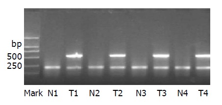
RT-PCR analysis showed that 16 (48.5%) of 33 HCC cases were positive for HPA mRNA, whereas only one case of liver paracancerous tissue was positive for HPA mRNA, and all 9 cases of normal liver tissues were negative (Figure 1, Table 1).
Figure 1.

M: marker, DL2000; T1–T4: HCC cancer tissues of four cases, which display a positive band at 585 bp; N1–N4: paracancer tissues; interior reference is seen at 224 bp for all the specimens.
Table 1.
Relationship between HPA and nm23-H1 expression and clinicopathological data and metastasis and recurrence of HCC.
| Parameters | Case number | HPA (+) | P | nm-23-H1 (+) | P |
| Tumor size | |||||
| ≤5 cm | 8 | 3 | 0.25 | 5 | 0.176 |
| >5 cm | 25 | 13 | 10 | ||
| Tumor integument | |||||
| Intact | 14 | 5 | 0.13 | 8 | 0.146 |
| Unintact | 19 | 11 | 7 | ||
| Metastasis and recurrence | |||||
| High | 14 | 10 | 0.023 | 3 | 0.018 |
| Low | 19 | 6 | 12 | ||
| AFP level | |||||
| Positive | 21 | 12 | 0.125 | 9 | 0.262 |
| Negative | 12 | 4 | 6 | ||
| HBsAg | |||||
| Positive | 24 | 12 | 0.292 | 9 | 0.106 |
| Negative | 9 | 4 | 6 | ||
| Liver cirrhosis | |||||
| Yes | 18 | 8 | 0.241 | 9 | 0.235 |
| No | 15 | 8 | 6 | ||
| Edmondson grade | |||||
| Grade I, II | 13 | 3 | 0.019 | 8 | 0.096 |
| Grade III, IV | 20 | 13 | 7 | ||
| TNM stage | |||||
| I, II | 18 | 6 | 0.047 | 9 | 0.235 |
| III, IV | 15 | 10 | 6 | ||
| Follow-up | |||||
| Metastasis and recurrence (+) | 14 | 11 | 0.003 | 3 | 0.024 |
| Metastasis and recurrence (-) | 14 | 3 | 9 |
Immunohistochemical staining
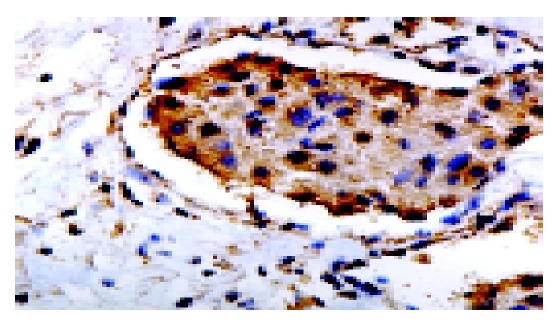
The positive staining for nm23-H1 protein appeared as yellow-brown particles in cytoplasm, but biliary ducts and blood vessels and liver sinusoidal endotheliocytes and interstitial cells had no positive staining. Well-distributed and diffuse strong staining could be seen in normal and paracancerous tissues. As for cancer tissue, some cases presented lamellar and focal staining (Figure 2). On the whole, the stainings of normal and paracancerous tissues were stronger and their relatively positive cell numbers were more than that of cancer tissues.
Figure 2.

Immunohistochemical staining (brown-yellow) for nm23-H1 in cytoplasm (SP, ×400). Interstitial cells did not show positive stainings.
The positive expressions of nm23-H1 in HCC, paracancerous tissue and normal tissue were 45.5% (15/33), 72.7% (24/33) and 88.9% (8/9), respectively. The positive rate of nm23-H1 in HCC was markedly lower than that of normal and paracancerous tissues (P<0.05).
Relation between the expression of HPA and nm23-H1 and clinicopathological data and metastasis and recurrence
As shown in Figure 1, there was no correlation between the expression of HPA mRNA and the size, envelope completeness, level of AFP, status of HBsAg and existence of liver cirrhosis (P>0.05). Among 16 cases with positive HPA mRNA expression, 10 cases (62.5%) showed high tendency of metastasis or recurrence occupying 62.5% (10/16), and only 6 cases showed low tendency of metastasis or recurrence, indicating a significant difference. Of the 28 cases, who were followed-up for 6-16 mo, 11 cases (78.6%) out of 14 patients with marked metastasis and recurrence were positive for HPA, and 3 cases of the other 14 patients without metastasis and recurrence expressed HPA, showing a significant correlation between HPA expression and metastasis and recurrence (P = 0.003). In addition, there existed correlation between the expression of HPA and tumor pathological grade (P = 0.019) and TNM stage (P = 0.047). The lower the Edmondson grade and the later the TNM stage were, the higher the expression of HPA was. The expression of nm23-H1 had no relation with the size of tumor, formation of integument, levels of AFP, status of HBsAg, existence of cirrhosis, Edmondson grade and TNM stage (P>0.05). However, there was obvious difference in the expression of nm23-H1 between the cases with high and low tendencies of metastasis and recurrence (P = 0.018).
Correlation between HPA and nm23-H1 in HCC
The positive rates of nm23-H1 expression in the cases with positive HPA and negative HPA were 31.3% (5/16) and 58.8% (10/17), respectively, without significant difference.
Prediction of postoperative metastasis and recurrence
Of the 28 followed-up cases, 14 showed HPA positivity and 14 HPA negativity, and 12 cases were positive and 16 were negative for nm23-H1, 9 cases showed simultaneous existence of HPA positivity and nm23-H1 negativity, the other 19 cases showed only HPA negativity, or only nm23-H1 positivity or both simultaneous positivities. Of the 14 cases with HPA positivity, 11 patients experienced metastasis and recurrence, the prediction rate was 78.6% (11/14); of the 16 cases with nm23-H1 negativity, 11 had metastasis and recurrence, the prediction rate was 68.8% (11/16). Thus, the prediction of postoperative metastasis and recurrence could be made by either HPA positivity or nm23-H1 negativity for HCC patients. Of the 9 cases with simultaneous appearance of HPA positivity and nm23-H1 negativity, 8 patients had metastasis and recurrence, the prediction rate of the combination of the two was 88.9% (8/9).
DISCUSSION
Our results showed that the expression of HPA was markedly higher in HCC tissues as compared to the normal and paracancerous tissues. Ten cases out of 14 patients with high tendency of metastasis and recurrence had HPA positive expression, whereas 6 cases out of 19 patients with low tendency of metastasis and recurrence had positive HPA expression, revealing an obvious relation between HPA and metastasis and recurrence of HCC. Eleven cases out of 14 followed-up patients with definite metastasis and recurrence showed positive expression for HPA, while only 3 cases in 14 patients without metastasis and recurrence had HPA positive expression, further suggesting that expression of HPA was positively correlated with the higher tendency of tumor progression and postoperative metastasis and recurrence. Hence it may be valuable to examine HPA expression for the clinical prediction of metastasis and recurrence of HCC. However, HPA expression had no relation with tumor- size, integument completeness, AFP level, HBsAg status and existence of cirrhosis, yet it showed relation with pathological grade and TNM stage of HCC. The poorer the tumor differentiation and the later the TNM stage were, the higher the positive expression of HPA. Our study demonstrated that the positive expression of nm23-H1 protein was obviously lower in HCC tissues compared to the normal and paracancerous tissues, indicating that there might exist loss expression of nm23-H1 gene in some HCCs, probably at transcription, or translation or post-translation levels. Our results also revealed that nm23-H1 expression had no relation with HCC tumor size, integument formation, AFP level, HBsAg status, existence of cirrhosis, Edmondson grade and TNM stage. Moreover, we observed that nm23-H1 had a relation with metastasis and recurrence of HCC, suggesting that defect of nm23-H1 can also predict HCC metastasis and recurrence.
HPA is mainly synthesized and secreted from cancer cells, and we found that nm23-H1 was also principally located in cytoplasm. But in cases with positive or negative HPA mRNA, the rates of positive expression of nm23-H1 were 31.2% (5/6) and 58.8% (10/7), respectively, with no significant difference. Therefore, the correlation could not be surely testified between the expressions of HPA and nm23-H1 genes in HCC, indicating that both of HPA and nm23-H1 might not have direct correlation in the suppression of metastasis. This can be interpreted by the different biochemical mechanisms of HPA and nm23-H1. HPA impels tumor to progress and metastasize by degradating HSPG and releasing angiogenesis factors. As for the mechanism of action of nm23-H1, many scholars have inferred through careful study that nm23-H1, by interacting with GAP protein, participates in cellular signal transduction, cell differentiation and metastasis.
In this study, the prediction rate of HPA positivity reached 78.6% (11/14), and that of nm23-H1 negativity was 68.8% (11/16). Therefore, the expression patterns of HPA and nm23-H1 might be used to predict the postoperative metastasis and recurrence of HCC. Eight cases out of 9 with simultaneous HPA positive and nm23-H1 negative expression showed metastasis and recurrence, with prediction rate of 88.9% (8/9), which was higher than that of either HPA positivity or nm23-H1 negativity, singly. It indicates that the combined application of HPA and nm23-H1 expression can improve the accuracy precision of HPA, particularly that of nm23-H1.
Footnotes
Edited by Wang XL and Kumar M Proofread by Zhu LH
References
- 1.Chen XP, Peng SY, Liu YB, Zhu XX. Function of heparinase and suppressant in metastasis. Zhongguo Waike Zazhi. 2002;40:153 155. [Google Scholar]
- 2.Shimada M, Taguchi K, Hasegawa H, Gion T, Shirabe K, Tsuneyoshi M, Sugimachi K. Nm23-H1 expression in intrahepatic or extrahepatic metastases of hepatocellular carcinoma. Liver. 1998;18:337–342. doi: 10.1111/j.1600-0676.1998.tb00815.x. [DOI] [PubMed] [Google Scholar]
- 3.Lin LI, Lee PH, Wu CM, Lin JK. Significance of nm23 mRNA expression in human hepatocellular carcinoma. Anticancer Res. 1998;18:541–546. [PubMed] [Google Scholar]
- 4.Vlodavsky I, Friedmann Y, Elkin M, Aingorn H, Atzmon R, Ishai-Michaeli R, Bitan M, Pappo O, Peretz T, Michal I, et al. Mammalian heparanase: gene cloning, expression and function in tumor progression and metastasis. Nat Med. 1999;5:793–802. doi: 10.1038/10518. [DOI] [PubMed] [Google Scholar]
- 5.Wang C, Wang Y, Zhang XF, Yang FD, Xu GW. Nm23-H1 gene expression and its correlation to lymphatic metastasis or proliferative activity in pancreatic carcinoma. Zhonghua Putong Waike Zazhi. 2000;15:460 462. [Google Scholar]
- 6.Hulett MD, Freeman C, Hamdorf BJ, Baker RT, Harris MJ, Parish CR. Cloning of mammalian heparanase, an important enzyme in tumor invasion and metastasis. Nat Med. 1999;5:803–809. doi: 10.1038/10525. [DOI] [PubMed] [Google Scholar]
- 7.Toyoshima M, Nakajima M. Human heparanase. Purification, characterization, cloning, and expression. J Biol Chem. 1999;274:24153–24160. doi: 10.1074/jbc.274.34.24153. [DOI] [PubMed] [Google Scholar]
- 8.Kussie PH, Hulmes JD, Ludwig DL, Patel S, Navarro EC, Seddon AP, Giorgio NA, Bohlen P. Cloning and functional expression of a human heparanase gene. Biochem Biophys Res Commun. 1999;261:183–187. doi: 10.1006/bbrc.1999.0962. [DOI] [PubMed] [Google Scholar]


