

A 14-year-old male patient presented with focal masses of thick, adherent, plate like, yellow-brown scales, attached to the hair shafts, predominantly affecting the fronto-parietal area and vertex of the scalp [Figure 1]. The underlying scalp had thick, erythematous plaques with fine, non greasy, silvery-white scaling with noncicatricial alopecia [Figure 2]. Potassium hydroxide examination of scales and hair and culture for fungus was negative. Biopsy of the scalp lesion showed moderate epidermal hyperplasia with markedly diminished granular layer and thin flat mounds of layered parakeratosis containing few neutrophils, with moderately dense perivascular lymphocytic infiltrate in the upper dermis [Figure 3]. The features were suggestive of psoriasis.
Figure 1.
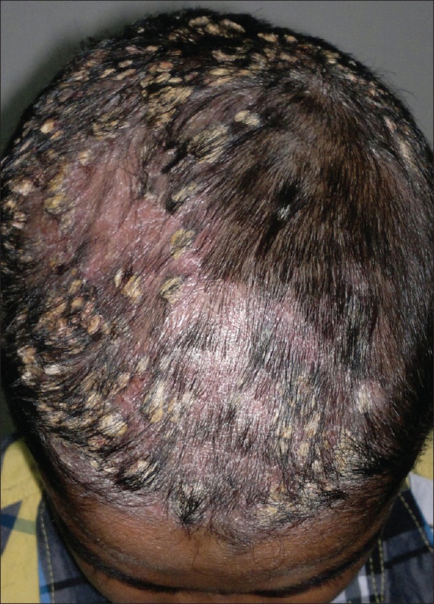
Focal masses of thick, adherent, asbestos like yellow brown scales over fronto-parietal and vertex region of scalp
Figure 2.

Psoriasis of scalp in the background with non-cicatricial alopecia affecting parietal region
Figure 3.

Histopathology showing epidermal hyperplasia, focal parakeratosis, diminished granular layer and moderately dense perivascular lymphocytic infiltrate in upper dermis (H and E, ×40)
Pityriasis amiantacea (synonyms: Tinea amintacea, asbestos scalp, porrigo amiantacea, tinea asbestina, keratosis follicularis amiantacea) is an inflammatory scaling reaction of the scalp, often without evident cause, that may occur at any age.[1] It usually occurs in pediatric patients and is characterized by large plates of scales firmly adherent to the hair and scalp. Focal hair loss, generally reversible but sometimes cicatricial, if associated with secondary infection may also occur. The condition usually begins during teenage years and progresses to more typical psoriasis in 2-15% of pediatric patients.[2] The essential features responsible for the asbestos like scaling are diffuse hyperkeratosis and parakeratosis together with follicular keratosis, which surrounds each hair with a sheath of horn.[3] It may also be observed as a sequel or complication of streptococcal infection, seborrheic dermatitis, atopic dermatitis, lichen simplex.[3]
Footnotes
Source of Support: Nil
Conflict of Interest: None declared.
REFERENCES
- 1.Plewig G, Jansen T. Pityriasis amiantacea. In: Wolff K, Goldsmith LA, Katz SI, Gilchrest BA, Paller SA, Leffell DJ, editors. Fitzpatrick's Dermatology in general medicine. 7th ed. New York: McGraw-Hill; 2008. pp. 219–24. [Google Scholar]
- 2.Abdel-Hamid IA, Agha SA, Moustafa YM, El-Labban AM. Pityriasis amiantacea: A clinical and etiopathologic study of 85 patients. Int J Dermatol. 2003;42:260–4. doi: 10.1046/j.1365-4362.2003.01755.x. [DOI] [PubMed] [Google Scholar]
- 3.Paller AS, Mancini AJ. Hurwitz Clinical Pediatric Dermatology. A Textbook of Skin Disorders of Childhood and Adolescence. 3rd ed. Philadelphia: Elsevier-Saunders; 2006. Papulosquamous and related disorders; pp. 85–106. [Google Scholar]