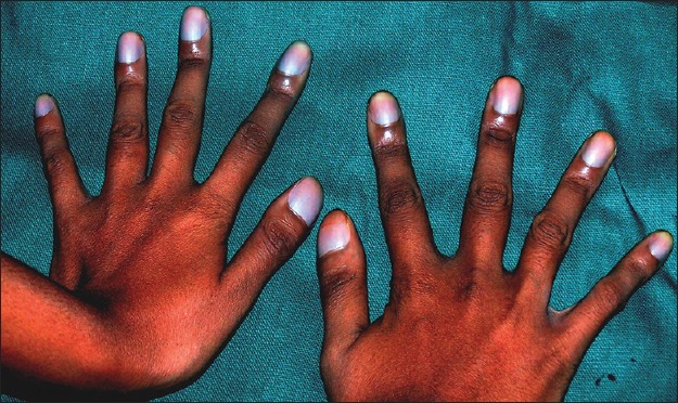
A 16-year-old male presented with white-colored fingernails since birth [Figure 1]. Toenails were normal. None of the family members were affected. Hematological investigations were normal. On cutaneous examination all finger nails were milky, porcelain white in color and smooth surfaced. No knuckle pads, palmoplantar keratoderma, or hearing loss was observed. General and systemic examination was normal. On clinical correlation, a diagnosis of idiopathic congenital true leukonychia totalis was made.
Figure 1.

Milky, porcelain, white-colored smooth-surfaced finger nails
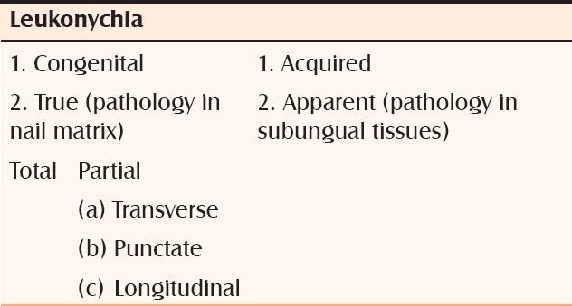
Leukonychia is the white discoloration of nail that loses its normal pink color, with disappearance of the lunula. It is the commonest chromatic abnormality of the nail apparatus. It is also referred to as white nail, milk spots, and fortune or gift spots. It is derived from the Greek word “leukos” means white and “onyx” means nail.[1,2] Leukonychia is classified as: [Table 1].
Table 1.
Classification of leukonychia

The proposed white discoloration of the nail is attributable to reflection of light from large keratohyaline granules present in the parakeratotic cells of the ventral portion of nail plate thus preventing the visualisation of normal vascular tissue underlying the nail plate.[2,3]
Total congenital leukonychia is very rare hereditary condition in which all nails are milky white with an autosomal dominant inheritance but it can be autosomal recessive in few cases. Hereditary leukonychia involves whole nail plate as against acquired one which shows transverse or punctate leukonychia. Mutations in PLCD1 gene on chromosome 3p21.3-p22 was identified as cause of hereditary leukonychia. This PLCD1 gene is localized to nail matrix and encodes phosphoinositide specific phospholipase C delta 1 subunit which is important enzyme in phosphoinositide metabolism required for molecular control of color and growth of nail.[4]
Leukonychia totalis is associated with multiple systemic diseases such as diabetes, liver cirrhosis, renal failure, cardiac failure, gall stones, renal calculi, duodenal ulcers, pili torti, trichilemmal cyst, sebaceous cyst, congenital hyperparathyroidism, Hodgkin's lymphoma, Leopard syndrome, epiphyseal dysplasia syndrome, Bart Pumphrey syndrome, mental retardation, etc., However, there are very few reports in the literature on sporadic congenital leukonychia totalis without any systemic abnormalities.[1,3]
Treatment is usually supportive with detailed investigation to check for association of any systemic or genetic disorders.
Footnotes
Source of Support: Nil
Conflict of Interest: Nil.
REFERENCES
- 1.De D, Handa S. Hereditary leukonychia totalis. Indian J Dermatol Venereol Leprol. 2007;73:355–7. doi: 10.4103/0378-6323.35746. [DOI] [PubMed] [Google Scholar]
- 2.Kwon NH, Kim JE, Cho BK, Jeong EG, Park HJ. Sporadic congenital leukonychia with koilonychia. Int J Dermatol. 2012;51:1400–2. doi: 10.1111/j.1365-4632.2010.04750.x. [DOI] [PubMed] [Google Scholar]
- 3.Lee YB, Kim JE, Park HJ, Cho BK. A case of hereditary leukonychia totalis and partialis. Int J Dermatol. 2011;50:233–4. doi: 10.1111/j.1365-4632.2010.04306.x. [DOI] [PubMed] [Google Scholar]
- 4.Kiuru M, Kurban M, Itoh M, Petukhova L, Shimomura Y, Wajid M, et al. Hereditary leukonychia, or porcelain nails, resulting from mutations in PLCD1. Am J Hum Genet. 2011;88:839–4. doi: 10.1016/j.ajhg.2011.05.014. [DOI] [PMC free article] [PubMed] [Google Scholar]


