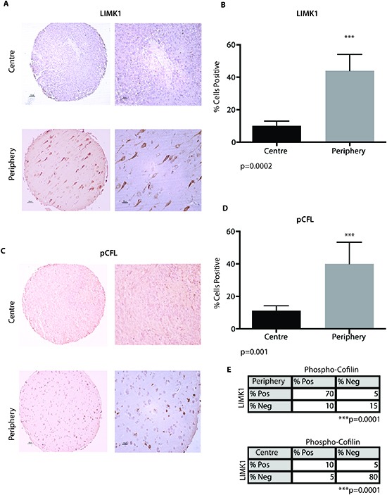
Figure 3. LIMK1 and pCFL are expressed in the periphery of GBM.

(A) A tissue microarray consisting (TMA) of patient matched centre and periphery samples (n=20) was stained for LIMK1 by immunohistochemical analysis (IHC). Samples were scored as positive or negative. (B) Ten field of views per sample were taken to quantify % percent of cells positive for LIMK1 in the centre portion of GBM or periphery of the tumour. (C) A TMA consisting of patient matched centre and periphery samples (n=16) was stained for pCFL by IHC. Samples were scored as positive or negative. (D) Ten field of views per sample were taken to quantify % percent of cells positive for pCFL in the centre portion of GBM or periphery of the tumour. ***p<0.001. (E) Co-expression analysis of pCFL and LIMK1 from serial IHC section of matching cores (n=5) in the centre and periphery of the tumour. pCFL and LIMK1 are positively co-expressed in tumor periphery cells (p=0.0001). pCFL and LIMK1 are negative in the same centre GBM tumor cells (p=0.0001).
