TO THE EDITOR
Telangiectasia macularis eruptive perstans (TMEP) was first described by Weber in 1930 based on a single case report of a 60 year old female with slightly raised, reddish telangiectatic macules of general distribution, although it was noted that individual telangiectasia could not be visualized. This original report did not include histological findings.1 Weber subsequently evaluated a second case with urticaria pigmentosa (UP) that closely resembled the index TMEP patient, and based on these two cases concluded that TMEP was a pigmentless variant of UP that presents primarily in adulthood.1,2 In subsequent reports, mast cell infiltration was noted histologically in both UP and TMEP; however the gross appearance and perivascular distribution of mast cells and association with dilated vessels was said to distinguish TMEP from UP. Thus, TMEP is now usually referred to as a rare form of cutaneous mastocytosis in which the skin has flat telangiactatic macules in a generalized distribution.3–6 The relative lack of information on the consequences of a diagnosis of TMEP has led to confusion in clinical management. Based on these observations, we conducted a survey of patients that were evaluated for mastocytosis who carried a diagnosis of TMEP in order to determine how the diagnosis of TMEP was being applied and if there were any consistent features to report.
Using the NIH Biomedical Translational Research Information System, we conducted a retrospective chart review of 299 subjects who were referred to the National Institute of Allergy and Infectious Diseases (NIAID) on IRB approved mastocytosis protocols. All subjects signed informed consent. We identified 24 subjects whose records included the diagnosis of TMEP (Table 1). We then analyzed clinical and laboratory data, including skin and bone marrow biopsies, as relevant to the diagnosis of mastocytosis in the skin (MIS), including TMEP and UP, and systemic mastocytosis.
Table 1.
Demographic and clinical characteristics of the patients referred with telangiectasia macularis eruptive perstans.
| Skin biopsy characteristics | ||||||||||
|---|---|---|---|---|---|---|---|---|---|---|
| Patient | Age (yrs) | Gender | Referral diagnosis | TMEP diagnosis | Telangiec tasia | Infiltrate | ↑mast cells | Perivascular | Dilated vessels | NIH diagnosis |
| 1 | 34 | F | TMEP | Bx | N | MC, MN | Y | Y | N | UP, ISM |
| 2 | 45 | F | TMEP | Both | Y | MC, E | Y | Y | - | UP |
| 3 | 28 | F | TMEP | Bx | - | MC, E | - | Y | - | UP, ISM |
| 4 | 51 | M | TMEP | Bx | - | MC, E | N | Y | - | UP |
| 5 | 42 | F | TMEP | Bx | - | MC | Y | Y | Y | UP |
| 6 | 62 | F | TMEP/UP | Clinical | NR | NR | NR | NR | NR | UP, ISM |
| 7 | 43 | F | UP | Bx* | NR | NR | NR | NR | NR | UP, ISM |
| 8 | 22 | F | CM | Bx | NR | NR | NR | NR | NR | UP, ISM |
| 9 | 48 | F | CM | Bx | Y | MC, E | Y | Y | Y | UP, ISM |
| 10 | 20 | F | CM | Bx | - | MC, MN | Y | Y | - | UP |
| 11 | 46 | F | SM | Both | - | MC | - | Y | - | UP, ISM |
| 12 | 22 | F | SM | Bx | NR | NR | NR | NR | NR | UP, ISM |
| 13 | 29 | F | SM | Bx | NR | NR | NR | NR | NR | UP, ISM |
| 14 | 28 | F | SM | Bx | - | MC | - | - | - | UP, ISM |
| 15 | 28 | M | SM | Bx | Y | MC | Y | - | - | UP, ISM |
| 16 | 40 | F | SM | Both | Y | MC, MN | Y | Y | NR | UP, ISM |
| 17 | 43 | M | SM | Bx | - | MC, MN | Y | Y | NR | UP, ISM |
| 18 | 32 | F | SM | Bx | - | MC, L | Y | Y | Y | UP, ISM |
| 19 | 42 | M | SM | Bx | NR | NR | NR | NR | NR | UP, ISM |
| 20 | 20 | M | SM | Bx | Y | MC, E, L | Y | Y | - | UP |
| 21 | 48 | M | SM | Both | Y | MC, E, L | - | Y | Y | UP |
| 22 | 2 | M | M | Bx | - | L | Y | Y | - | No M |
| 23 | 58 | M | M | Both | - | MC, E, L | Y | Y | - | UP, ISM |
| 24 | 54 | F | None | Both | Y | MC, MN | Y | Y | - | UP |
Legend and abbreviations: F, female; M, male; TMEP, telangiectasia macularis eruptive perstans; UP, urticaria pigmentosa; M, mastocytosis; SM, systemic mastocytosis; CM, cutaneous mastocytosis; ISM, indolent systemic mastocytosis; Bx, skin biopsy; Clinical, clinical observation on physical exam; Both, by both skin biopsy and clinical observation; NR, not reported; Y, yes; N, no; NR, no report available; -, no comment; MC, mast cells, E, eosinophils, L, lymphocytic; MN, mononuclear;
UP seen on skin biopsy
Of the 24 subjects identified with a diagnosis of TMEP, 8 (33%) were male and 16 (67%) were female. The median age (interquartile range [IQR]) at initial visit to the NIH was 41 years (19.5). Six subjects had “TMEP” specified in the referring diagnosis, while the remaining 18 subjects were referred for “cutaneous or systemic mastocytosis” with TMEP reported by skin biopsy or clinical observation.
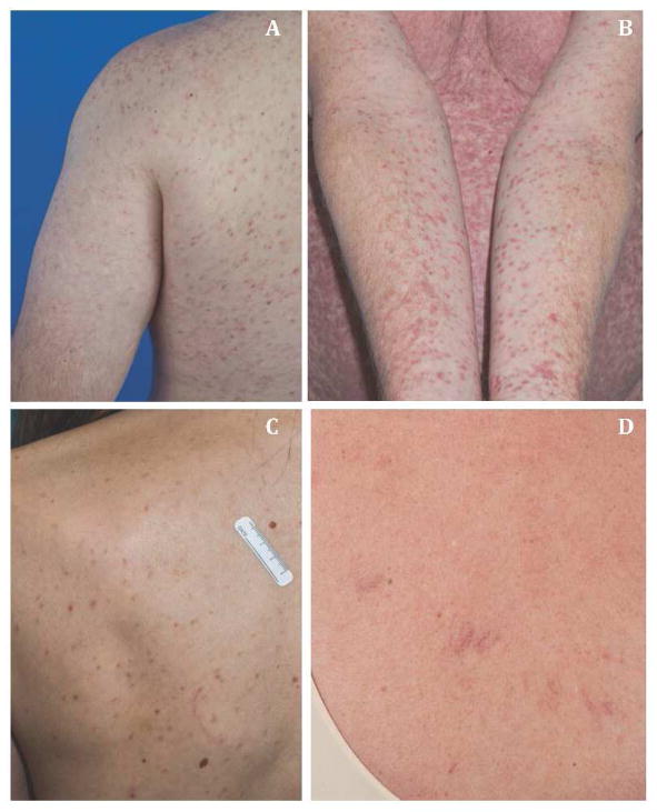
TMEP is generally described with onset in adulthood, distribution on the chest and extremities, and association with a negative Darier’s sign.4–7 In contrast, UP is often found in a generalized and random distribution, has the highest concentration of lesions on the trunk, is associated with a positive Darier’s sign, and is commonly reported in both children and adults.4,5 In our TMEP group, the rash was most frequently distributed on the chest and extremities and resembled in appearance that of classic UP (Figure 1A–C). TMEP is shown in Figure 1D. Darier’s sign was positive in 6/15 patients (40%).
Figure 1.
TMEP or UP? Three pictures of patients referred with a diagnosis of TMEP but have cutaneous lesions consistent with UP. A) 48 year old male diagnosed with UP and no evidence of systemic mastocytosis; B) 58 year old male with UP and indolent systemic mastocytosis; C) 42 year old female with UP and no evidence of systemic mastocytosis; D) 52 year old female with TMEP, and an elevated tryptase of 36.0 ng/mL. Her bone marrow biopsy shows increased mast cells with > 25% appearing spindle-shaped but no mast cell aggregates. Macular telangiectasia are present.
Of the 24 patients identified with TMEP, 9 (24%) reported a history of anaphylaxis and 4 (17%) with syncope. All patients (100%) reported cutaneous manifestations, such as urticaria, pruritus or flushing. Eighteen patients (75%) reported gastrointestinal symptoms, such as abdominal pain, cramping, nausea, vomiting, or diarrhea. Cardiovascular symptoms, such as tachycardia or palpitations, were reported in nine patients (38%). Two patients (8%) had palpable hepatosplenomegaly. Dual-energy X-ray absorptiometry (DEXA) had been performed at outside facilities on 12 patients and yielded 5 (42%) with osteopenia and 1 (8%) with osteoporosis, based on the World Health Organization (WHO) definitions.8 Thus, the majority of patients identified with TMEP had extracutaneous findings.
All 24 patients underwent a laboratory evaluation. The white blood cell counts, hemoglobin, platelets, PT, PTT, C-reactive protein, and serum B12 levels were normal. The median (IQR) serum tryptase and serum IgE level in the TMEP group were 24.2 ng/mL (34) and 8.85 IU/mL (12.1) respectively. Serum tryptase and IgE levels were comparable among the patients ultimately diagnosed with mastocytosis in the skin (all UP) and those with indolent systemic mastocytosis (tryptase 22.6 vs. 32.4, p=0.18; IgE 8.7 vs. 6.8, p=0.67 respectively).
TMEP is said to be histopathologically identified by the presence of mast cell infiltrates around venules with dermal dilated capillaries of the superficial venous plexus.4 Upon review of 18 TMEP skin biopsy reports, all reports noted mast cell infiltration, of which 89% were distributed perivascularly. Dilated vessels were noted in 4 patients (22%). The presence of telangiectasias are an important defining characteristic of TMEP and a distinguishing feature from UP; yet few of our patients had evidence of telangiectasias on observation (Table 1). Thus, in this group of patients, a definite diagnosis of TMEP was not substantiated by the presence of characteristic skin lesions and distinctive histopathology.
Twenty-one patients underwent bone marrow biopsy for evaluation of possible systemic mastocytosis. Sixteen patients (67%) met the WHO diagnostic criteria for indolent systemic mastocytosis, 7 (29%) had mastocytosis limited to the skin (MIS, all UP), and 1 patient did not meet criteria for MIS or systemic disease (4%). 9 While the diagnosis of TMEP was not clinically substantiated in any of these patients, the majority of our cohort was ultimately diagnosed with indolent systemic mastocytosis. The high correlation in our cohort with systemic mastocytosis (typically found in adults with UP) suggests it is more appropriate to reclassify TMEP as a highly vascularized form of UP. For symptomatic relief both patients with TMEP and UP may benefit from treatment with oral antihistamines, montelukast and psoralen with ultraviolet A (PUVA).10,11 While both cutaneous forms of mastocytosis can be limited to the skin, bone marrow evaluation should be pursued if clinical history and findings suggests systemic involvement. Prognosis depends on whether the patient meets WHO criteria for systemic mastocytosis.
Based on our findings, the diagnosis of TMEP appears to be over-used, has little clinical utility and is confusing to physicians and patients. Highly vascularized UP more accurately describes these pigmented macular lesions that localize with observable telangiectasia. We suggest that in the majority of instances where the diagnosis of TMEP is used, the lesions are more accurately described as UP.
Clinical Implication.
Telangiectasia macularis eruptive perstans (TMEP) is a rare form of mastocytosis in the skin characterized by flat dermal telangiactatic macules. Our analysis indicates it is being over diagnosed in patients where a more accurate diagnosis would be urticaria pigmentosa.
Acknowledgments
Funding Statement: This work was supported by the Division of Intramural Research, National Institute of Allergy and Infectious Diseases, National Institutes of Health
Footnotes
Publisher's Disclaimer: This is a PDF file of an unedited manuscript that has been accepted for publication. As a service to our customers we are providing this early version of the manuscript. The manuscript will undergo copyediting, typesetting, and review of the resulting proof before it is published in its final citable form. Please note that during the production process errors may be discovered which could affect the content, and all legal disclaimers that apply to the journal pertain.
References
- 1.Weber FP, Hellenschmied R. Telangiectasia macularis eruptive perstans. British Journal of Dermatology and Syphilis. 1930;42:374–382. [Google Scholar]
- 2.Weber FP. Telangiectasia macularis eruptiva perstans – probably a telangiectatic variety of urticaria pigmentosa in an adult. Proceedings of the Royal Society of Medicine. 1930;24:96–97. doi: 10.1177/003591573002400204. [DOI] [PMC free article] [PubMed] [Google Scholar]
- 3.Nickel WR. Urticaria Pigmentosa. JAMA Arch Dermatology. 1957;76:476–498. doi: 10.1001/archderm.1957.01550220084017. [DOI] [PubMed] [Google Scholar]
- 4.Soter NA. The skin in mastocytosis. Journal of Investigative Dermatology. 1991;96:32S–39S. [PubMed] [Google Scholar]
- 5.Sarkany RPE, et al. Telangiectasia macularis eruptive perstans: a case report and review of the literature. Clinical and Experimental Dermatology. 1998;23:38–39. doi: 10.1046/j.1365-2230.1998.00273.x. [DOI] [PubMed] [Google Scholar]
- 6.Akin C, Valent P. Diagnostic criteria and classification of mastocytosis in 2014. Immunology and Allergy Clinics of North America. 2014;34:207–18. doi: 10.1016/j.iac.2014.02.003. [DOI] [PubMed] [Google Scholar]
- 7.Costa DL, et al. Telangiectasia macularis eruptive perstans: a rare form of adult mastocytosis. Journal of Clinical and Aesthetic Dermatology. 2011;4:52–54. [PMC free article] [PubMed] [Google Scholar]
- 8.World Health Organization. Assessment of fracture risk and its application to screening for postmenopausal osteoporosis: Report of a WHO Study Group. World Health Organization technical report series. 1994;843:1–129. [PubMed] [Google Scholar]
- 9.Valent P, et al. Diagnostic criteria and classification of mastocytosis: a consensus proposal. Leukemia Research. 2001;25:603–625. doi: 10.1016/s0145-2126(01)00038-8. [DOI] [PubMed] [Google Scholar]
- 10.Sotiriou E, et al. Telangiectasia macularis eruptive perstans successfully treated with PUVA. Photodermatology, Photoimmunology, and Photomedicine. 2010;26:46–47. doi: 10.1111/j.1600-0781.2009.00480.x. [DOI] [PubMed] [Google Scholar]
- 11.Cengizlier R, et al. Treatment of telangiectasia macularis eruptive perstans with montelukast. Allergologia et Immunopathologia. 2009;37:334–336. doi: 10.1016/j.aller.2009.03.010. [DOI] [PubMed] [Google Scholar]


