

Abstract
Studies have demonstrated that electromagnetic waves, as the one of the most important physical factors, may alter cognitive and non-cognitive behaviors, depending on the frequency and energy. Moreover, non-ionizing radiation of low energy waves e.g. very low frequency waves could alter this phenomenon via alterations in neurotransmitters and neurohormones. In this study, short, medium, and long-term exposure to the extremely low frequency electromagnetic field (ELF-EMF) (1 and 5 Hz radiation) on behavioral, hormonal, and metabolic changes in male Wistar rats (250 g) were studied. In addition, changes in plasma concentrations for two main stress hormones, noradrenaline and adrenocorticotropic hormone (ACTH) were evaluated. ELF-EMF exposure did not alter body weight, and food and water intake. Plasma glucose level was increased and decreased in the groups which exposed to the 5 and 1Hz wave, respectively. Plasma ACTH concentration increased in both using frequencies, whereas nor-adrenaline concentration showed overall reduction. At last, numbers of rearing, sniffing, locomotor activity was increased in group receiving 5 Hz wave over the time. In conclusions, these data showed that the effects of 1 and 5 Hz on the hormonal, metabolic and stress-like behaviors may be different. Moreover, the influence of waves on stress system is depending on time of exposure.
Keywords: Low-frequency electro-magnetic field, Corticosterone, Adrenaline, Adrenocorticotropic Hormone (ACTH), Stress
INTRODUCTION
People nowadays due to technological advances and the increasing use of electronic equipment are constantly exposed to electromagnetic radiation. In addition to man-made sources, the radiation from natural sources like the sun and the earth are released regularly (Zhang et al., 1995; Hayakawa, 2004). The body of studies showed that electromagnetic radiations have several important effects on different bodyregions depending on their energy (Grundler et al., 1992). For instance, increase in the permeability of the cell membrane, chromosome structural changes and chemical changes in DNA structure are shown to be occurred after electromagnetic wave exposure (Grundler et al., 1992).
Legally, spectrum of electromagnetic waves between 1 Hz to 300 Hz is called Extremely Low Frequency Electro-Magnetic Fields (ELF-EMF) (Wilson et al., 1990; Pesce et al., 2013). In fact, these wave ranges can be found in everywhere in modern societies (Haarala et al., 2003; Avendano et al., 2012). Due to increasing concern about the modernization of society, people are more concerned with the effects of radio waves (Feychting et al., 2005). These type of non-ionizing electromagnetic waves of radiation also emitted from industrial productive sources of waves can be used to power generation plants, transmission lines and electrical equipment all electrical work with the city. In addition, electrical appliances such as the notebooks and mobile phones can be found in human living environment as electromagnetic waves generators and its generated ELF waves are available in this way on biological systems.
ELF-EMFs can alter growth, morphology, differentiation, death program and nerve impulse transmission in the cells (Kerr et al., 1972; Pirozzoli et al., 2003; Grassi et al., 2004). Szemerszky and colleagues have shown that chronic exposure to a 50 Hz ELF-EMF can increase proopiomelanocortine (POMC) mRNA in the rat anterior pituitary gland which was in relation to an increase in ACTH and corticosterone hormones as well (Szemerszky et al., 2010). It showed that extremely-ELF-EMFexposure leads to increased oxidative stress in chick embryonic cells and humans erythrocytes (Lahijani et al., 2009). On the other hand, oxidative stress induced DNA damage and lipid peroxidation, systemic disorders, and finally,cell death (Fernie and Bird, 2001; Kovacic and Edwards, 2010). Another study showed that extremely ELF-EMF exposure can stimulate the immune system via the reduction of serum levels of ACTH and cortisol (Michal and Marta, 2004).
ELF-EMFs waves with different modulation frequencies have been widely used for treatment of several disease such as epilepsy, fracture and wound healing, because of their low energy (Athanasiou et al., 2007; Santini et al., 2009). On contrary, some studies demonstrated that ELF-EMFs may increase carcinogenic risk in the childhood leukemia and sclerosis (Coleman and Bera, 1988; Kheifets et al., 2005).
Given that the special activity in the CNS system produce the concealer waves with specific frequencies such as 1 Hz and 5 Hz that related to different behaviors including calm sleep and emotional stress respectively (Yamamoto, 1998).
However, previous studies have focused on the parameters and behaviors which directly addressed the activity of stress system. For example, POMC, ACTH, and corticosterone as the major stress hormones as well as anxiety, forced swimming test, situational and social anxiety as well as locomotor activity pattern as most behavioral tests in this regard was evaluated in some studies (Balassa et al., 2009; Szemerszky et al., 2010). It must be noted that in addition to the above mentioned hormones and behaviors, other stress hormones namely epinephrine and norepinephrine (Karatsoreos and McEwen, 2011) also may be affected during electromagnetic field exposure. In addition, stress can directly affect the brain dopamine system via direct interaction between corticosterone and D1 dopamine receptors the enzyme tyrosine hydroxylase activity (Czyrak et al., 2003). Investigators however are believed that dopamine-related behaviors such as locomotor activity, rearing, and sniffing may be evaluate as indirect stress behaviors instead of direct behaviors (Czyrak et al., 2003). Based on these facts, investigation in this regard may help us to understanding more precisely on how exposure to the ELF-EMF waves can interact with the brain stress system. Predictions to be applied waves with the specific frequencies such as theta hippocampal, delta, beta and alpha brain waves can be used to the induces of a special behavior. Because of this, we want to investigate the effects and side effects of application of these waves on rat animal models, with this goal that these waves can be used for the treatment of stress and other behavioral abnormality. In this study, using 1 and 5 Hz ELF-EMF waves with frequencies proportional to the frequencies of brain, behaviors and stress hormone secretion after Acute and Chronic exposure were assessed.
MATERIAL AND METHODS
Animals
Male Wistar rats (Pasture Institute, Tehran, Iran) weighing 250 ± 10 g at the time of experiments were used (n=8/group). The animals were housed four per cage, in a room under a 12 h light: 12 h dark cycle (lights on 07:00 h) and controlled temperature (23 ± 1°C) with free access to food and water. Animals were allowed to adapt to the laboratory conditions for at least 1 week before radiation. All experiments were performed between 12:00 and 14:00 h and each rat was tested only once. All procedures in this study are in accordance with the guide for the Care and Use of Laboratory Animals as adopted by the Ethics Committee of Baqiyatallah (a.s.) University (357: November 2000).
Device for electromagnetic field exposure and shielded room
The radiation were carried out using the ELF electromagnetic field generator depicted in Fig. 1 on six animals were placed in a cage made by Plexiglas (60×60×60 cm) once a day with 75 mW and 0.1 mT. For possible intervention of external interference to testingwave a spatial room has been designed in as much as the whole room parts, including the ceiling and walls, windows, even the smallest openings fully covered by aluminum foil (0.4 mm diameter). For further conformation this step had been checked by a wave detectorand lack of effective radiation in shielded room was confirmed. Then electromagnetic generating device antenna was fitted to the symmetrical (for waves uniformly irradiation) in the top boxes. All conditions have been done for control group except the irradiation.
Fig. 1.

ELF electromagnetic field generator.
Experimental design
The testing process is divided into two phases, and each phase lasting for 21 days. In this study, two frequencies of 1 and 5 Hz were used (once a day, 75 mW and 0.1 mT). In each phase, on days 1, 3, 7, 14 and 21 the animals were sacrifice for biological assessments. Blood samples, brain, adrenal gland were collected for further analysis. Eight animals were used in each experimental group. The time-line of the experiments procedure is shown in Fig. 2.
Fig. 2.
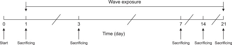
The time-line of the experiments as described in the text.
Measuring body weight
Before beginning of experiment in each day, rats were weighed by the scales with ± 0.1 g accuracy and then turnedto their home cages.
Measurement of water and food intake
During the experiment days 400 g rat chew and 150 ml tap water were placed in each cage. Twenty four hours before and 24 hours after the irradiation the reminder food and water were calculated as water and food intake index.
Anorexia
The time elapsed between rats replacement in the home cage and beginning food intake was calculated as the anorexia. After anorexic testing the rats move to animal house, expected those whom underwent sacrifice procedure.
Measurement of locomotor activity, rearing and sniffing
The animals were placed in an open field container (30×30×40 cm high), which its floor was divided to 16 equal-sized squares. A video camera was placed on the top of the apparatus at 120 cm heights for video typing. Each animal was placed in the apparatus and after 5 min for habituation, its activity was recorded for 10 min. The types then were analyzed for locomotion (number of line crossing) sniffing and rearing off line.
Hormonal analysis
ACTH and corticosterone, and adrenaline as major stress hormones were determined by an ELISA method. Blood samples were collected in the Ependorf tubes with 5% EDTA and were centrifuged in 4°C for 5 min in 3000 RPM. The supernatant was collected for ELISA assay using the appropriate kites (corticosterone, ACTH, adrenaline; all from CUSABIO CO., Japan).
Statistical analysis
Two-Way Analysis of (Two-Way ANOVA) using time and frequency as the factors was applied. Further analyses for individual between-groups comparisons were carried out with post hoc Tukey’s test. Data were displayed as Mean ± SEM. In all comparisons, p<0.05 was considered to be statistically significant.
RESULTS
The effects of exposure to 1 Hz and 5Hz frequency waves on food and water intake, body weight, anorexia, and exploratory-like behavior
Two-way ANOVA analysis revealed that exposure to an extremely Low-Frequency electroMagnetic Field (1 Hz) did not alter weight, water and food intake (Fig. 3) in the animals. However, anorexia was extremely high in these animals. Locomotor activity, rearing, and sniffingalso were not changed (Fig. 4).
Fig. 3.

The effects of acute and chronic exposure to the ELF-EMF (1 and 5 Hz) on body weight (A), anorexia (B), and water (C) and food (D) intake. Data are expressed as mean ± S.E.M of eight animals per group. +++p<0.001 different from respective control group.
Fig. 4.

The effects of acute and chronic exposure to the ELFEMF on dopamine-related behaviors (locomotor activity, panel A), (numbers of rearing, panel B) and (sniffing, panel C). Data are expressed as mean ± S.E.M of eight animals per group. +p<0.05, ++p<0.01, +++p<0.001 different from respective control group.
Analysis for 5 Hz wave revealed that although food and water intake, anorexia and body weight also were not changed in the animals (Fig. 3), but locomotor activity, sniffing and rearing were dramatically increased when the animals were exposed to the 5 Hz wave (Fig. 4). Further analysis indicated the interaction between time and wave frequency in this regard.
The effects of exposure to 1 Hz and 5Hz frequency waves on stress hormones in rats
Our results indicated that plasma corticosterone was reduced in animals from both wave lengths over the time even though the plasma ACTH was elevated in these animals (Fig. 5). In addition, plasma adrenaline was increased in the animals exposed to 1 Hz but decreased in the animals exposed to 5 Hz waves respectively (Fig. 5). Results of two-way ANOVA analyses with p values for all different experiments showed in Table 1.
Fig. 5.

The effects of acute and chronic exposure to the ELF-EMF on plasma ACTH (panel A), adrenaline (panel B), and corticosterone (panel C) concentrations. Data are expressed as mean ± S.E.M of eight animals per group. +p<0.05, ++p<0.01, +++p<0.001 different from respective control group.
Table 1.
Results of two-way ANOVA analyses with p values for different experiments
| Intera-group | Inter-group | Inter-Intra groups interactions | ||||
|---|---|---|---|---|---|---|
| ACTH | F(4,70) | p | F(1,70) | p | F(4,70) | p |
| 1 Hz | 2.31 | 0.084 | 15.13 | 0.000 | 6.25 | 0.000 |
| 5 Hz | 2.54 | 0.075 | 12.21 | 0.000 | 4.03 | 0.000 |
| Adrenaline | F(4,70) | p | F(1,70) | p | F(4,70) | p |
| 1 Hz | 5.14 | 0.01 | 11.24 | 0.000 | 3.28 | 0.042 |
| 5 Hz | 2.04 | 0.164 | 3.12 | 0.051 | 3.14 | 0.061 |
| Corticosterone | F(4,70) | p | F(1,70) | p | F(4,70) | p |
| 1 Hz | 4.18 | 0.041 | 7.08 | 0.000 | 3.94 | 0.041 |
| 5 Hz | 6.35 | 0.045 | 6.35 | 0.047 | 3.87 | 0.041 |
| Weight | F(4,70) | p | F(1,70) | p | F(4,70) | p |
| 1 Hz | 3.454 | 0.012 | 37.226 | 0.000 | 0.731 | 0.574 |
| 5 Hz | 1.280 | 0.286 | 2.215 | 0.141 | 2.658 | 0.040 |
| Anorexia | F(4,70) | p | F(1,70) | p | F(4,70) | p |
| 1 Hz | 336.445 | 0.000 | 1217.449 | 0.000 | 316.186 | 0.000 |
| 5 Hz | 4.677 | 0.002 | 390.000 | 0.000 | 8.635 | 0.000 |
| Water | F(3,56) | p | F(1,56) | p | F(3,56) | p |
| 1 Hz | 0.559 | 0.645 | 1.351 | 0.250 | 0.411 | 0.746 |
| 5 Hz | 0.367 | 0.777 | 2.500 | 0.119 | 1.260 | 0.297 |
| Food Intake | F(3,56) | p | F(1,70) | p | F(3,56) | p |
| 1 Hz | 6.436 | 0.001 | 0.210 | 0.648 | 1.562 | 0.209 |
| 5 Hz | 1.914 | 0.138 | 2.974 | 0.090 | 0.679 | 0.568 |
| Locomotor Activity | F(4,70) | p | F(1,70) | p | F(4,70) | p |
| 1 Hz | 0.752 | 0.560 | 6.885 | 0.011 | 0.164 | 0.956 |
| 5 Hz | 1.476 | 0.219 | 46.385 | 0.000 | 0.594 | 0.668 |
| Rearing | F(4,70) | p | F(1,70) | p | F(4,70) | p |
| 1 Hz | 2.547 | 0.047 | 0.114 | 0.737 | 4.167 | 0.004 |
| 5 Hz | 16.613 | 0.000 | 133.402 | 0.000 | 15.621 | 0.000 |
| Sniffing | F(4,70) | p | F(1,70) | p | F(4,70) | p |
| 1 Hz | 4.013 | 0.005 | 1.159 | 0.285 | 2.630 | 0.041 |
| 5 Hz | 16.588 | 0.000 | 195.883 | 0.000 | 11.587 | 0.000 |
DISCUSSION
The key founding of this study showed that exposure to the ELF-EMFat 1 Hz and 5 Hz frequencies did not alter body weight, and food and water intake, while increased ACTH concentration level in both using frequencies (1 and 5 Hz). These interventions have different effect on stress related behaviors including numbers of rearing and sniffing (1 Hz did not alter these behaviors, while 5 Hz did), locomotor activity (1 Hz and 5 Hz did not and increased this behavior, respectively), anorexic time (1 Hz increase but 5 Hz decrease), plasma adrenaline (1 Hz increased and 5 Hz decreased), corticosterone (both 1 Hz and 5 Hz decreased) concentrations.
These changes have been shown to be occurred after electromagnetic field exposure might be due to changes in the ability of the field to change protein activity both inside and/or outside the cell (Turro, 1991; Kula et al., 1999; Levitt, 2008). The biological effects of electromagnetic fields may lead to following changes: a: the exposure, and b: alternation in biological molecules structure (Adey, 1993). Whenever these changes over pass from the range of normal variations, or physiological compensatory mechanisms,can alter cell behaviors, while it seems that if these alternations are in the range of natural variation they have no effects (Adey, 1993).
Results obtained from the present study indicated that despite the effects of electromagnetic field exposure on anorexia, which is time dependent as well, no significant change were observed in body weight, or food and water intake in the animals. This finding may indicate that the mechanism (s) which control the body fuel cannot be affected by such frequencies. We suggest that other frequencies should be test in this regard. Moreover, the time of exposure also should be test. Next part of these data may explain the observed results. Our findings suggest that plasma ACTH levels elevated after exposure to the both 1 Hz and 5 Hz electromagnetic field over the time. These finding indicate that brain stress system is activated after field exposure and it can be postulated that corticotropin releasing factor (CRF) may be released from its sources (hypothalamic paraventricullar nucleus magnocelular cells) and involving in anorexia observed in the present study. The anorexia observed in the first part of our results is suggested by the above explanation. It must be noted that brain CRF rising is suggested for anorexia as a consequence of stress exposure (Ciccocioppo et al., 2001). The ability of these electromagnetic fields for activation of brain stress system is one of the most prominent findings. However, we did not trace the changes in the CRF activity directly in the present study, which can be conducted in the future studies. An interesting finding is that despite the ACTH plasma rising, anorexia is lowered in the animals exposed to 5 Hz electromagnetic field frequency. The explanation may be that this frequency may activate several mechanisms in the animal’s brain other that stress system which can overcome to the effects of stress system. In this regard, our data indicated that all of the dopamine-related behaviors including sniffing, rearing and locomotor activity are increased in the animals after field exposure. The above mentioned behaviors can be observed only after brain dopamine increment (Richardson and Gratton, 1996), which is postulated for involvement in animal feeding behavior (Salamone et al., 1997). As mentioned in the introduction section, stress can affect the brain dopamine system. And investigators believed that dopamine-related behaviors such as locomotor activity, rearing, and sniffing can be measured as indirect stress behaviors. According to these data one can postulated that after 5 Hz electromagnetic field exposure, brain stress and brain dopamine reward systems may be activate simultaneously, and the negative effect of stress on feeding behavior is over shad by the brain reward (dopamine) over activity. In this regard, the functional and anatomical interactions between brain reward and brain stress systems are explained (Bullmore and Sporns, 2009). In the case of 1 Hz electromagnetic field exposure, it can be demonstrated that since the field exposure just can activates brain stress system and cannot activate brain dopamine system (since none of the dopamine-related behaviors changed), the stress system over activity is profound and anorexia observed in the animals as well. The plasma corticosterone level was decreased in the animals exposed to the both frequencies. This data may indicate that exposure to these electromagnetic fields can induced adrenal gland hypoactivty. One explanation is that plasma ACTH concentration is increased in the animals after the prolong fields’ exposure and these increments may reduce adrenal gland activity. In this regard, data indicated that plasma corticosterone level may decline after chronic ACTH release (Reul et al., 1993). Another explanation is that exposure to the field can affects all parts of the body indifferently and hyper or hypo activity of different organs may be observed despite of their biologic activity and their natural role. In the last part of the experiments, results indicated that plasma adrenaline level also affected by the field exposure in 1 Hz frequency. Adrenaline is considered as a stress hormone which is released especially after a physical stress exposure (Gunnar and Quevedo, 2007). Our data may indicate that field exposure also may play as a physical stressor. However, these data also indicated that different frequencies exposure may leads to different brain and body responses. This finding also explains the hazardous of the fields for normal body function over the time.
Our data demonstrated that the responses observed in the animals after exposure to the different frequencies of ELFEMF (1 and 5 Hz) may differ according to the frequency, and time of exposure. These data also indicated that one of the most prominent organs which can be activate after field exposure is at least some parts of the brain, which manifested by changes in behavior and hormonal level in the animals. In other word, our data explained the hazardous of the field exposure which can destabilize all body organs by the mechanisms which are not fully understand.
Acknowledgments
This work is supported by the Neuroscience Research Center, Baqiyatallah University of Medical Sciences.
REFERENCES
- Adey WR. Biological effects of electromagnetic fields. J Cell Biochem. 1993;51:410–416. doi: 10.1002/jcb.2400510405. [DOI] [PubMed] [Google Scholar]
- Athanasiou A, Karkambounas S, Batistatou A, Lykoudis E, Katsaraki A, Kartsiouni T, Papalois A, Evangelou A. The effect of pulsed electromagnetic fields on secondary skin wound healing: An experimental study. Bioelectromagnetics. 2007;28:362–368. doi: 10.1002/bem.20303. [DOI] [PubMed] [Google Scholar]
- Avendano C, Mata A, Sanchez Sarmiento CA, Doncel GF. Use of laptop computers connected to internet through Wi-Fi decreases human sperm motility and increases sperm DNA fragmentation. Fertil Steril. 2012;97:39–45. e2. doi: 10.1016/j.fertnstert.2011.10.012. [DOI] [PubMed] [Google Scholar]
- Balassa T, Szemerszky R, Bárdos G. Effect of short-term 50 Hz electromagnetic field exposure on the behavior of rats. Physiol Hung. 2009;96:437–448. doi: 10.1556/APhysiol.96.2009.4.4. [DOI] [PubMed] [Google Scholar]
- Bullmore E, Sporns O. Complex brain networks: graph theoretical analysis of structural and functional systems. Nat Rev Neurosci. 2009;10:186–198. doi: 10.1038/nrn2575. [DOI] [PubMed] [Google Scholar]
- Ciccocioppo R, Martin-Fardon R, Weiss F, Massi M. Nociceptin/orphanin FQ inhibits stress-and CRF-induced anorexia in rats. Neuroreport. 2001;12:1145–1149. doi: 10.1097/00001756-200105080-00019. [DOI] [PubMed] [Google Scholar]
- Coleman M, Bera V. A review of epidemiological studies of the health effects of living near or working with electricity generation and transmission equipment. Int J Epidemiol. 1988;17:1–13. doi: 10.1093/ije/17.1.1. [DOI] [PubMed] [Google Scholar]
- Czyrak A, Mackowiak M, Chocyk A, Fijal K, Wêdzony K. Role of glucocorticoids in the regulation of dopaminergic neurotransmission. Pol J Pharmacol. 2003;55:667–674. [PubMed] [Google Scholar]
- Fernie KJ, Bird DM. Evidence of oxidative stress in American kestrels exposed to electromagnetic fields. Environ Res. 2001;86:198–207. doi: 10.1006/enrs.2001.4263. [DOI] [PubMed] [Google Scholar]
- Feychting M, Ahlbom A, Kheifets L. EMF and health. Annu. Rev. Public Health. 2005;26:165–189. doi: 10.1146/annurev.publhealth.26.021304.144445. [DOI] [PubMed] [Google Scholar]
- Grassi C, D’Ascenzo M, Torsello A, Martinotti G, Wolf F, Cittadini A, Azzena GB. Effects of 50Hz electromagnetic fields on voltage-gated Ca2+ channels and their role in modulation of neuroendocrine cell proliferation and death. Cell Calcium. 2004;35:307–315. doi: 10.1016/j.ceca.2003.09.001. [DOI] [PubMed] [Google Scholar]
- Grundler W, Kaiser F, Keilmann F, Walleczek J. Mechanisms of electromagnetic interaction with cellular systems. Naturwissenschaften. 1992;79:551–559. doi: 10.1007/BF01131411. [DOI] [PubMed] [Google Scholar]
- Gunnar M, Quevedo K. The neurobiology of stress and development. Annu Rev Psychol. 2007;58:145–173. doi: 10.1146/annurev.psych.58.110405.085605. [DOI] [PubMed] [Google Scholar]
- Haarala C, Björnberg L, Ek M, Laine M, Revonsuo A, Koivisto M, Hämäläinen H. Effect of a 902 MHz electromagnetic field emitted by mobile phones on human cognitive function: A replication study. Bioelectromagnetics. 2003;24:283–288. doi: 10.1002/bem.10105. [DOI] [PubMed] [Google Scholar]
- Hayakawa M. Electromagnetic phenomena associated with earthquakes: A frontier in terrestrial electromagnetic noise environment. Recent Res. Devel. Geophysics. 2004;6:81–112. [Google Scholar]
- Karatsoreos IN, McEwen BS. Psychobiological allostasis: resistance, resilience and vulnerability. Trends Cogn Sci. 2011;15:576–584. doi: 10.1016/j.tics.2011.10.005. [DOI] [PubMed] [Google Scholar]
- Kerr JF, Wyllie AH, Currie AR. Apoptosis: a basic biological phenomenon with wide-ranging implications in tissue kinetics. . Br. J. Cancer. 1972;26:239–257. doi: 10.1038/bjc.1972.33. [DOI] [PMC free article] [PubMed] [Google Scholar]
- Kheifets L, Repacholi M, Saunders R, Van Deventer E. The sensitivity of children to electromagnetic fields. Pediatrics. 2005;116:e303–e313. doi: 10.1542/peds.2004-2541. [DOI] [PubMed] [Google Scholar]
- Kovacic P, Edwards C. Integrated approach to the mechanisms of thyroid toxins: electron transfer, reactive oxygen species, oxidative stress, cell signaling, receptors, and antioxidants. J Recept Signal Transduct Res. 2010;30:133–142. doi: 10.3109/10799891003702678. [DOI] [PubMed] [Google Scholar]
- Kula B, Sobczak A, Grabowska-Bochenek R, Piskorska D. Effect of electromagnetic field on serum biochemical parameters in steelworkers. J. Occup. Health. 1999;41:177–180. [Google Scholar]
- Lahijani MS, Tehrani DM, Sabouri E. Histopathological and ultrastructural studies on the effects of electromagnetic fields on the liver of preincubated white leghorn chicken embryo. Electromagn Biol Med. 2009;28:391–413. doi: 10.3109/15368370903287689. [DOI] [PubMed] [Google Scholar]
- Levitt MH. Spin dynamics: basics of nuclear magnetic resonance. John Wiley & Sons, England 2008 [Google Scholar]
- Michal K, Marta WO. Electromagnetic fields and human endocrine system. Sci World J. 2004;4:23–28. doi: 10.1100/tsw.2004.175. [DOI] [PMC free article] [PubMed] [Google Scholar]
- Pesce M, Patruno A, Speranza L, Reale M. Extremely low frequency electromagnetic field and wound healing: implication of cytokines as biological mediators. Eur Cytokine Netw. 2013;24:1–10. doi: 10.1684/ecn.2013.0332. [DOI] [PubMed] [Google Scholar]
- Pirozzoli M, Marino C, Lovisolo G, Laconi C, Mosiello L, Negroni A. Effects of 50 Hz electromagnetic field exposure on apoptosis and differentiation in a neuroblastoma cell line. Bioelectromagnetics. 2003;24:510–516. doi: 10.1002/bem.10130. [DOI] [PubMed] [Google Scholar]
- Reul J, Stec I, Söder M, Holsboer F. Chronic treatment of rats with the antidepressant amitriptyline attenuates the activity of the hypothalamic-pituitary-adrenocortical system. Endocrinology. 1993;133:312–320. doi: 10.1210/endo.133.1.8391426. [DOI] [PubMed] [Google Scholar]
- Richardson NR, Gratton A. Behavior-relevant changes in nucleus accumbens dopamine transmission elicited by food reinforcement: an electrochemical study in rat. J Neurosci. 1996;16:8160–8169. doi: 10.1523/JNEUROSCI.16-24-08160.1996. [DOI] [PMC free article] [PubMed] [Google Scholar]
- Salamone J, Cousins M, Snyder B. Behavioral functions of nucleus accumbens dopamine: empirical and conceptual problems with the anhedonia hypothesis. Neurosci Biobehav Rev. 1997;21:341–359. doi: 10.1016/s0149-7634(96)00017-6. [DOI] [PubMed] [Google Scholar]
- Santini MT, Rainaldi G, Indovina PL. Cellular effects of extremely low frequency (ELF) electromagnetic fields. Int J Radiat Biol. 2009;85:294–313. doi: 10.1080/09553000902781097. [DOI] [PubMed] [Google Scholar]
- Szemerszky R, Zelena D, Barna I, Bárdos G. Stress-related endocrinological and psychopathological effects of short-and long-term 50Hz electromagnetic field exposure in rats. Brain Res Bull. 2010;81:92–99. doi: 10.1016/j.brainresbull.2009.10.015. [DOI] [PubMed] [Google Scholar]
- Turro NJ. Modern molecular photochemistry. University Science Books; USA: 1991. [Google Scholar]
- Wilson BW, Stevens RG, Anderson LE. Extremely low frequency electromagnetic fields. Battelle Press; USA: 1990. [Google Scholar]
- Yamamoto J. Relationship between hippocampal theta-wave frequency and emotional behaviors in rabbits produced with stresses or psychotropic drugs. Jpn J Pharmacol. 1998;76:125–127. doi: 10.1254/jjp.76.125. [DOI] [PubMed] [Google Scholar]
- Zhang XR, Kobayashi H, Hayakawa A, Ishigaki T. An evaluation of the biological effects of three different modes of magnetic fields on cultured mammalian cells. J Med Sci. 1995;58:157–164. [PubMed] [Google Scholar]