Fig. 7.

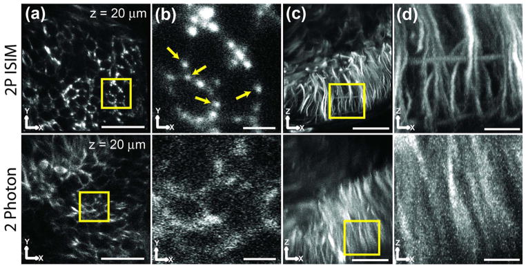
2P ISIM provides better resolution and SNR than conventional 2P microscopy. The same brain region in a zebrafish embryo was imaged in 2P ISIM (top row) and on a conventional, point-scanning 2P system (the Leica SP5, bottom row). (a) XY slices ~20 μm from the coverslip. Scale bar: 20 μm. (b) Higher-magnification views of region marked by the yellow square in (a). Scale bar: 3 μm. Yellow arrows indicate individual microtubule bundles. (c) XZ maximum intensity projections of the volumes. Scale bar: 20 μm. (d) Higher-magnification views of the region marked by the yellow square in (c). Scale bar: 5 μm. Images are raw, i.e., they have not been deconvolved. See also Media 6 and Media 7.
