Description
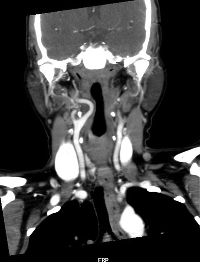
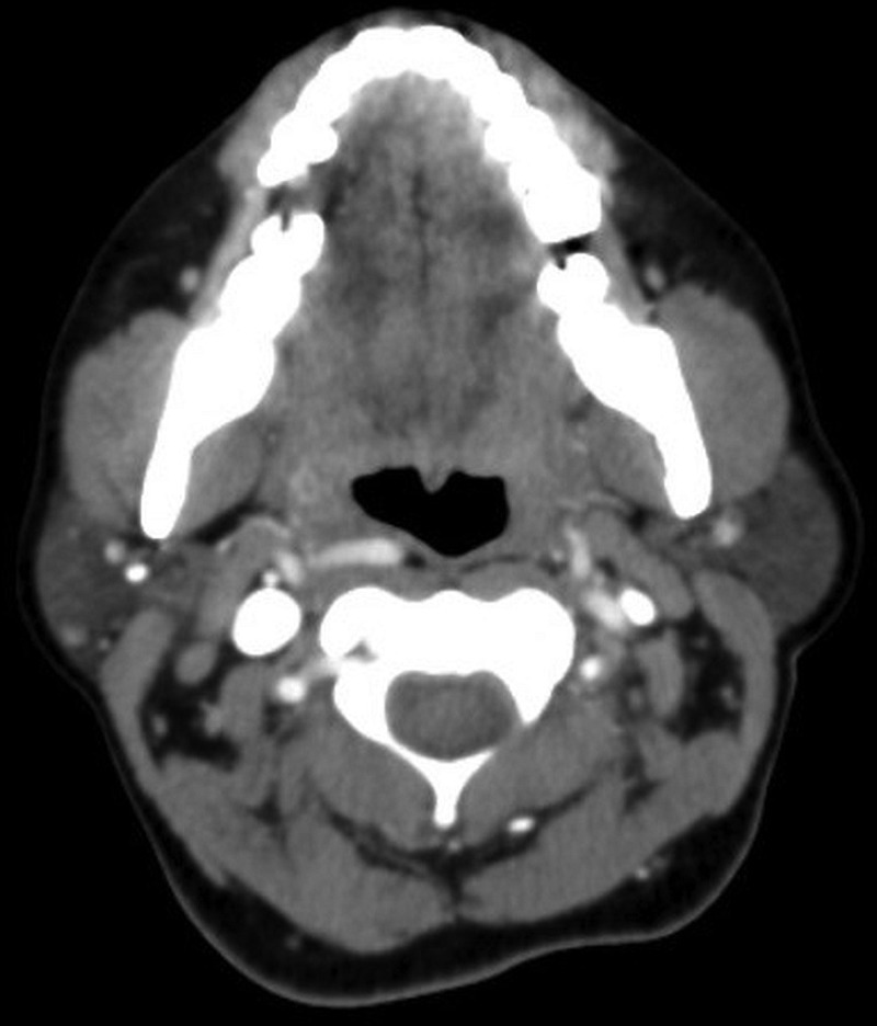
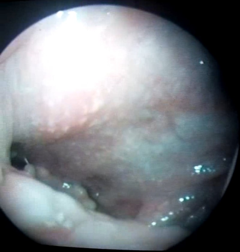
A 45-year-old non-hypertensive woman presented with a foreign body sensation in the throat of 3 months duration. Endoscopic examination showed a pulsating swelling in the right oropharynx with intact oropharyngeal mucosa (video 1). Contrast-enhanced CT (CECT) revealed an aberrant medial course of the right internal carotid artery (ICA) 2 cm long in the lateral pharyngeal space with 3 mm of interspersed soft tissue between ICA and oropharyngeal mucosa (figures 1 and 2).
Figure 1.

Coronal contrast-enhanced CT of the neck showing an aberrant medial course of the right internal carotid artery 2 cm long in the lateral pharyngeal space.
Figure 2.

Axial contrast-enhanced CT of the neck showing aberrant internal carotid artery in proximity to the oropharyngeal wall with 3 mm of interspersed soft tissue.
Endoscopic video demonstrating a pulsatile mass in the right oropharyngeal wall synchronous with heart pulsations.

ICA after bifurcation from the common carotid artery extends to the skull base without branching in the neck.1 2 It lies posterolateral to the tonsillar fossa approximately 2.5 cm from the mucosal surface.2 Aberrant ICA is an uncommon variant that accounts for 5% of incidental occurrences in the neck.1 It has been classified based on its ectopic position or presence of neoplasms.3 The ectopic variation is commonly seen in the temporal bone and is either tortuous or kinking. A sharp bend denotes kinking whereas curving, elongation, undulation and redundancy denote tortuosity.2 3 Aberrant ICA develops either as a congenital deformity or due to degenerative vessel wall changes such as hypertension, fibromuscular dysplasia and atherosclerosis.2 3 Patients may be asymptomatic or may present with a foreign body sensation in the throat, dysphagia or change of voice.2 3 CECT, contrast MRI or angiography are gold standards in diagnosis.3
Aberrant ICA can be misdiagnosed as an abscess or a tumour, the manipulation of which can result in torrential uncontrolled haemorrhage. Hence it is absolutely essential for clinicians to be aware of this rare but important clinical variation of ICA.
Learning points.
Aberrant internal carotid artery (ICA) can be due to congenital malformation or degenerative changes in the vessel wall.
Contrast MRI and contrast-enhanced CT or angiography form the gold standard in diagnosis.
Aberrant ICA can be misdiagnosed as an abscess or tumour in the oropharynx, manipulation of which can cause life-threatening haemorrhage.
Acknowledgments
The authors thank Dr Dipak Ranjan Nayak and Dr Balakrishnan for their constant support.
Footnotes
Contributors: AMB was responsible for preparing the manuscript. RN collected the literature. NC collected the case data. DG collected the multimedia files and images.
Competing interests: None.
Patient consent: Obtained.
Provenance and peer review: Not commissioned; externally peer reviewed.
References
- 1.Srinivasan S, Ali SZ, Chwan LT. Aberrant retropharyngeal (submucosal) internal carotid artery: an underdiagnosed, clinically significant variant. Surg Radiol Anat 2013;35:449–50. [DOI] [PubMed] [Google Scholar]
- 2.Pfeiffer J, Ridder GJ. A clinical classification system for aberrant internal carotid arteries. Laryngoscope 2008;118:1931–6. [DOI] [PubMed] [Google Scholar]
- 3.Prokopakis EP, Bouralias CA, Bizaki AJ et al. . Ectopic internal carotid artery presenting as an oropharyngeal mass. Head Face Med 2008;4:20. [DOI] [PMC free article] [PubMed] [Google Scholar]
Associated Data
This section collects any data citations, data availability statements, or supplementary materials included in this article.
Supplementary Materials
Endoscopic video demonstrating a pulsatile mass in the right oropharyngeal wall synchronous with heart pulsations.


