

Abstract
Stereognosis has been defined as the appreciation of the form of objects by palpation. Whilst this definition holds good for the manual exploration of objects, it is possible for the shape of objects to be explored intra orally referred to as oral stereognosis. To better understand patients’ relative satisfaction with complete dentures, differences in oral stereognostic perception, based on the identification of 6 edible objects was analyzed in a group of 30 edentulous individuals at 3 stages, namely, just before (pre-treatment), 30 min after (30 min post-treatment) and 1 month after (1 month post-treatment) the insertion of new dentures. The time required to identify each object was recorded and the correctness of identification of each object was scored using oral stereognostic score. Descriptive statistics, Wilcoxon signed rank test, Spearman’s rank correlation test, Pearson Chi square test was used to statistically analyze the data obtained. OSA scores was significantly increased 1 month post-treatment compared to 30 min post-treatment (p < 0.05). It was found that Oral stereognostic test is reliable for measuring patients’ oral stereognostic perception and may be used as one of the clinical aids in appreciating the functional limitations imposed by the prostheses.
Keywords: Oral stereognosis (OS), Complete dentures, Oral stereognostic score, OSA (oral stereognostic ability)
Introduction
Stereognosis has been defined as the appreciation of the form of objects by palpation. This definition refers to manual exploration of objects, it is also possible to extend the tactile perception of objects intra orally which is referred to as Oral stereognosis (OS). It was first introduced by Berry and Mahood [1] in 1966.
Various authors have worked on the concept of oral stereognosis by using variety of test shapes and materials.
The National Institute of Dental Research has developed a range of 20 shapes and suggested to use test forms within this range for the assessment of oral stereognosis [2]. Stereognosis testing is not designed to assess specific group of receptors but is as indicator of overall sensory ability of the patient [3].
One of the major concerns of denture wearers is adaptation to new or replacement dentures, regardless of their experience of wearing dentures. This study aimed to see whether there was difference between stereognostic abilities before and after rehabilitation with complete dentures in non-experienced denture wearers and secondly, hypothesized that evaluating oral stereognostic ability in patients after rehabilitation with complete denture prosthesis may provide useful information about the sensory abilities of denture patients and aid in interpreting the role of adaptation and adjustment to the complete denture prosthesis.
So the aim and objectives of the present study was to:
To determine and compare the oral stereognostic ability just before (denture insertion), 30 min after (30 min post denture insertion) and 1 month after (1 month post-treatment) insertion of complete dentures.
To compare the duration of time taken for sensory perception of edible objects before and after insertion of complete dentures.
Materials and Methods
Following institutional ethical committee approval and obtaining informed consent from the experimental subjects the oral stereognosis tests (OST) was conducted on thirty edentulous patients in the age group of 55–60 years, edentulous for a period of more than 3 months, reporting to the department of Prosthodontics JSS Dental College and Hospital, JSS University, Mysore, Karnataka. The patients were non-experienced denture wearers.
Inclusion Criteria
Patients who will visit to the college for follow up check-up after 1 month.
Patients would receive new dentures and he or she would not have any past experience of denture usage.
All the patients involved in this study would be free from oral symptoms and pathologies and significant medical conditions.
No history of temporomandibular joint disorders.
Exclusion Criteria
Uncooperative patients.
Patients with oral lesions, pathologies and TMJ disorders.
Those who do not or not willing to sign the consent form.
Any past experience of denture usage.
Six shapes were chosen from 20 items used at the National Institute of Dental Research for stereognostic tests (Fig. 1) [2]. Square, rectangle, circle, oval, triangle and star were the shapes included in the study as some shapes shared similar characteristics to increase the difficulty of discrimination. Test specimens were made from raw carrots (Fig. 3) to permit free oral manipulation by participants and to eliminate the need for handles on test items, as used by some investigators [4–6]. Stainless steel punches were fabricated by Micro Excellers, Mysore, for cutting 5 mm thick slices of carrot and diameter varying from 10–14 mm to obtain the six standardized shapes (Fig. 2).
Fig. 1.

NIDR shapes for evaluation of OS
Fig. 3.
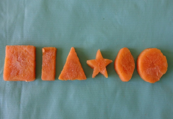
Test pieces of six forms of square, rectangle, triangle, star, oval, circle—5 mm thick, 10 mm diameter
Fig. 2.
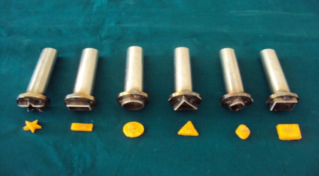
Stainless steel metal punches
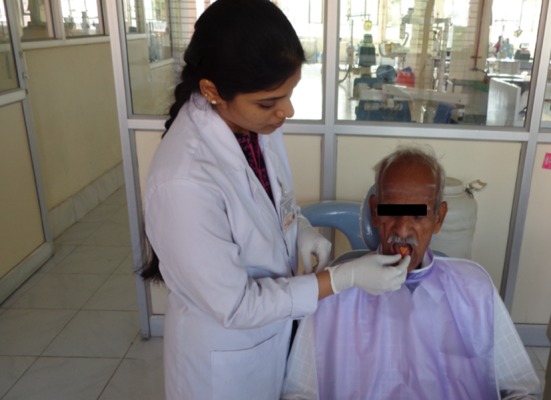
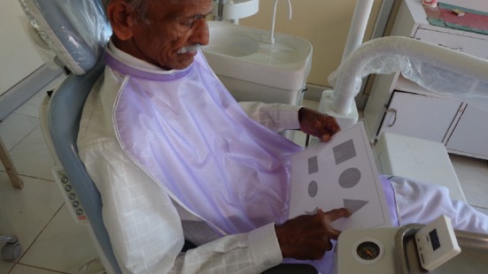
The test was carried out in a quiet environment in the hospital where the subject was seated comfortably in an upright position. The test was conducted before denture insertion; 30 min after denture insertion and after 1 month of denture insertion (Fig. 6). Pictures of all six test specimens were shown to the subject in the chart and the corresponding picture was pointed out for each shape by the subject. To prevent learning effect, no practical trials was held. For each of the tests, the six shapes were used in random order, and each shape was presented once to the patient.
Fig. 6.
Oral manipulation of test samples with dentures
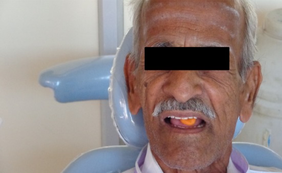
The test specimen was placed on the mid dorsum of the tongue, the patient being blindfolded and a digital stopwatch was started as soon as the test piece made contact with the tongue (Fig. 4). Subjects were allowed to freely manipulate the test piece in the mouth (Fig. 5). Subjects then pointed to the shape on the laminated sheet which they thought was the one they had explored in their mouth (Fig. 7). Participants were not informed of the correct answers at any point during testing. The six test specimens were presented in the random order. The time taken for recognizing the shape was noted in seconds and the answers were recorded using a three point rating scale [6].
Fig. 4.
Edible object being placed in the mouth
Fig. 5.
Free oral manipulation of test samples
Fig. 7.
Test samples being identified with dentures
The scoring system was according to Smith and McCord [7, 8].
A correct identification of the test specimen was scored two points.
An incorrect identification within the same group of forms was scored as one point.
An incorrect identification of similar form was scored as zero.
For example, when a circle form will be presented, the correct answer of ‘circle’ was scored as two points; that of an ‘ellipse’ was scored as one point; and the other four answers (square, rectangle, triangle and star) was scored as zero. Possible range of scores would be from 12 (all correct) to 0 (all incorrect and dissimilar). Thus, higher the score, better is an individual’s stereognostic ability.
The data gathered were analyzed by means of SPSS software (version 17). Initial analysis of mean and standard deviation involved the use of descriptive statistics. Comparison of OSA scores before and after rehabilitation with complete dentures was analyzed using Wilcoxon signed rank test. To analyze the correlation between OSA scores and time taken to identify the test specimens Spearman’s rank correlation test was used. Chi square test was used to compare the OSA scores between the groups. p < 0.05 was set for statistical significance.
Results
The present study evaluated OSA using edible objects before denture insertion, 30 min after denture insertion and after 1 month of insertion of dentures in non-experienced denture wearers.
The mean OSA scores value before denture insertion was found to be 9.33 and it increased to 11.86 after 1 month of denture fabrication (Table 1). The comparison of OSA scores before denture insertion and after denture insertion was found to be statistically highly significant. Further, Wilcoxon signed rank test was performed to compare OSA scores before denture insertion, after 30 min and 1 month post insertion of dentures. Results obtained were significant proving that edentulous individuals without dentures scored low when compared with 30 min of insertion and after 1 month post insertion of complete dentures (Table 2). Similarly, the time taken to identify the test specimens before denture insertion and after 1 month of denture insertion was decreased from 13.38 (8.21) to 5.57 (2.91) s (Tables 3, 4).
Table 1.
Descriptive statistics of OSA score identify the objects
| Time | n | Mean | SD | Minimum | Maximum |
|---|---|---|---|---|---|
| OSA WOa | 15 | 9.33 | 1.63 | 6.00 | 12.00 |
| OSA WDb | 15 | 11.33 | 0.92 | 10.00 | 12.00 |
| OSA WD1c | 15 | 11.86 | 0.92 | 11.00 | 12.00 |
aWithout dentures (WO)
bWith dentures (WD)
cWith dentures after 1 month (WD1)
Table 2.
Descriptive statistics of time taken to identify the objects
| Time | n | Mean | SD | Minimum | Maximum |
|---|---|---|---|---|---|
| T-WOa | 90 | 13.39 | 8.23 | 32.00 | 2.00 |
| T-WDb | 90 | 8.11 | 4.98 | 25.00 | 2.00 |
| T-WD1c | 90 | 5.58 | 2.92 | 13.00 | 2.00 |
aTime taken to identify objects WO
btime taken to identify objects WD
ctime taken to identify objects WD1
Table 3.
Comparison of OSA scores between groups using Wilcoxon signed rank test
| Groups | Mean difference | z value | p value |
|---|---|---|---|
| OSA-WO/OSA-WD | −1.80 | −3.31 | 0.001 HS |
| OSA-WO/OSA-WD1 | −2.53 | −3.01 | 0.002 HS |
| OSA-WD/OSA-WD1 | −0.73 | −2.37 | 0.018 S |
Table 4.
Comparison of time between groups using Wilcoxon signed rank test
| Groups | Mean difference | z value | p value |
|---|---|---|---|
| TWO vs TWD | 5.28 | −5.686 | 0.001 HS |
| TWO vs TWD1 | 7.81 | −7.156 | 0.001 HS |
| TWD vs TWD1 | 2.53 | −6.830 | 0.001 HS |
Results of Pearson Chi square test showed that the percentage of identification of shapes of shapes before denture insertion was 73.3 %, on insertion of dentures increased (76.7 %) and was still higher after 1 month post insertion of dentures (85.6 %). The findings suggested that there was a significant improvement in the OSA of non-experienced denture wearers after wearing new dentures for 1 month (Tables 5, 6).
Table 5.
Results of Pearson Chi square test
| Groups | Shapes not identified (0) | Shapes identified with similar forms (1) | Correctly identified shapes (2) |
|---|---|---|---|
| OSA (WO) | 36 (20 %) | 12 (6.7 %) | 132 (73.3 %) |
| OSA (WD) | 12 (6.7 %) | 30 (16.7 %) | 138 (76.7 %) |
| OSA (WD1) | 2 (1.1 %) | 24 (13.3 %) | 154 (85.6 %) |
Table 6.
Pearson Chi square test between groups
| Comparison | OSA(WO)/OSA(WD) | OSA(WO)/OSA(WD1) | OSA(WD)/OSA(WD1) |
|---|---|---|---|
| Chi square test | 19.848 | 36.113 | 8.686 |
| p value | 0.001 HS | 0.001 HS | 0.013 HS |
Results of Spearman’s rank correlation (Table 7) were significant thus proving that as OSA scores increased the time taken to identify the shapes had decreased.
Table 7.
Correlation of OSA scores with time taken to identify the objects using Spearman’s rank correlation test
| Groups | Correlation values | p value |
|---|---|---|
| OSA(WO)/TWO | −0.449 | 0.093 |
| OSA(WD)/TWD | −0.342 | 0.212 |
| OSA(WD1)/TWD1 | −0.409 | 0.131 |
Discussion
The physiologic function of the masticatory system is primarily dependent upon the integration of sensory feedback and motor neuron response. Proprioception and perception are the sensory responses that act to program and monitor the motor responses [8]. A defect of the perceptive input may result in pathologic changes to parts of the masticatory system. The tactile discrimination or recognition of form without the aid of vision is referred to as stereognosis—which is a psycho neurological ability and is an important indicator of central nervous system integrity [9, 10].
Oral stereognosis involves some motor activity and manipulation of test pieces inserted into the oral cavity and their interactions with lips, tongue and teeth. The oral exploration phenomenon involves elaborate functions of the parietal cortex. Received sensations are synthesized in the cortex and compared with previous sensorial memories [6].
Oral cavity has a very high ratio of sensory innervations—through trigeminal nerve and its innervations in the oral cavity has a very large representation in the cerebrum compared to the innervations of body peripheral areas [11]. The receptors present in the dentofacial region send information to CNS through afferent pathways, which could be information regarding the consistency of food to be masticated. Efferent impulses from CNS are conveyed to the muscles of mastication to bring about motor response—masticatory activity in a co-ordinated manner [12].
A fundamental concern of sensory motor function coupled with a general interest in oral sensation and perception has led investigators to extend the exploration of tactile perception of form intra orally—which is oral stereognosis. The major change observed in the oral status of an edentulous individual is loss of teeth resulting in the complete loss of proprioception which has helped to program the masticatory system. The loss of sensory ability related to age may co-relate with the willingness of elderly patients to swallow larger food boluses—implying that they cannot accurately estimate the bolus size and shape [13, 14].
Various authors have analyzed oral stereognostic ability. The types of test used differed, essentially, in the number, shape, size and material of the objects used, but the studies had in common, the principle of identifying the shape of an object placed inside the mouth without using the eye-sight. The present study used test specimens made out of raw carrots supposedly; participants were able to manipulate these items freely without fear of swallowing foreign objects.
The mean correct identification of various acrylic test forms was 63 % rate as reported in a study conducted by van Aken et al. [4]; it was 44.9 % using metallic forms as reported by Litvak et al. [3]. In the present study, mean correct identification of edible test forms by 30 patients was 85.6 %. The studies conducted by Litvak et al. [3], Hochberg and Kabcenell [9], Garret et al. [14] evaluated OSA using metallic and acrylic test forms by concentrating mainly on previous denture wearers. In our study, participants were mainly non-experienced denture wearers and OSA was evaluated before, after 30 min and after 1 month of denture insertion using edible object (carrot) to permit free oral manipulation, eliminate the need for handles and floss for acrylic and metallic forms as used by some investigators.
In a study conducted by Garret et al. [14] edible object was used to evaluate OSA in dentate persons and experienced denture wearers and was concluded that OSA increased on insertion of dentures. However, our study concentrated on evaluating OSA in non-experienced denture wearers at three stages: pre treatment, 30 min post treatment and after 1 month of insertion of dentures and concluded that OSA increased immediately on insertion of complete dentures and was still higher after 1 month post insertion of dentures. It may thus be suggested that the increase in stereognostic ability in subjects tested with new dentures, as opposed to without dentures, may in part be attributed to their increased ability to manipulate objects, due to the presence of the correctly fabricated upper and lower complete denture.
As the OSA score was statistically significant the results of this study supports the hypothesis that the presence of dentures improves stereognostic ability. The total time required to correctly identify the shape of the objects by patients after 1 month post insertion of dentures was statistically significant when compared to patients before insertion of dentures.
Subjects with high scores in the stereognosis test presumably receive more complete and accurate sensorial data about objects inside their mouth, and in consequence have a better ability to appreciate the functional limitations imposed by the presence of a complete removable denture, as well as noticing more clearly any defects in the artifact, compared to subjects with lower stereognostic ability. This would mean that the oral stereognosis test is a valid aid in predicting adaptability to complete removable dentures and their acceptance by the patient.
Clinical Implications
The results of this study appear to show that oral perception, evaluated through the oral stereognosis test, might play a role in the complex process of incorporating and accepting rehabilitation with complete removable dentures.
The success of prosthodontic treatment is dependent on sensory–motor relationships. The clinician must consider the learning skill of training the patient to wear the dentures. Patient performance could be improved by giving transitional appliances which may serve as a training device to improve oral manipulative and perceptive skills. The patient with high level of oral perception may be more responsive to clinical procedures.
Limitations
The sample size selected in the study is limited. Further studies should be conducted to recognize size differences rather than shape differences.
Conclusions
The results of this study lead to the following conclusion that covering the palatal mucosa with a denture does not reduce oral stereognostic ability; oral stereognostic test is a reliable test to measure patients’ oral stereognostic perception—which would enable the patient to appreciate the functional limitations of the denture. The dentist can be more aware of what he/she can expect in the form of patient response during and after treatment.
The critical evaluation of prosthesis should not only include dentist’s objective skills of treatment and the patient’s subjective judgment about oral health, function and esthetics but also the patient’s oral perceptive skills.
Acknowledgments
The research project has received JSS Grants for completion of the project. The authors sincerely thank JSS Dental College and Hospital, JSS University, Mysore, for all the support.
Conflict of interest
Nil.
References
- 1.Berry DC, Mahood M. Oral stereognosis and oral ability in relation to prosthetic treatment. Br Dent J. 1966;120:179–185. [PubMed] [Google Scholar]
- 2.Jacobs R, van Steenberghe D. Role of periodontal ligament receptors in the tactile function of teeth: a review. J Periodontal Res. 1994;29:153–157. doi: 10.1111/j.1600-0765.1994.tb01208.x. [DOI] [PubMed] [Google Scholar]
- 3.Litvak H, Silverman SI, Garfinkel L. Oral stereognosis in dentulous and edentulous subjects. J Prosthet Dent. 1971;25:139–150. doi: 10.1016/0022-3913(71)90100-4. [DOI] [PubMed] [Google Scholar]
- 4.Van Aken AAM, Van Waas MAJ, Van Rossum GM. Differences in oral stereognosis between complete denture wearers. Int J Prosthodont. 1994;4:75–79. [PubMed] [Google Scholar]
- 5.Mantecchini G, Bassi F, Pera P, Preti G. Oral stereognosis in edentulous subjects rehabilitated with complete removable dentures. J Oral Rehabil. 1998;25:185–189. doi: 10.1046/j.1365-2842.1998.00221.x. [DOI] [PubMed] [Google Scholar]
- 6.Crum RJ, Loiselle RJ. Oral perception and proprioception: a review of the literature and its significance to prosthodontics. J Prosthet Dent. 1972;28:215–230. doi: 10.1016/0022-3913(72)90141-2. [DOI] [PubMed] [Google Scholar]
- 7.van Steenberghe D. The structure and function of periodontal innervations. A review of the literature. J Periodont Res. 1979;14:185–203. doi: 10.1111/j.1600-0765.1979.tb00223.x. [DOI] [PubMed] [Google Scholar]
- 8.Smith PW, McCord JF. Oral stereognostic ability in edentulous and dentate individuals. Eur J Prosthodont Restor Dent. 2002;10:53–56. [PubMed] [Google Scholar]
- 9.Hochberg I, Kabcenell J. Oral stereognosis in normal and cleft palate individuals. Cleft Palate J. 1967;4:47. [PubMed] [Google Scholar]
- 10.Rossetti PHO, Bonachela WC, Nunes LMO. Oral stereognosis related to the use of complete dentures: a literature Review. Int J Oral Med Sci. 2004;2:57–60. doi: 10.5466/ijoms.2.57. [DOI] [Google Scholar]
- 11.Brill N, Schubeler, Tryde G. Aspects of occlusal sense in natural and artificial teeth. J Prosthet Dent. 1962;12:123–128. doi: 10.1016/0022-3913(62)90018-5. [DOI] [Google Scholar]
- 12.Meyer G, Fanghänel J, Proff P. Morphofunctional aspects of dental implants. Ann Anat. 2012;194:190–194. doi: 10.1016/j.aanat.2011.09.006. [DOI] [PubMed] [Google Scholar]
- 13.Jacobs R, Bou Serhal C, Van Steenbergh D. Oral stereognosis: a review of literature. Clin Oral Investig. 1998;2:3–10. doi: 10.1007/s007840050035. [DOI] [PubMed] [Google Scholar]
- 14.Garret NR, Kapur KK, Jochen DG. Oral stereognostic ability and masticatory performance in denture wearers. Int J Prosthodont. 1994;7:567–572. [PubMed] [Google Scholar]