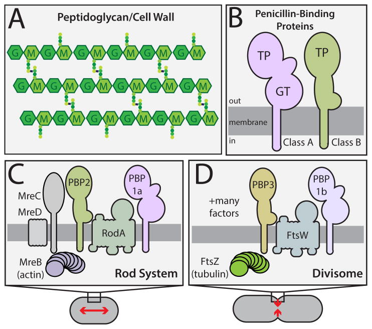
Figure 1. Peptidoglycan structure and the machines that synthesize it.
A. The PG matrix consists of glycan chains with the repeating unit of N-acetylmuramic acid (MurNAc, M) and N-acetylglucosamine (GlcNAc, G). Attached to the MurNAc sugars are peptides (colored circles) used to form crosslinks between adjacent glycans. B. Domain structure of the PG synthases. Both classes of PBPs have a single transmembrane domain with a large catalytic domain in the periplasm. C–D. Schematics highlighting the components of the two main PG synthetic systems in rod-shaped bacteria. Both systems require a dedicated bPBP (PBP2 or PBP3) and a SEDS (shape/elongation/division/sporulation) family protein (RodA or FtsW) for proper function. See text for details.

