

Abstract
Background:
Considering the importance of improvement of endothelial function in patients with type 1 diabetes mellitus (DM) and to prevent its related micro- and macro-vascular complications; in this study, the effect of vitamin C administration on improving endothelial function of children with type 1 diabetes was investigated.
Methods:
In this analytic-experimental study, children with type 1 DM aged 6-18 years and a group of healthy children enrolled. Vitamin C (250 mg/daily) administrated for the two studied groups for 1-month. Endothelial function evaluated by flow-mediated dilatation (FMD) and intima-media thickness (IMT) measurement using vascular Doppler ultrasonography, before and after trial.
Results:
In this study, 18 patients with type 1 diabetes (DM) and 19 normal children as the control group were studied. After vitamin C administration IMT reduced in all studied groups (P < 0.05). FMD increased in all studied groups, but it was significant only in the control group (P = 0.02 in the control group and P = 0.07 in patients with DM). Mean differences of IMT 2 – IMT 1, FMD 2 – FMD 1 and left ventricular (LV) mass 2 – LV mass 1 and blood pressure (BP) were not significantly different in two studied groups (P > 0.05). Mean differences of IMT 2 – IMT 1, FMD 2 – FMD 1, LV mass 2 – LV mass 1 and BP were not significantly different in patients with HbA1c ≤ 7 g/dl and those with HbA1c >7 g/dl and control group (P > 0.05).
Conclusions:
The findings of the current study indicated that vitamin C may have a protective effect on endothelial dysfunction, but regarding its effectiveness among the high-risk population such as diabetic patients with and without appropriate glycemic control the study was not sufficiently powered due to its small sample size.
Keywords: Endothelium, type 1 diabetes, vitamin C
INTRODUCTION
Type 1 diabetes mellitus (DM) is one of the most common pediatric chronic diseases, which is characterized by immune-mediated destruction of pancreatic β cells.[1] It is associated with an increased rate of morbidity and mortality due to its related micro- and macro-vascular complication. Atherosclerosis and cardiovascular disease are considered as its most common complications with an estimated two- to ten-fold increased risk.[2,3]
Evidences suggested that endothelial dysfunction has an important pathophysiological role in the early development of atherosclerosis among patients with type 1 diabetes.[4]
Endothelial dysfunction is characterized by vasomotor dysfunction and consequently vasodilatation impairment. Nitric oxide (NO) has a crucial role in this process. NO release and endothelial nitric oxide synthase expression are reduced both in endothelial dysfunction and atherosclerosis.[5,6]
The generation of oxidative stress and production of oxygen-derived free radicals is considered as one of the most important mechanism involved in endothelial dysfunction.[6] In patients with type 1 diabetes, it is mentioned that oxygen-derived free radicals are produced from mitochondrial, enzymatic, and nonenzymatic sources. Some in vivo and in vitro studies indicated that adequate scavenging of these radicals by antioxidant agents such as vitamin C could restore endothelium dependent vasodilation and consequently normal endothelial function.[7]
Endothelial function could be measured by different biochemical factors, but there are also some noninvasive methods for this purpose. Brachial artery flow-mediated dilatation (FMD) and carotid artery intima-media thickness (IMT) measurement with high-resolution ultrasound is one of the noninvasive methods, which evaluate both endothelial function and structural arterial changes.[8]
Accordingly, increased IMT and decreased FMD have been demonstrated in high-risk population with atherosclerosis such as type 1 diabetic patients.[9,10] There are also a few studies which investigated the effectiveness of antioxidant agents such as vitamin C in improvement of endothelial dysfunction. However, the results were not conclusive enough.[11,12]
Hence, considering the importance of improvement of endothelial function in this group of patients and to prevent its related micro-and macro-vascular complications, in this study, the effect of vitamin C administration on improving endothelial function of children with type 1 diabetes was investigated.
METHODS
In this analytic-experimental study, a group of children aged 3-18 years with diagnosed type 1 diabetes, and a group of healthy children who were referred to Imam Hossein Children's Hospital, the only pediatric referral center in Isfahan, affiliated to Isfahan University of Medical Sciences were enrolled.
Patients with diabetes were selected by nonrandomized convenience method from patients who were referred to endocrinology clinics of the hospital. Children in the control group were selected from outpatient children without appreciable cardiovascular risk factors who were referred for a routine visit or from healthy brothers and sisters of selected patients.
All subjects were nonsmokers, nonpregnant and without any history of systemic disease.
Those with a history of recent use of vitamin C, not cooperation and diagnosis of new disorders and any type of congenital heart disease, which was found during echocardiography, were excluded.
The protocol of the study was approved by the Regional Ethics Committee of Isfahan University of Medical Sciences. Written informed consent was obtained from all selected patients or their parents.
Basal characteristics of the studied patients were recorded from their medical files. Selected patients recalled, a pediatric cardiologist examined their clinically. Endothelial function of studied subjects during the first visit was evaluated by FMD and IMT measurement using vascular Doppler ultrasonography (EKO 7 Machine by Samsung Medison Company and by a vascular transducer 7 MHz).
Vitamin C (Osvah Pharmaceutical Company, Tehran, Iran) was administrated for all studied groups with a dose of 250 mg, daily for 1-month. After that period studied subjects underwent vascular Doppler ultrasonography for second FMD and IMT measurement. Mean of the obtained endothelial parameters before and after vitamin C administration was compared in each studied groups.
Endothelial function measurement
Weight, height, and blood pressure (BP) of the studied children were measured before the procedure. The subjects were recommended to do not exercise or use caffeine, folic acid, nitrate, high fat diet, vitamin C (both as a supplement or dietary) for at least 24 h before the procedure.
The procedure was performed in the morning after 8 h of fasting, in a temperature-controlled room at 25°C in a supine position.
All subjects were examined by the same physician, who was blinded to their clinical conditions at the time of examination.
Flow-mediated dilatation measurement
Studied subjects were examined in the supine position with their forearm placed in a semi open splint. The high-frequency (7 MHz) vascular transducer (EKO 7 by Samsung Medison Company) was fixed with a stereotactic probe-holding device. In order to make a flow stimulus by reactive hyperemia a pediatric BP cuff was fixed on the wrist of the subjects and radial artery was imaged in a longitudinal plane 5 cm distal from the antecubital fossa. After performing a baseline rest image, the blood flow velocity was estimated by time averaging the Doppler signal from a mid-artery sample volume. Cuff deflation was followed by a brief high-flow state after a 5 min interval of ischemia. After cuff deflation, the image of the radial artery and the Doppler signal recorded alternatively for 5 min with 20 s intervals. After the procedure, obtained images saved on the EKO 7 hard disk and analyzed. Distance measurements of radial artery provided at maximum systolic extension. FMD analyzed by a pediatric cardiologist.[13]
Intima-media thickness measurement
Carotid arteries imaged using a high-frequency (7 MHz) vascular linear transducer. Subjects were in the supine position with the head turned 45° away from the scanner. Two segments including the distal 1 cm of the common carotid artery and its bifurcation were evaluated on each side. Measurements of the two segments were performed at 2-mm intervals at near and far wall from the transducer and maximum and mean of IMT were calculated for them. Sonography and reading were assessed by a pediatric cardiologist.[14]
Statistical analysis
Obtained data analyzed using SPSS version 18 (SPSS Inc., Chicago, IL, USA) software.
Data were presented as the mean ± standard deviation (SD). The comparison of quantitative data between the two studied groups was done with the independent t-test. The comparison of such data before and after intervention, in each group was done by intention to treat analysis using mixed effect model (within group). P < 0.05 were considered as statistically significant.
RESULTS
In this study, 18 patients with type 1 DM and 19 normal children as control group were studied.
Demographic characteristics, mean ± SD of endothelial function markers and BP of patients in the studied population are presented in Table 1.
Table 1.
Demographic characteristics and mean±SD of endothelial function parameters of studied patients with diabetes and control group
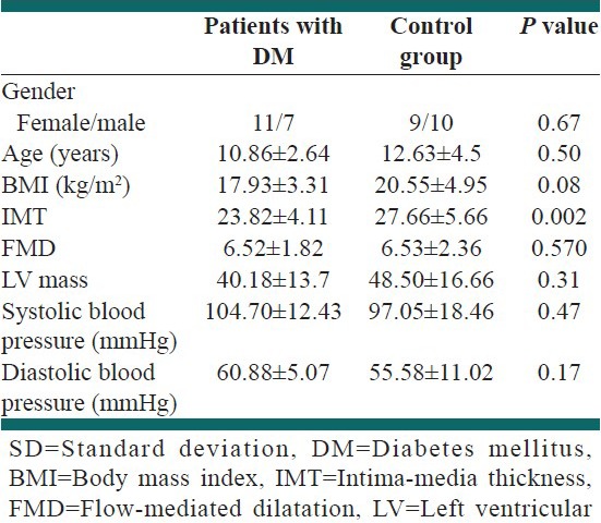
Mean ± SD of HbA1c and duration of diabetes were 8.46 ± 2.31 g/dl and 2.3 ± 1.5 years, respectively. Mean dose of insulin was 25.27 ± 9.1 units.
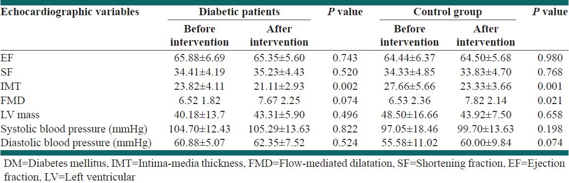
Echocardiographic findings and endothelial function markers and blood pressure of patients in the two studied groups before and after intervention are presented in Table 2.
Table 2.
Echocardiographic findings of patients with DM and control group before and after vitamin C administration
Mean differences (after intervention-before intervention) of IMT 2 – IMT 1, FMD 2 – FMD 1 and left ventricular (LV) mass 2 – LV mass 1 were not significantly different among male and female (P > 0.05).
Mean differences of IMT 2 – IMT 1, FMD 2 – FMD 1 and LV mass 2 – LV mass 1 and BP were not significantly different in two studied groups (P > 0.05).
In 6 (33.3%) patients with type 1 DM HbA1c ≤7 g/dl. Mean differences of IMT 2 – IMT 1, FMD 2 – FMD 1, LV mass 2 – LV mass 1 and BP were not significantly different in patients with HbA1c ≤7 g/dl and those with HbA1c >7 g/dl and control group (P > 0.05).
DISCUSSION
In this study, we evaluated the effectiveness of vitamin C on endothelial function of type 1 DM patients and a group of healthy children by ultrasound assessment. The findings of our study indicated that vitamin C could improve IMT in both control and type 1 DM patients. IMT improvement was not different in diabetic patients with poor and good glycemic control. FMD had trend to be increased in two groups, but the difference was statistically significant in the control group.
The endothelial impairment in type 1 diabetic patients had been reported in several reports. Accordingly, it was considered as an early event in the atherosclerotic process and it is developed before the clinical manifestation of atherosclerotic complications.[15,16] There are also evidences that some intervention could improve this process and consequently delay the early development of atherosclerosis and other related micro- and macro-vascular complications.[12]
Vitamin C or ascorbic acid is an essential antioxidant in humans and its utility in improving endothelial dysfunction was determined in many studies, so in this study, we evaluated its effect on endothelial function parameters, which assessed by noninvasive methods and ultrasonography that is, IMT and FMD. As reported by previous studies using such noninvasive methods could be useful in early diagnosis of subclinical atherosclerosis in clinical practice and consequently they might be used for planning preventive strategies in high-risk patients for cardiovascular event.[17]
Increased IMT has a predictive value for the development of cardiovascular morbidity and mortality and FMD decrease in early phase of atherosclerosis.[18,19] Many studies have reported increased IMT and decreased FMD among type 1 diabetic patients.[20,21]
Gül et al. in Turkey have showed that increased IMT in type 1 diabetic patients was associated with microvascular complications.[22] Odermarsky et al. in Denmark have indicated that patients with type 1 DM with low plasma vitamin C concentrations could be more prone to microvascular dysfunction and increased IMT.[23]
In the current study, mean of IMT at baseline was significantly higher in healthy children than type 1 DM patients, it may be due that the BMI of diabetic patients was lower than the control group and as IMT increased with higher BMI, and so it could explain the obtained data.
Flow-mediated dilatation was not significantly different in two studied groups at baseline. It may be due to small sample size. Another explanation was the duration of DM in the studied population. Mean duration of disease in DM patients was 2.3 years. It seems that endothelial dysfunction was not developed during the early years of the disease and it could be detected in patients with at least 5-6 years history of the disease.[24]
Though after administration of vitamin C, IMT decreased and FMD increased in both diabetic and control group, but the mean difference of IMT and FMD after intervention was not significantly different. The short duration of the disease or short duration of study could explain the findings.
There were few reports regarding the effectiveness of vitamin C on improvement of endothelial function among children with type 1 diabetes and the results were also controversial.
Timimi et al. have indicated that vitamin C selectively could restores the impaired endothelial function among patients with insulin dependent DM.[25]
It seems that its improvement in DM patients would be more significant if the glycemic control of DM patients would be appropriate also. Mean of HbA1c in the studied population was 8.6 and only 33.4% of patients had appropriate glycemic control.
Ceriello et al. in Italy have reported that that long-lasting hyperglycemia in type 1 diabetic patients induces permanent alterations in endothelial cells, which may contribute to endothelial dysfunction by increased oxidative stress even when hyperglycemia is normalized. They concluded that normalization of both endothelial dysfunction and oxidative stress can be achieved in type 1 diabetic patients, with a combination of near-normalization of hyperglycemia and antioxidant treatment. They indicated that vitamin C could be effective in improvement of endothelial function in cases with appropriate glycemic control.[26]
In our study, mean differences of endothelial markers in diabetic patients with poor and good glycemic control was not different significantly. It may be due to the small sample size of subgroups.
In addition, it seems that the result would be more conclusive if other CVD risk factors such as lipid profile as well as insulin level have also been evaluated. However, there are evidences that insulin could induce vasodilatation.[27]
CONCLUSIONS
The findings of the current study indicated that vitamin C may have a protective effect on endothelial dysfunction, but regarding its effectiveness among high-risk populations, such as diabetic patients with and without appropriate glycemic control the study was not sufficiently powered due to its small sample size.
Footnotes
Source of Support: Nil
Conflict of Interest: None declared.
REFERENCES
- 1.American Diabetes Association. Diagnosis and classification of diabetes mellitus. Diabetes Care. 2013;(36 Suppl 1):S67–74. doi: 10.2337/dc13-S067. [DOI] [PMC free article] [PubMed] [Google Scholar]
- 2.Dorman JS, Tajima N, LaPorte RE, Becker DJ, Cruickshanks KJ, Wagener DK, et al. The Pittsburgh Insulin-Dependent Diabetes Mellitus (IDDM) Morbidity and Mortality Study: Case-control analyses of risk factors for mortality. Diabetes Care. 1985;(8 Suppl 1):54–60. doi: 10.2337/diacare.8.1.s54. [DOI] [PubMed] [Google Scholar]
- 3.Laing SP, Swerdlow AJ, Slater SD, Botha JL, Burden AC, Waugh NR, et al. The British Diabetic Association Cohort Study, II: Cause-specific mortality in patients with insulin-treated diabetes mellitus. Diabet Med. 1999;16:466–71. doi: 10.1046/j.1464-5491.1999.00076.x. [DOI] [PubMed] [Google Scholar]
- 4.Sibal L, Aldibbiat A, Agarwal SC, Mitchell G, Oates C, Razvi S, et al. Circulating endothelial progenitor cells, endothelial function, carotid intima-media thickness and circulating markers of endothelial dysfunction in people with type 1 diabetes without macrovascular disease or microalbuminuria. Diabetologia. 2009;52:1464–73. doi: 10.1007/s00125-009-1401-0. [DOI] [PubMed] [Google Scholar]
- 5.Calles-Escandon J, Cipolla M. Diabetes and endothelial dysfunction: A clinical perspective. Endocr Rev. 2001;22:36–52. doi: 10.1210/edrv.22.1.0417. [DOI] [PubMed] [Google Scholar]
- 6.Creager MA. Flow-mediated vasodilation is mediated by endothelium-derived nitric oxide. J Appl Physiol (1985) 2005;99:1625. doi: 10.1152/japplphysiol.00829.2005. [DOI] [PubMed] [Google Scholar]
- 7.De Vriese AS, Verbeuren TJ, Van de Voorde J, Lameire NH, Vanhoutte PM. Endothelial dysfunction in diabetes. Br J Pharmacol. 2000;130:963–74. doi: 10.1038/sj.bjp.0703393. [DOI] [PMC free article] [PubMed] [Google Scholar]
- 8.Lorenz MW, Markus HS, Bots ML, Rosvall M, Sitzer M. Prediction of clinical cardiovascular events with carotid intima-media thickness: A systematic review and meta-analysis. Circulation. 2007;115:459–67. doi: 10.1161/CIRCULATIONAHA.106.628875. [DOI] [PubMed] [Google Scholar]
- 9.Singh TP, Groehn H, Kazmers A. Vascular function and carotid intimal-medial thickness in children with insulin-dependent diabetes mellitus. J Am Coll Cardiol. 2003;41:661–5. doi: 10.1016/s0735-1097(02)02894-2. [DOI] [PubMed] [Google Scholar]
- 10.Harrington J, Peña AS, Gent R, Hirte C, Couper J. Aortic intima media thickness is an early marker of atherosclerosis in children with type 1 diabetes mellitus. J Pediatr. 2010;156:237–41. doi: 10.1016/j.jpeds.2009.08.036. [DOI] [PubMed] [Google Scholar]
- 11.Ceriello A, Piconi L, Esposito K, Giugliano D. Telmisartan shows an equivalent effect of vitamin C in further improving endothelial dysfunction after glycemia normalization in type 1 diabetes. Diabetes Care. 2007;30:1694–8. doi: 10.2337/dc07-0318. [DOI] [PubMed] [Google Scholar]
- 12.Beckman JA, Goldfine AB, Gordon MB, Garrett LA, Keaney JF, Jr, Creager MA. Oral antioxidant therapy improves endothelial function in Type 1 but not Type 2 diabetes mellitus. Am J Physiol Heart Circ Physiol. 2003;285:H2392–8. doi: 10.1152/ajpheart.00403.2003. [DOI] [PubMed] [Google Scholar]
- 13.Corretti MC, Anderson TJ, Benjamin EJ, Celermajer D, Charbonneau F, Creager MA, et al. Guidelines for the ultrasound assessment of endothelial-dependent flow-mediated vasodilation of the brachial artery: A report of the International Brachial Artery Reactivity Task Force. J Am Coll Cardiol. 2002;39:257–65. doi: 10.1016/s0735-1097(01)01746-6. [DOI] [PubMed] [Google Scholar]
- 14.Meyer AA, Kundt G, Steiner M, Schuff-Werner P, Kienast W. Impaired flow-mediated vasodilation, carotid artery intima-media thickening, and elevated endothelial plasma markers in obese children: The impact of cardiovascular risk factors. Pediatrics. 2006;117:1560–7. doi: 10.1542/peds.2005-2140. [DOI] [PubMed] [Google Scholar]
- 15.Järvisalo MJ, Raitakari M, Toikka JO, Putto-Laurila A, Rontu R, Laine S, et al. Endothelial dysfunction and increased arterial intima-media thickness in children with type 1 diabetes. Circulation. 2004;109:1750–5. doi: 10.1161/01.CIR.0000124725.46165.2C. [DOI] [PubMed] [Google Scholar]
- 16.Jin SM, Noh CI, Yang SW, Bae EJ, Shin CH, Chung HR, et al. Endothelial dysfunction and microvascular complications in type 1 diabetes mellitus. J Korean Med Sci. 2008;23:77–82. doi: 10.3346/jkms.2008.23.1.77. [DOI] [PMC free article] [PubMed] [Google Scholar]
- 17.Abdelghaffar S, El Amir M, El Hadidi A, El Mougi F. Carotid intima-media thickness: An index for subclinical atherosclerosis in type 1 diabetes. J Trop Pediatr. 2006;52:39–45. doi: 10.1093/tropej/fmi071. [DOI] [PubMed] [Google Scholar]
- 18.Bots ML, Hoes AW, Koudstaal PJ, Hofman A, Grobbee DE. Common carotid intima-media thickness and risk of stroke and myocardial infarction: The Rotterdam Study. Circulation. 1997;96:1432–7. doi: 10.1161/01.cir.96.5.1432. [DOI] [PubMed] [Google Scholar]
- 19.Shivalkar B, Dhondt D, Goovaerts I, Van Gaal L, Bartunek J, Van Crombrugge P, et al. Flow mediated dilatation and cardiac function in type 1 diabetes mellitus. Am J Cardiol. 2006;97:77–82. doi: 10.1016/j.amjcard.2005.07.111. [DOI] [PubMed] [Google Scholar]
- 20.Rabago Rodriguez R, Gómez-Díaz RA, Tanus Haj J, Avelar Garnica FJ, Ramirez Soriano E, Nishimura Meguro E, et al. Carotid intima-media thickness in pediatric type 1 diabetic patients. Diabetes Care. 2007;30:2599–602. doi: 10.2337/dc07-0922. [DOI] [PubMed] [Google Scholar]
- 21.Poredos P, Kek Ljubec A, Poredos P, Visnovic Poredos A. Endothelial dysfunction predictor of structural changes of arterial wall in type I diabetes. Int Angiol. 2006;25:280–6. [PubMed] [Google Scholar]
- 22.Gül K, Ustün I, Aydin Y, Berker D, Erol K, Unal M, et al. Carotid intima-media thickness and its relations with the complications in patients with type 1 diabetes mellitus. Anadolu Kardiyol Derg. 2010;10:52–8. doi: 10.5152/akd.2010.012. [DOI] [PubMed] [Google Scholar]
- 23.Odermarsky M, Lykkesfeldt J, Liuba P. Poor vitamin C status is associated with increased carotid intima-media thickness, decreased microvascular function, and delayed myocardial repolarization in young patients with type 1 diabetes. Am J Clin Nutr. 2009;90:447–52. doi: 10.3945/ajcn.2009.27602. [DOI] [PubMed] [Google Scholar]
- 24.Zhumanova BM, Asfendiiarova SZh, Zaslavskaia RM, Berishev RN. Factors affecting intima-media thickness in carotid arteries in diabetes mellitus of the young. Klin Med (Mosk) 2010;88:31–3. [PubMed] [Google Scholar]
- 25.Timimi FK, Ting HH, Haley EA, Roddy MA, Ganz P, Creager MA. Vitamin C improves endothelium-dependent vasodilation in patients with insulin-dependent diabetes mellitus. J Am Coll Cardiol. 1998;31:552–7. doi: 10.1016/s0735-1097(97)00536-6. [DOI] [PubMed] [Google Scholar]
- 26.Ceriello A, Kumar S, Piconi L, Esposito K, Giugliano D. Simultaneous control of hyperglycemia and oxidative stress normalizes endothelial function in type 1 diabetes. Diabetes Care. 2007;30:649–54. doi: 10.2337/dc06-2048. [DOI] [PubMed] [Google Scholar]
- 27.Sundell J, Laine H, Nuutila P, Rönnemaa T, Luotolahti M, Raitakari O, et al. The effects of insulin and short-term hyperglycaemia on myocardial blood flow in young men with uncomplicated type I diabetes. Diabetologia. 2002;45:775–82. doi: 10.1007/s00125-002-0819-4. [DOI] [PubMed] [Google Scholar]