
Figure 7. Reduction of IgG inhibited LPS-induced proinflammatory cytokine production through downregulating TLR4 expression in cervical cancer cells.
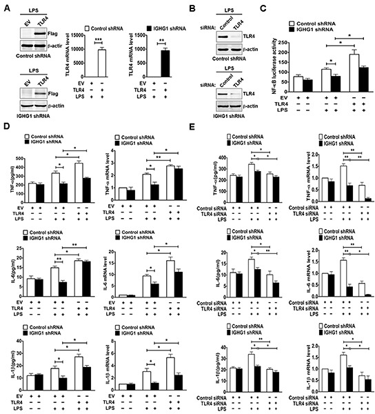
(A) HeLa cells stably expressing IGHG1 shRNA or control shRNA were transiently transfected with empty vector or Flag-tagged TLR4 plasmids respectively. Overexpression of TLR4 was measured with immunoblot (left panel) or RT-qPCR (right panel). (B) The above stably transformed cells were transfected transiently with TLR4 siRNA or control siRNA. TLR4 protein levels were detected. (C) The above stably transformed cells concomitantly overexpressing TLR4 were transiently cotransfected with pTK–Renilla-luciferase and NF-κB luciferase reporter plasmids. After 24 h of culture, the cells were treated with or without 100 ng/ml LPS for 20 h. NF-κB luciferase activity was measured using the Dual-Luciferase Reporter Assay System normalized against Renilla luciferase activity. The above stably transformed cells concomitantly overexpressing TLR4 (D) or downregulating TLR4 expression (E) were treated with or without 100 ng/ml LPS for 20 h. The proinflammatory cytokines (TNF-α, IL-6, and IL-1β) were measured with ELISA (left panel) and RT-qPCR (right panel). Data are presented as means±S. D. of three independent experiments (*P<0.05; **P< 0.01; ***P< 0.001).