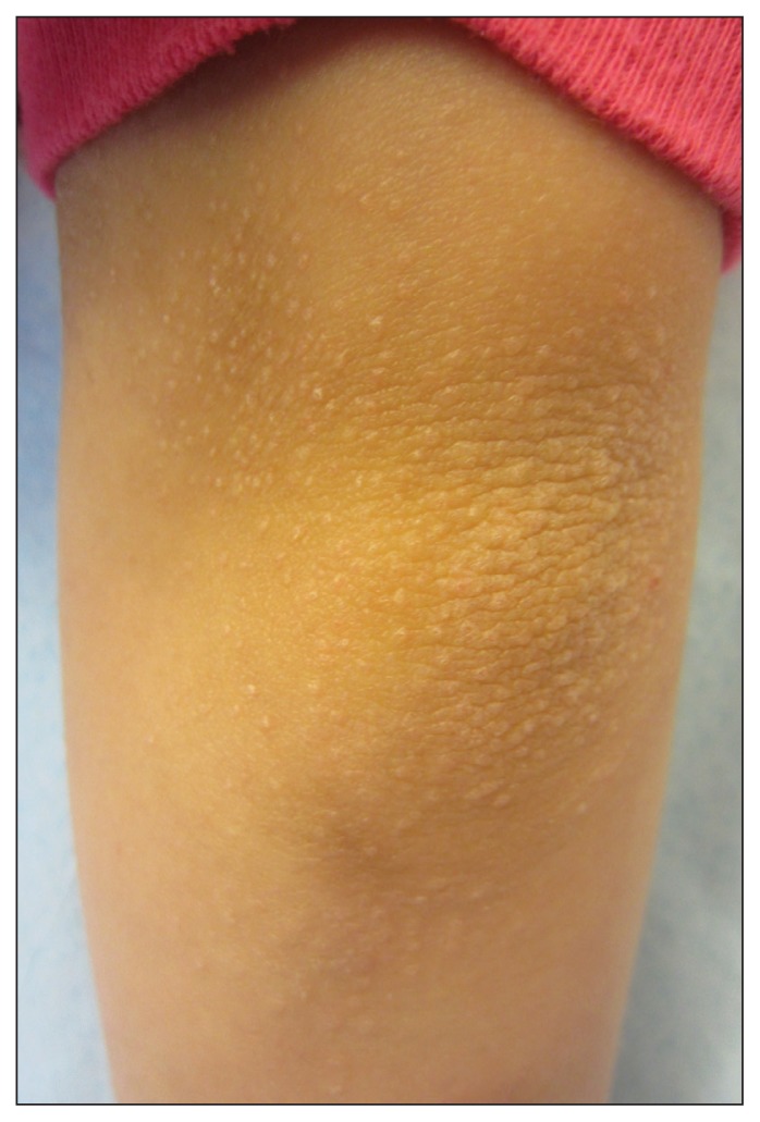
A previously healthy eight-year-old girl presented with a 9- to 12-month history of a papular eruption on her arms and back that was slightly pruritic. Her medical and family histories were otherwise unremarkable. Physical examination showed multiple flat-topped papules over the extensor surfaces of her back and her arms (Figure 1). The patient had normal nails. Based on the clinical findings, lichen nitidus was diagnosed.
Figure 1:
Multiple flat-topped, skin-coloured pinhead-sized papules on the arm of an eight-year-old girl.
Lichen nitidus is an uncommon inflammatory skin eruption of unknown cause that typically affects children and young adults, without sex or racial bias. The characteristic shiny, flat-topped, skin-coloured, pinhead-sized papules are usually arranged in groups on the chest, abdomen, genitalia and upper extremities. Lesions can appear along lines of trauma due to the Koebner phenomenon. In rare instances, the distribution is generalized.1,2 Examination of the oral cavity can occasionally show minute grey-white flat papules on the buccal mucosa. Nail thickening, ridging, pitting or detachment may be seen.2,3 Although affected individuals are usually asymptomatic, pruritus or concern about the cosmetic appearance may complicate the condition.
Diagnosis of lichen nitidus is usually based on the clinical presentation. However, histologic findings show distinctive features, including downward enlargement of the epidermal rete ridges surrounding a focal inflammatory infiltrate (resembling a claw clutching a ball).1
Lichen nitidus resolves spontaneously in a few years without treatment.1,2 As the papules clear, they are often replaced by postinflammatory hyperpigmented macules that tend to resolve in months.2 Treatment with a topical corticosteroid, topical calcineurin inhibitor, oral antihistamine or ultraviolet light should be reserved for symptomatic or generalized lichen nitidus. The success rates of these treatments are variable.1 In the above case, the patient was reassured, and no treatment was prescribed.
Clinical images are chosen because they are particularly intriguing, classic or dramatic. Submissions of clear, appropriately labelled high-resolution images must be accompanied by a figure caption and the patient’s written consent for publication. A brief explanation (250 words maximum) of the educational significance of the images with minimal references is required.
Footnotes
Competing interests: None declared.
This article has been peer reviewed.
The authors have obtained patient consent.
References
- 1.Arizaga AT, Gaughan MD, Bang RH. Generalized lichen nitidus. Clin Exp Dermatol 2002;27:115–7. [DOI] [PubMed] [Google Scholar]
- 2.Tilly JJ, Drolet BA, Esterly NB. Lichenoid eruptions in children. J Am Acad Dermatol 2004;51:606–24. [DOI] [PubMed] [Google Scholar]
- 3.Bettoli V, De Padova MP, Corazza M, et al. Generalized lichen nitidus with oral and nail involvement in a child. Dermatology 1997;194:367–9. [DOI] [PubMed] [Google Scholar]


