Abstract
Hyperpigmentation of gingiva becomes more pronounced if it is associated with “gummy smile.” Correction of gummy smile and depigmentation together are key to complete patient satisfaction. An 810 nm (1.5 W, pulsed) GaAlAs diode laser was used to achieve the desired results in a 22-year-old female patient. The 6-month follow-up results showed excellent color and contour of the gingiva. Mere depigmentation without correcting gummy smile may look cosmetically good but esthetically unacceptable. Diode laser was used as it is known to be an excellent tool as compared with other conventional surgical procedures in terms of patient and operator comfort.
Keywords: Crown lengthening, depigmentation, diode laser, gummy smile
INTRODUCTION
Hyperpigmentation of the gingiva has always been a concern to certain patients, especially young females. Although melanin pigmentation of the gingiva is completely benign and does not present a medical problem, its unaesthetic appearance as “black gums” is more pronounced in patients having a high smile line (gummy smile). Such a combination always demands management of both the problems simultaneously.
“Gummy smile” can be due to incomplete passive eruption, maxillary protrusion, hyperactive muscle of lips, short lip, gingival enlargement, etc., Differential diagnosis for gummy smile should be considered before short-listing gingivectomy. Gingival recontouring can be performed by surgical blade, electrosurgery or lasers. Gingival depigmentation can also be performed by surgical blade, coarse diamond bur, electrosurgery, cryosurgery or lasers. Considering the advantages of lasers over other modalities of treatment, this article will focus on the management of such a case using GaAlAs diode lasers of 810 nm.
CASE REPORT
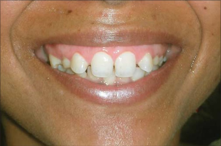
A 22-year-old female patient was referred with a complaint of gummy smile and hyperpigmented gingiva [Figure 1]. On examination, pseudopockets (approximately 2–3 mm) were detected in the maxillary and mandibular anteriors along with generalized excessive amount of melanin pigmentation on the gingiva [Figure 2]. The patient was explained about the treatment options and informed that, after depigmentation, melanin pigmentation tends to recur and, as gingivectomy is also performed simultaneously, the overall appearance would be worth doing it.
Figure 1.
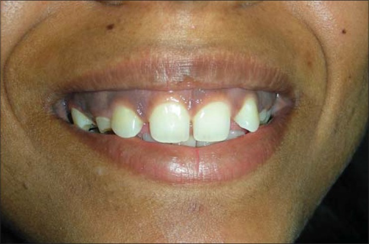
Profile view of gummy smile with hyper melanin pigmentation of the gingiva
Figure 2.
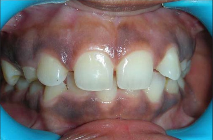
Intraoral view of gummy smile with hyper melanin pigmentation of the gingiva
Treatment plan
Gingivectomy and depigmentation were planned in the 15–25 region and the 34–44 region using a 810 nm diode laser. Although procedures like this can be performed under topical anesthesia, infiltration was planned as the patient was anxious and sensitive.
Procedure
Under infiltration anesthesia with 2% lignocaine (with 1:100,000 adrenaline), an 810 nm GaAlAs diode laser (Picasso™)*(Picasso, by AMD lasers) was used in contact mode with the gingival tissues. The procedure was performed at 1.5 W in pulsed mode. Initially, gingivectomy was performed followed by gingivoplasty and depigmentation [Figure 3]. The patient was extremely cooperative and comfortable during the procedure. No dressing was applied after the surgery. Postoperatively, a vit. E gel was prescribed for local application -three to four times a day for 2 days. Because a large area of the raw wound was kept exposed, antibiotic Cap. Amoxicillin 250 mg three times a day for 3 days and an analgesic ibuprofen–paracetamol was prescribed twice a day for 1 day and later SOS.
Figure 3.
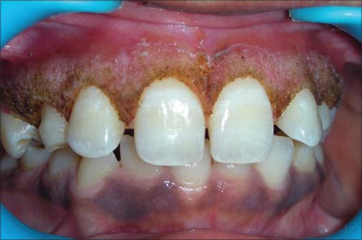
Immediate postoperative view of gingivectomy and depigmentation using an 810 nm diode laser
The patient reported no adverse effects or discomfort during the postoperative healing phase. She was happy and satisfied with 6-month postoperative results [Figures 4 and 5].
Figure 4.
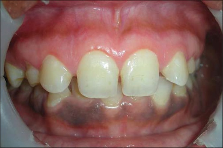
Six-month postoperative intraoral view
Figure 5.
Six-month postoperative profile view
DISCUSSION
Diode laser is a versatile laser and has been successfully used at three wavelengths: 980 nm, 810 nm and, more recently, 940 nm. It has numerous soft tissue applications, viz frenectomy, crown lengthening, gingival depigmentation, troughing, etc. In a diode laser, the substance stimulated to produce a coherent light beam is a semiconductor. This technology has produced a laser that is compact and economic.
A diode laser has become an important tool in the dental armamentarium due to its exceptional ease of use and affordability. It also has key advantages with regard to periodontal treatment. The diode laser is well absorbed by melanin, hemoglobin and other chromophores that are present in periodontal disease.[1] This provides the diode laser with broad clinical utility: It cuts precisely, coagulates, ablates and vaporizes the target tissue with much less trauma, improved postoperative healing and faster recovery time.[2,3,4]
Melanin pigmentation in gingiva is present only when melanin granules synthesized by melanocytes are transferred to the keratinocytes.[5] Therefore, recurrence of the pigmentation is inevitable and has been reported from 24 h to 8 years following surgery.[6] Conventional procedures for corrections of pigmented gingiva consist of surgical stripping with scalpel, use of electrosurgery, cryosurgery or a coarse diamond bur. However, the laser is the least-invasive and predictable method of performing this procedure.[7] It has a high level of precision, requires minimal anesthesia and causes very low collateral tissue damage.[6] Cases where only gingivectomy is required can be performed without anesthesia or under topical anesthesia, but because both gingivectomy and depigmentation were planned together in this patient and a significantly larger area needs to be operated upon, infiltration anesthesia was given.
CONCLUSION
Given the incredible ease of use and its versatility in treating soft tissue, the diode laser becomes the “soft tissue handpiece” in the dentists’ armamentarium. Patients with complaints of hyperpigmented gingiva should also be assessed for the presence of gummy smile. Once the amount of gingiva displayed is reduced by gingivectomy, the patient's concern for hyperpigmented gingiva will also reduce. As a result, in most of the cases, a repeat surgery for depigmentation is not required even after the recurrence of melanin pigmentation.
Footnotes
Source of Support: Nil.
Conflict of Interest: None declared.
REFERENCES
- 1.Raffetto N. Lasers for initial periodontal therapy. Dent Clin North Am. 2004;48:923–36. doi: 10.1016/j.cden.2004.05.007. [DOI] [PubMed] [Google Scholar]
- 2.Goharkhay K, Moritz A, Wilder-Smith P, Schoop U, Kluger W, Jakolitsch S, et al. Effects on oral soft tissue produced by a diode laser in vitro. Laser Surg Med. 1999;25:401–6. doi: 10.1002/(sici)1096-9101(1999)25:5<401::aid-lsm6>3.0.co;2-u. [DOI] [PubMed] [Google Scholar]
- 3.Walinski C. Irritation fibroma removal: A comparison of two laser wavelengths. Gen Dent. 2004;52:236–8. [PubMed] [Google Scholar]
- 4.Adams TC, Pang PK. Lasers in aesthetic dentistry. Dent Clin North Am. 2004;48:833–60. doi: 10.1016/j.cden.2004.05.010. [DOI] [PubMed] [Google Scholar]
- 5.Lerner AB, Fitzpatric TB. Biochemistry of melanin formation. Physio Rev. 1950;30:91–126. doi: 10.1152/physrev.1950.30.1.91. [DOI] [PubMed] [Google Scholar]
- 6.Kher U. Clinical applications of soft tissue diode lasers. Gonion. 2010;1:56–60. [Google Scholar]
- 7.Rowson JE. The contact diode laser. Br J Oral Maxillofac Surg. 1995;33:123. [Google Scholar]


