Abstract
Tracheal rupture is a rare complication of endotracheal intubation. Risk factors include short neck, repeated attempts due to failed intubation, inappropriate stylus, over-inflation of the cuff, poor positioning of the tube, inappropriate tube size, weakened membrane structure due to steroid use, chronic obstructive pulmonary disease, tracheomalacia, kyphosis, and use of nitric oxide during the operation. In this article, we suggest that high-volume, low-pressure tubes may not always provide a low-pressure effect and could rupture due to reduced tracheal perfusion pressure and ischemic damage upon over-inflation.
Keywords: Tracheal rupture; High-volume, low-pressure endotracheal tube, Nitrous oxide
Özet
Trakea rüptürü, endotrakeal entübasyonun nadir bir komplikasyonudur. Risk faktörleri kısa boyun, başarısız entübasyondan dolayı işlemin tekrarlanması, uygunsuz stile, kafın fazla şişirilmesi, uygunsuz tüp boyutu, uzun süreli steroid kullanımına bağlı zayıflamış membran varlığı, kronik obstrüktif pulmoner hastalık ve operasyonda nitrik oksit kullanılmasıdır. Bu yazıda yüksek volüm-düşük basınçlı tüplerle her zaman düşük basınç etkisi sağlanmadığı ve tüpün aşırı şişirilmesi durumunda trakeal perfüzyon basıncının azalması ve trakeal mukozada iskemik hasar sonucu rüptüre neden olduğu tartışıldı.
Introduction
Tracheal rupture is a rare complication of endotracheal intubation. Its incidence is less than 0.1%. Iatrogenic tracheal injuries usually occur after thoracic surgeries and bronchoscopic procedures [1]. In this article we present two cases with tracheal rupture due to diffusion of nitrous oxide to the cuffs of high-volume, low-pressure intubation tubes.
Case Reports
Case 1:
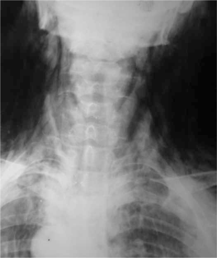
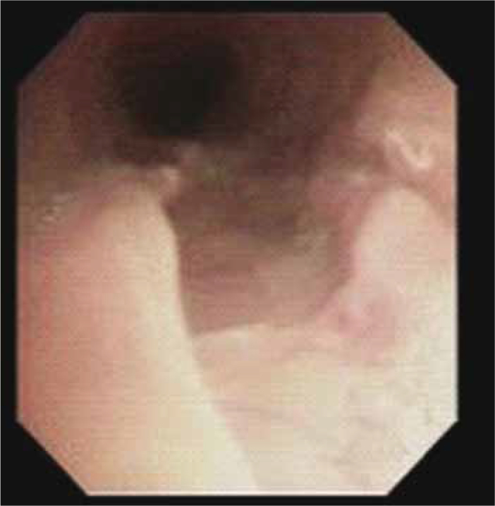
Surgery was planned for a 68 year-old female patient because of retinal detachment. Negative T-waves at V1-V4 were noticed in the pre-anesthesia visit and effort capacity was determined to be group 2. Laboratory examination showed pH 7.4, PaO2 65 mmHg, and PaCO2 31 mm Hg, thus theophylline ethylendiamine 240 mg i.v. (Aminocardol, NOVARTIS), n-acetylcysteine (Asist BİLİM) i.v, and inhaled ipratropium bromide (Combivent Boehringer Ingelheim) were administered preoperatively. The patient was taken into the operation room and pre-oxygenation was performed with 100% oxygen for 5 minutes before induction. Propofol i.v. 2 mg/kg (Propofol, Fresenius Kabi) and recuronium bromide i.v 50 mg (Esmeron, Organon) were administered. The patient was intubated on the first attempt using a A-7.5 mm ID (Well Lead, China) spiral tube and size 4 blade. Maintenance was achieved using sevoflurane 1.5 MAC (Sevorane, Abbott) and nitrous oxide (N2O) 65%. Pulse, blood pressure, oxygen saturation and end-tidal carbon dioxide levels were monitored. MAP was between 100–110 mmHg, EtCO2 was between 35–45 mmHg, and SpO2 was 98%. Inhaled agents were stopped at the end of the surgical procedure. The patient was decurarized using neostigmine and atropine; the endotracheal tube was removed after recovery of spontaneous respiration. She was observed in the PACU for two hours and then transferred to the ophthalmology department when her vital signs remained stable. On the first postoperative day, extensive subcutaneous emphysema developed in the face and neck (Figure 1). Emergency bronchoscopy revealed a 2.5 cm-wide rupture in the lateral part of the posterior wall of the trachea (Figure 2) that was treated by primary repair. The patient was discharged on the 5th postoperative day when no pathology was observed in posteroanterior chest X-rays and the mediastinal emphysema regressed.
Fig. 1.

Radiographic view of the first case.
Fig. 2.

Bronchoscopic view of the first case.
Case 2:
Surgery in the ENT department was planned for a 48 year-old female patient because of otosclerosis. Depression diagnosed by the psychiatry department and treated using an antidepressant drug (serotonin reuptake inhibitor) was the only pathology noticed in the pre-operative assessment by the anesthesiologists. Laboratory examinations were normal. The patient had no pulmonary disease or collagen tissue disease, and did not use any drug. Propofol i.v. 2 mg/kg (Propofol, Fresenius Kabi) and recuronium bromide i.v 50 mg (Esmeron, Organon) were administered. The patient was intubated using an A-7.5mm ID (Well Lead, China) tube with a spiral cuff and size 4 blade on the first laryngoscopy. Maintenance was achieved using desflurane 1.5 MAC (Suprane, Baxter) and nitrous oxide 60%. Pulse, blood pressure, oxygen saturation and end-tidal carbon dioxide levels were monitored. MAP was between 95–100 mmHg, EtCO2 was between 35–45 mmHg, and SpO2 was 100%. Inhaled agents were stopped at the end of the surgical procedure. The patient was decurarized using neostigmine and atropine; the endotracheal tube was removed after recovery of spontaneous respiration. We did not have any problems during the operation. Two hours later, the patient was extubated at the end of the surgery and sent to the clinic. A swelling was noticed in her neck at the end of the day, and progressed to swelling and crepitation that involved her whole neck and face on the next day. The patient was evaluated by chest physicians, and emergency bronchoscopy was carried out. Bronchoscopy revealed a 1.5 cm-wide membranous rupture in the trachea. Surgical treatment was not considered. The patient was followed in the thoracic surgery clinic and discharged on the 5rd day.
Discussion
Tracheal rupture is an uncommon and potentially life-threatening event. Iatrogenic tracheal injuries usually occur after thoracic surgeries and bronchoscopic procedures, but tracheal rupture secondary to endotracheal intubation is very rare. Management of tracheal rupture in patients with difficult airways is a challenging problem. Risk factors for tracheal rupture include short neck, repeated attempts due to failed intubation, inexperienced anesthesiologist, inappropriate stylus, over-inflation of the cuff, poor positioning of the tube, inappropriate tube size, coughing or movement of the patient, weakened membrane structure due to steroid use, chronic obstructive pulmonary disease, tracheomalacia, kyphosis and use of nitric oxide during the operation [2]. Our two cases were both intubated for the first time and they had no risk factors for tracheal rupture except for N2O administration. They had no collagen tissue disease, obstructive pulmonary disease or corticosteroid use. The ID of the tube was 8 mm in the first case, who was 68 years old. The 8 mm ID might have been too large for her trachea, and because of her old age the trachea have been fragile. However, the ID of the tube in the second case was 7.5 mm and the patient was 48 years old; thus the tube size and patient age may have been adequate for the procedure. Ruptures are 4 to 6 cm wide and usually located at the junction of the cartilaginous and membranous trachea [3]. In our first case, a 2.5 cm-wide rupture was seen in the lateral part of the posterior wall of the trachea. In the second case a 1.5 cm-wide membranous rupture was seen in the trachea. Our two cases were both female. Females are more prone to these injuries as their tracheas are narrower than those of males [2]. Our anesthesia team was experienced. These intubations were not difficult, thus we did not use a stylet. We did not use PEEP and also did not change the position of the patient during the operation. Before extubation, the tube cuffs were deflated. We controlled the tubes and cuffs before intubation. The cuffs of the tubes were inflated when the sound of air leakage from trachea stopped. We used high-volume, low-pressure cuffed intubation tubes.
There are two major types of cuffs: high-pressure, low-volume and low-pressure, high-volume. High-pressure cuffs are associated with greater ischemic damage to the tracheal mucosa and are less suitable for intubations of long duration [4,5]. Low pressure cuffs may increase the likelihood of sore throat (larger mucosal contact area), aspiration, spontaneous extubation, and difficult insertion (because of the floppy cuff). Nonetheless, because of the lower incidence of mucosal damage, low-pressure cuffs are more commonly recommended [5]. Cuff pressure depends on several factors: inflation volume, the diameter of the cuff in relation to the trachea, tracheal and cuff compliance, and intrathoracic pressure (cuff pressures increase with coughing) [4]. Cuff pressure may rise during general anesthesia as a result of the diffusion of nitrous oxide from the tracheal mucosa into the cuff [5]. If the cuff of the endotracheal tube (TT) is overinflated and its pressure exceeds the tracheal perfusion pressure, tracheal blood flow may be compromised. If these pressures exceed the capillary-arteriolar blood pressure, tissue ischemia occurs. This can lead to a sequence of inflammation, ulceration, granulation, and stenosis. Inflation of a TT cuff to the minimum pressure that creates a seal during routine positive-pressure ventilation (usually at least 20 mmHg) reduces tracheal blood flow by 75% at the cuff site. Further cuff inflation or induction of hypotension can totally eliminate mucosal blood flow.
The development of high-volume, low-pressure endotracheal tube cuffs has reduced the incidence and severity of such complications by permitting a relatively low-pressure, nonleaking seal to be established [6]. However, these cuffs can be easily overinflated, generating excessive cuff pressure that can result in mucosal injury. This effect may also occur during N2O administration in air-filled cuffs. We used N20 as an anesthesic agent in our two cases. N2O diffuses more rapidly into the cuff than nitrogen diffuses out of the cuff, thus creating excessive pressure even when the initial sealing pressure is satisfactory [7]. A number of measures, such as use of high-volume, low-pressure endotracheal tube cuffs, balloon-limiting pressure, inflation of the cuff with a N2O/oxygen gas mixture or isotonic saline, gas barrier cuff, a new ultrathin-walled endotracheal tube, and continuous monitoring of the tracheal cuff pressure [6,7] have been proposed in order to minimize this hyperpressure and thereby prevent tracheal injury.
Filling of the cuff with an anesthetic gas mixture is a simple and reliable method for maintaining stable cuff pressure during anesthesia. In patients anesthetized with nitrous oxide, the inflation of the tracheal tube cuff with a gas mixture of the same composition as the inhaled mixture can prevent excessive cuff pressure and reduce the incidence of tracheal injury. After tracheal intubation in anesthetized patients, the cuff can be inflated with an inhaled gas mixture, because this may prevent cuff hyperpressure [7].
In conclusion, the development of high-volume, low-pressure endotracheal tube cuffs has reduced the incidence of tracheal injury. However, these cuffs can be easily overinflated, generating excessive cuff pressure that can result in mucosal injury. We recommend tracheal tube cuff monitoring during surgery to prevent fatal overinflation of the cuff, which is permeable to nitrous oxide.
Footnotes
Conflict interest statement The authors declare that they have no conflict of interest to the publication of this article.
References
- 1.Moschini V, Losappio S, Dabrowska D, Iorno V. Tracheal rupture after tracheal intubation: effectiveness of conservative treatment. Minerva Anestesiol. 2006;72:1007–12. [PubMed] [Google Scholar]
- 2.Stannard K, Wells J, Cokis C. Tracheal rupture following endotracheal intubation. Anaesth Intensive Care. 2003;31:588–91. doi: 10.1177/0310057X0303100518. [DOI] [PubMed] [Google Scholar]
- 3.Jougon J, Ballester M, Choukroun E, Dubrez J, Reboul G, Velly JF. Conservative treatment for postintubation tracheobronchial rupture. Ann Thorac Surg. 2000;69:216–20. doi: 10.1016/s0003-4975(99)01129-7. [DOI] [PubMed] [Google Scholar]
- 4.Erolçay H, Yüceyar L, Aykaç B. Kardiyopulmoner by-pass’ın trakeal tüp balon basıncına etkisi. Cerrahpaşa Tıp Dergisi. 2002;33:28–32. [Google Scholar]
- 5.Edward Morgan G, Michail MS, Muray MJ. Clinical Anesthesiology. 4th Edn. Lange; New York: 2006. pp. 206–8. [Google Scholar]
- 6.Bernhard WN, Yost L, Turndorf H, Danziger F. Cuffed tracheal tubes: physical and behavioral characteristics. Anesth Analg. 1982;61:36–41. [PubMed] [Google Scholar]
- 7.Raeder JC, Borchgrerink PC, Sellevold OM. Tracheal tube cuff pressures: the effects of different gas-mixtures. Anaesthesia. 1985;40:444–7. [PubMed] [Google Scholar]


