Abstract
Absent left main coronary artery (LMCA) is a rare congenital cardiac malformation. We present a case report of a 65-year-old woman with anomalous origin of the left anterior descending (LAD) and circumflex (LCx) artery separated from the left sinus of Valsalva that was diagnosed by multi-detector row computed tomography (MDCT) coronary angiography. Our case indicates that MDCT plays an important role in the diagnosis of some rare coronary anomalies.
Keywords: Absent Left Main Coronary Artery, Coronary variation, MDCT
Özet
Sol ana koroner arter yokluğu konjenital kardiyak malformasyonlar arasında oldukça nadir görülmektedir. Biz bu vakada çok kesitli bilgisayarlı tomografi (ÇKBT) ile tanısını koyduğumuz, sol sinüs valsalvadan ayrı çıkım gösteren sirkümfleks arter ve sol anterior inen arter anomalisi olan 65 yaşında bayan hastayı sizlerle paylaşmak istedik. Bizim vakamızda olduğu gibi ÇKBT, nadir görülen bazı koroner arter anomalilerinin tanısında da oldukça başarıyla yer almaktadır.
Introduction
Anomalous coronary arteries can be benign or life threatening. Identification of anomalous coronary anatomy is important because of its complications (e.g., myocardial ischemia and sudden cardiac death). Absent LMCA is a rare congenital cardiac malformation. We report a similarly rare case of anomalous origin of the LAD artery and Cx artery separated from the left sinus of Valsalva. The origins of coronary arteries are easily identified with MDCT.
Case Report
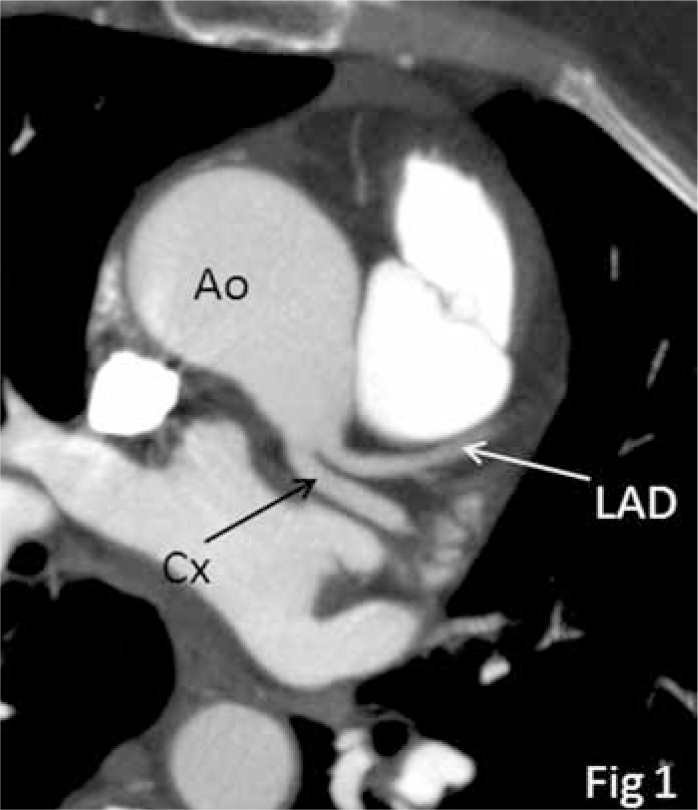
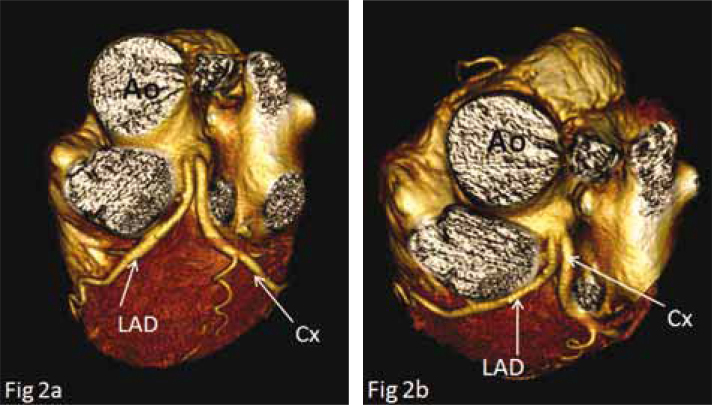
A 65-year-old woman was referred to our hospital because of episodic chest pain during exercise, which had started 1 year earlier. Biochemical, blood and urine test values were within normal limits. An MDCT coronary angiography was acquired. Axial images of the heart revealed a separate origin of the LAD and LCx arteries. The LMCA was absent (Fig. 1). The volume-rendered images of the anterior view (Fig. 2a) and superior view (Fig. 2b) show the LAD and LCx arteries, which bifurcate from the left sinus of Valsalva separately.
Fig. 1.

Axial MDCT angiogram of the heart shows separate origin of LAD and LCX. (LAD: left anterior descending, LCX: left circumflex artery).
Fig. 2.
A, B. Volume-rendered images from the superior view (A) and anterior view (B) show absent left main coronary artery with separate origins of LCx and LAD arteries in a 65-year-old woman presenting with episodes of chest pain.
Discussion
The LMCA, which originates from the left coronary sinus, is 5–10 mm in length [1]. It passes leftward, posterior to the pulmonary trunk, and bifurcates into the LAD and LCx arteries [2]. Coronary artery anomaly has been reported at a rate of 0.6% to 1.3% in routine angiographic series. The incidence of LMCA anomaly is between 0.02% and 0.07% [3, 4]. In 0.41% of cases, the left main artery is absent. In this condition, the LAD and LCx arteries bifurcate separately [5, 6]. This anomaly is 30.4% of all coronary anomalies [7]. Ectopic origin of the LAD and LCx coronary artery arising separately from the left sinus of Valsalva is a rare anomaly of the coronary arteries.
Typically, echocardiography is the first modality chosen for this diagnosis. However, conventional angiography is accepted as the gold standard in vascular imaging and is used wisely. Also, a treadmill test and Thallium test (scintigraphy) may be used, despite their low sensitivity rates. Catheterization of anomalous origin coronary arteries, and thus detecting their abnormal courses, is quite difficult. During catheter angiography (CA), LAD and LCx arising separately from the left sinus of Valsalva are easily misdiagnosed as occlusion or atresia of coronary vessel if the clinician operator evaluates only one orifice simultaneously. On the other hand, CA is an invasive tool that provides only luminal information. Compared with MDCT, CA takes long longer to acquire and has higher radiation exposure. Maddux et al. showed that the amount of contrast agent used and the radiation exposure time were reduced by 33% and 28%, respectively, for MDCT compared to rotational coronary angiography [8]. Nevertheless, it is possible with MDCT to acquire three-dimensional cardiac images that visualize all the vascular structures and their courses. Also, MDCT can accurately provide important diagnostic information about the origins, courses and anatomical relations of the coronary arteries, and thus, it can precisely detect the congenital anomalies of coronary vessels. For these reasons, MDCT has changed the clinical approach to vascular anomalies and raises the question if conventional angiography should still be the gold standard modality [9].
Due to new advances in CT technology, coronary anatomy is being demonstrated with MDCT [10]. We detected the absence of LMCA and separate coursing of LAD and LCx from the left sinus Valsalva in a 65-year-old female patient with episodic chest pain who was referred to our clinic to investigate possible coronary artery disease.
MDCT is a better modality than catheter angiography in such cases because it is non-invasive, provides three-dimensional images and demonstrates all vascular structures at the same time. In terms of detecting anomalous and variation coronary arteries, MDCT is superior to angiography.
Footnotes
Conflict interest statement The authors declare that they have no conflict of interest to the publication of this article.
References
- 1.Miller SW. Cardiac Angiography. Boston, MA: Little, Brown The Little Brown Library of Radiology; 1984. Normal Angiographic Anatomy and Measurements; pp. 51–71. [Google Scholar]
- 2.Duran C, Kantarci M, Durur Subasi I, et al. Remarkable anatomic anomalies of coronary arteries and their clinical importance: a multi-detector computed tomography angiographic study. Comput Assist Tomogr. 2006;30:939–48. doi: 10.1097/01.rct.0000230004.38521.8e. [DOI] [PubMed] [Google Scholar]
- 3.Mavi A, Ayalp R, Serçelik A, et al. Frequency in the anomalous origin of the left main coronary artery with angiography in a Turkish population. Acta Med Okayama. 2004;58:17–22. doi: 10.18926/AMO/32117. [DOI] [PubMed] [Google Scholar]
- 4.Iniguez Romo A, Macaya Miquel C, Alfonso Monterola F. Congenital anomalies of coronary artery origin: a diagnostic challenge. Rev Esp Cardiol. 1991;44:161–7. [PubMed] [Google Scholar]
- 5.Baim DS, Grossman W. Coronary angiography Cardiac Catheterization Angiography, and Intervention. 5th ed. Baltimore, MD: Williams & Wilkins; 1996. pp. 183–208. [Google Scholar]
- 6.Danias PG, Stuber M, McConnell MV. The diagnosis of congenital coronary anomalies with magnetic resonance imaging. Coronary Artery Dis. 2001;12:621–6. doi: 10.1097/00019501-200112000-00005. [DOI] [PubMed] [Google Scholar]
- 7.Bergman RA, Afifi AK, Miyauchi R. Coronary Arteries. Illustrated Encyclopedia of Human Anatomic Variation: Opus II: Cardiovascular System: Arteries: Head, Neck, and Thorax. http://www.anatomyatlases.org/AnatomicVariants/Cardiovascular/Text/Arteries/Coronary.Shtml.
- 8.Rigattieri S, Ghini AS, Silvestri P, et al. A randomized comparison between rotational and standart coronary angiography. Minerva Cardioangiol. 2005;53:1–6. [PubMed] [Google Scholar]
- 9.Lee EY, Siegel MJ, Hildebolt CF, Gutierrez FR, Bhalla S, Fallah JH. MDCT evaluation of thoracic aortic anomalies in pediatric patients and young adults: comparison of axial, multi-planar, and 3D images. AJR Am J Roentgenol. 2004;182:777–84. doi: 10.2214/ajr.182.3.1820777. [DOI] [PubMed] [Google Scholar]
- 10.Kantarci M, Ceviz N, Durur I, et al. Effect of the reconstruction window obtained at the isovolumic relaxation period on the image quality in electrocardiographic-gated 16-multi-detector-row computed tomography coronary angiography studies. J Comput Assist Tomogr. 2006;30:258–61. doi: 10.1097/00004728-200603000-00017. [DOI] [PubMed] [Google Scholar]


