Abstract
PCI-24781 is a novel histone deacetylase inhibitor that inhibits tumor proliferation and promotes cell apoptosis. However, it is unclear whether PCI-24781 inhibits Enhancer of Zeste 2 (EZH2) expression in malignant gliomas. In this work, three glioma cell lines were incubated with various concentrations of PCI-24781 (0, 0.25, 0.5, 1, 2.5 and 5 μM) and analyzed for cell proliferation by the MTS [3-(4,5-dimethylthiazol-2-yl)-5-(3-carboxymethoxyphenyl)-2-(4-sulfophenyl)-2H-tetrazolium] assay and colony formation, and cell cycle and apoptosis were assessed by flow cytometry. The expression of EZH2 and apoptosis-related proteins was assessed by western blotting. Malignant glioma cells were also transfected with EZH2 siRNA to examine how PCI-24781 suppresses tumor cells. EZH2 was highly expressed in the three glioma cell lines. Incubation with PCI-24781 reduced cell proliferation and increased cell apoptosis by down-regulating EZH2 in a concentration-dependent manner. These effects were simulated by EZH2 siRNA. In addition, PCI-24781 or EZH2 siRNA accelerated cell apoptosis by down-regulating the expression of AKT, mTOR, p70 ribosomal protein S6 kinase (p70s6k), glycogen synthase kinase 3A and B (GSK3a/b) and eukaryotic initiation factor 4E binding protein 1 (4E-BP1). These data suggest that PCI-24781 may be a promising therapeutic agent for treating gliomas by down-regulating EZH2 which promotes cell apoptosis by suppressing the phosphatidylinositol 3-kinase (PI3K)/Akt/mammalian target of the rapamycin (mTOR) pathway.
Keywords: EZH2, malignant gliomas, PCI-24781, PIK3K/Akt/mTOR signaling pathway
Introduction
Malignant gliomas are the most common and deadly brain tumor, accounting for ∼80% of malignant tumors that affect the central nervous system (Dolecek et al., 2012). Recently, the survival of patients with malignant gliomas has improved from an average of 10 months to 14 months because of advances in surgery, radiotherapy and chemotherapy (Tarbell et al., 2008). Nevertheless, the overall five-year survival rate for malignant gliomas is still < 5% (Huse and Holland, 2010). Consequently, there is an urgent need to explore and develop new anticancer agents.
Enhancer of Zeste 2 (EZH2) is a histone N-methyltransferase component of the Polycomb Repressive Complex 2 (PRC2) that mediates repression of tumor-suppressor gene activity via trimethylation of lysine 27 or lysine 9 of histone H3 (Simon and Lange, 2008). EZH2, which is frequently over-expressed in various cancers, including gliomas (Orzan et al., 2011), promotes cell proliferation and cell cycle progression by up-regulating cell division cycle 2 (Cdc2) and cyclin B1(Xia et al., 2014), blocks apoptosis by down-regulating the proapoptotic factors Puma and Bad (Hubaux et al., 2013), accelerates cell infiltration and metastasis by inhibiting Raf-1 kinase inhibitor protein (RKIP) (Ren et al., 2012) and activates tumor angiogenesis by silencing vasohibin 1 (Lu et al., 2010). These multiple actions ultimately result in poor prognosis in cancer patients (Liu et al., 2014). Therefore, the development of EZH2 inhibitors is a promising therapeutic approach for treating cancer, especially gliomas.
Transcriptional repression mediated by the PRC2 protein complex embryonic ectoderm development (EED)-EZH2 involves histone deacetylation. Translational repression mediated by PRC2 can be relieved by the histone deacetylase inhibitor (HDACI) trichostatin A (van der Vlag and Otte, 1999). In addition, other HDACIs, such as LBH589, LBH589 and suberoylanide hydroxamic acid (SAHA) have also been shown to deplete the EZH2 level in acute myeloid leukemia (Bhalla et al., 2007, Fiskus et al., 2009) and malignant gliomas (Orzan et al., 2011).
PCI-24781 is a novel hydroxamate-based inhibitor that can inhibit all HDAC isoforms (Banuelos et al., 2007). Assessment of the activity of PCI-24781 against tumor cell lines in vitro showed that this compound inhibited the growth of various solid tumor lines, including soft tissue sarcoma (Lopez et al., 2009), Hodgkins lymphoma (Bhalla et al., 2009), gallbladder carcinoma (Kitamura et al., 2012), neuroblastoma (Zhan et al., 2013). In contrast, little is known about the activity of PCI-24781 in malignant gliomas, particularly with regard to whether this compound inhibits EZH2 expression in these cancers and the underlying mechanisms involved.
In this work, we examined the effects of PCI-24781 in three malignant glioma cell lines and used short interfering RNA (siRNA) to assess the influence of down-regulating EZH2 expression.
Materials and Methods
Cell lines and culture
The human malignant glioma cell lines LN18 and LN229 were kindly provided by the School of Life Sciences at Fudan University and cell line U87MG was a kind gift of the Shanghai Institutes for Biological Sciences. Normal human astrocytes (NHA) were purchased from ScienCell Research Laboratories (Carlsbad, CA, USA). All cell lines were cultured in Dulbecco’s modified Eagle’s medium (DMEM) containing 10% fetal bovine serum, penicillin (100 U/mL) and streptomycin (100 mg/mL) (Lonza, Walkersville, MD, USA) and incubated at 37 °C in a humidified atmosphere of 5% CO2. The culture medium was renewed every two days.
Cell viability and colony formation assay
All cell lines were seeded in 96-well plates at a density of 5×10³ cells per well and treated with different concentrations (0, 0.25, 0.5, 1, 2.5 and 5 μM) of PCI-24781 (Selleckchem Chemicals, Houston, TX, USA). After 48 h, cell viability was assessed with 3-(4,5-dimethylthiazol-2-yl)-5-(3-carboxymethoxyphenyl)-2-(4-sulfophenyl)-2H-tetrazolium (MTS) (CellTiter 96 AQueous Non-Radioactive Cell Proliferation Assay solution, 20 μL/well; Promega Corporation, Madison, WI, USA). The absorbance of each well at 490 nm was measured in a SpectraMAX® M5 multidetection microplate reader (Molecular Devices, Sunnyvale, CA, USA). The growth rate was calculated using the following equation: growth rate = mean optical density (OD) of samples / mean OD of controls. In addition, after 48 h, the treated cells were cultured in culture medium containing G418 (400 μg/mL) for 14 days. Surviving colonies were stained with gentian violet after methanol fixation and visible colonies (≥ 50 cells) were counted. Photographs of culture plates were taken with a Sony NEX-7 camera (Sony Corporation, Japan).
Cell apoptosis and cycle analysis
Cell cycle distribution was detected by staining with propidium iodide (PI) followed by flow cytometry. Briefly, cells (2×10⁵) incubated with 0.5 and 1 μM PCI-24781 for 48 h were collected by centrifugation at 1,000 rpm for 5 min and washed twice in ice-cold phosphate-buffered saline (PBS). The cells were then fixed with 70% ice-cold ethanol at 4 °C overnight followed by staining with PI (BD Biosciences, San Diego, CA, USA).
Cell apoptosis was assessed using annexin V/PI kits (Invitrogen, Carlsbad, CA, USA) in conjunction with flow cytometry. Cells were washed twice with PBS and stained with 195 μL of Annexin V-FITC conjugated solution for 10min at room temperature followed by staining with 10 μL of PI. Stained cells were analyzed with a FACSCalibur flow cytometer (BD Biosciences, Franklin Lakes, NJ, USA).
RT-PCR analyses
Total RNA was isolated using Trizol® reagent and cDNA was generated by reverse transcription using the protocol included in the Reverse transcription system kit (TIANGEN Biotech, Beijing, China). The primers for EZH2 and β-actin were those described by Zhang et al. (2012): EZH2, forward primer 5′-GCCAGACTGGGAAGAAATCTG-3′ and reverse primer 5′-TGTGCTGGAAAATCCAAGTCA-3′; β-actin, forward primer 5′-CTGGGACGACATGGAGAAAA-3′ and reverse primer 5′-AAGGAAGGCTGGAAGAGTGC-3′. The PCR parameters for relative quantification were as follows: 30 s at 95 °C, followed by 40 cycles of 30 s at 60 °C and then 30 s at 72 °C. The β-actin gene was used as an internal control.
Western blotting
Cells were harvested 48 h after culture and rinsed with PBS three times followed by lysis in ice-cold lysis buffer for 30 min. The cells were then centrifuged (12,000 rpm, 10 min, 4 °C) and the supernatant was collected to determine the protein concentration by the bicinchoninic acid (BCA) assay (Pierce, Thermo Scientific, Rockford, IL, USA). Twenty micrograms of each cellular lysate was subjected to a sodium dodecyl sulfate-polyacrylamide gel electrophoresis (SDS-PAGE) and then transferred to a polyvinylidene fluoride (PVDF) membrane (Bio-Rad, Hercules, CA, USA). The membrane was blocked in Tris-buffered saline (TBS) containing 5% non-fat milk (Wyeth Pharmaceuticals Inc., Philadelphia, PA, USA) for 2 h, and subsequently incubated with the desired primary antibody (1:1000) overnight at 4 °C, followed by incubation with secondary antibody (1:5000) at room temperature for 1 h. Immunoreactive bands were detected by enhanced chemiluminescence using a chromogenic substrate and exposure to X-ray film. β-Actin served as a loading control. Cleaved PARP, cleaved caspase 3 antibodies and the secondary anti-rabbit IgG conjugated to horseradish peroxidase were purchased from Santa Cruz Biotechnology (Santa Cruz, CA, USA). The EZH2 and β-actin antibodies were purchased from Cell Signaling Technology (Beverly, MA, USA). AKT, mammalian target of rapamycin (mTOR), p70 ribosomal protein S6 kinase (p70s6k), glycogen synthase kinase 3A and B (GSK3a/b) and eukaryotic initiation factor 4E binding protein 1 (4E-BP1) antibodies were purchased from Jackson Immuno Research (West Grove, PA, USA).
siRNA knock-down of EZH2
For silencing experiments, cells were seeded at a density of 2×10⁵ cells per well in 6-well plates with 2 mL of DMEM without serum and antibiotics, and allowed to reach ∼70% confluence on the day of transfection. siRNA and Lipofectamine 2000 (Invitrogen) were dissolved in 50 μL of OPTIMEM (GIBCO) and then the two solutions were mixed for 5 min. Cells were transfected with the mixture of solutions according to the manufacturer’s transfection protocol. The transfection groups were as follows: (1) EZH2 siRNA (SIR), 5′-AAGACTCTGAATGCAGTTGCT-3′ (Wagener et al., 2008), (2) scrambled siRNA control (siRNA-NC), purchased from Dharmacon (Lafayett, CO, USA) and (3) blank control (mock). The transfection process lasted for 4 h, after which the cells were cultured in medium with serum and analyzed by flow cytometry and western blotting to detect cell apoptosis and the expression of related proteins.
Statistical analysis
The experiments were done in triplicate and the results were expressed as the mean ± standard deviation (SD). Time-dependent variations within the same group were analyzed using analysis of variance (ANOVA) for repeated measures followed by the Bonferroni test. Differences among groups were analyzed by one-way ANOVA and Fisher’s LSD test. A value of p < 0.05 indicated significance. All data analyses were done using SPSS 17.0 software (SPSS Inc., Chicago, IL, USA).
Results
PCI-24781 inhibits the growth of malignant glioma cells
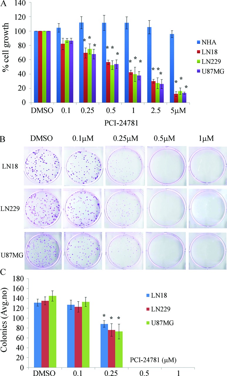
Figure 1 shows the effect of treating normal astrocytes and three malignant glioma cell lines with PCI-24781 for 48 h followed by the MTS and colony formation assays to assess cell proliferation. All three malignant glioma cell lines were sensitive to PCI-24781 and cell growth was inhibited in a concentration-dependent manner, whereas PCI-24781 had no significant effect on NHA. Similarly, PCI-24781 significantly inhibited the formation of malignant glioma cells colonies compared to untreated cells (p < 0.05) (Figure 1B,C). Together, these data show that PCI-24781 inhibited the proliferation of malignant glioma cells.
Figure 1.
PCI-24781 inhibits the growth of malignant glioma cells. (A) MTS analysis of normal astrocytes and three malignant glioma cell lines. (B) Formation of malignant glioma cell colonies. (C) Number of surviving colonies. In panels (A) and (C) the columns are the mean ± SD of 3 determinations. *p < 0.05 compared with the DMSO control.
PCI-24781 induces G2/M cell cycle arrest and apoptosis in malignant glioma cells
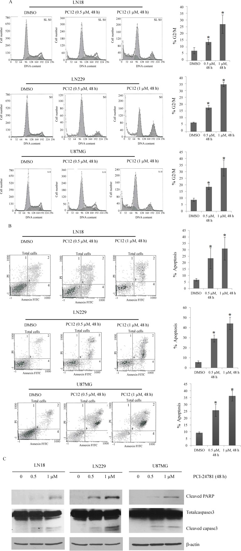
Annexin-V/PI staining and flow cytometry were used to assess whether PCI-24781 inhibited the growth of malignant glioma cells by preventing cell cycle progression and promoting apoptosis. Treatment of malignant glioma cells with 0.5 μM PCI-24781 increased the G2/M checkpoint activation from 7.2 ± 2.3% to 13.7 ± 2.0% in LN18 cells, from 6.7 ± 0.3% to 17.5 ± 1.5% in LN229 cells and from 8.6 ± 1.3% to 18.5 ± 1.9% in U87MG cells (p < 0.05 in each case) (Figure 2A). Similarly, PCI-24781 also induced apoptosis in a concentration-dependent manner (Figure 2B). The expression of poly ADP-ribose polymerase (PARP) and cysteine-protease P-3 (caspase 3), both of which are markers of cell apoptosis (Tutt et al., 2010; Steert et al., 2010), was assessed by western blotting. As anticipated, these two proteins were detected in the three malignant glioma cell lines, especially when they were treated with 1 μM PCI-24781 (Figure 2C). These findings indicate that PCI-24781 promoted G2 cell cycle arrest and induced apoptosis in malignant glioma cells.
Figure 2.
PCI-24781-induced G2 cell cycle inhibition and apoptosis in malignant glioma cells. Note the concentration-dependent increase in (A) G2/M checkpoint activation and (B) malignant glioma cell apoptosis. (C) PARP and caspase 3 cleavage was detected in three malignant glioma cell lines treated with 0.5 or 1 μM PCI-24781. In panels (A) and (C) the columns are the mean ± SD of 3 determinations. *p < 0.05 compared with the DMSO control.
PCI-24781-induced G2 cell cycle inhibition and apoptosis in malignant glioma cells. Note the concentration-dependent increase in (A) G2/M checkpoint activation and (B) malignant glioma cell apoptosis. (C) PARP and caspase 3 cleavage was detected in three malignant glioma cell lines treated with 0.5 or 1 μM PCI-24781. In panels (A) and (C) the columns are the mean ± SD of 3 determinations. *p < 0.05 compared with the DMSO control.
PCI-24781 inhibits EZH2 expression
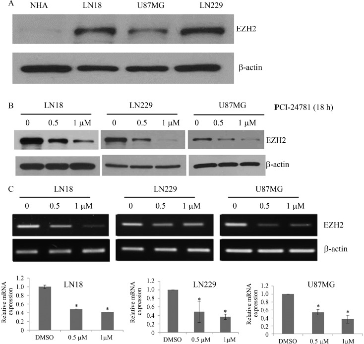
Previous work showed that EZH2 is highly expressed in high-grade glioma tissue and glioma stem-like cells and that treatment with the histone deacetylase inhibitor SAHA can decrease the expression of EZH2 (Orzan et al., 2011. We therefore examined the expression of EZH2 in the three malignant glioma cell lines and investigated the role of PCI-24781 in inhibiting EZH2 expression. EZN2 protein was expressed in the malignant glioma cell lines compared to virtually no expression in NHA (Figure 3A). Incubation with 0.5 μMor1 μM PCI-24781 for 48 h gradually reduced the levels of EZN2 mRNA and protein expression (Figure 3B,C).
Figure 3.
PCI-24781 inhibits EZN2 expression. (A) EZN2 protein expression in normal astrocytes and three malignant glioma cell lines. (B) EZN2 protein expression in three malignant glioma cell lines after treatment with PCI-24781. (C) EZH2 mRNA expression in three malignant glioma cell lines treated with PCI-24781. The columns are the mean ± SD of 3 determinations. *p < 0.05 compared with the DMSO control.
EZH2 gene silencing promotes cell apoptosis
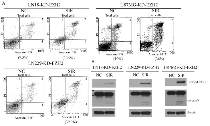
To explore whether PCI-24781 promoted apoptosis by down-regulating EZH2, an RNA-interference-mediated knock-down of EZH2 was done. As anticipated, the extent of apoptosis in the three malignant glioma cell lines transfected with EZH2 siRNA was significantly higher than in the negative control group (Figure 4A). Apoptosis-related proteins were also highly expressed after knock-down of EZH2 (Figure 4B). These effects mimicked those of PCI-24781, suggesting that our hypothesis was credible.
Figure 4.
EZH2 gene silencing promotes cell apoptosis. (A) Flow cytometry results showing apoptosis in three malignant glioma cell lines transfected with EZH2 siRNA. (B) Enhanced expression of the apoptosis-related proteins cleaved PARP and caspase 3 after knockdown of EZH2. KD – knockdown, NC – normal control (cells with scrambled siRNA), SIR – cells treated with specific siRNA.
EZH2 gene silencing reduces the role of the PI3K/Akt signaling pathway
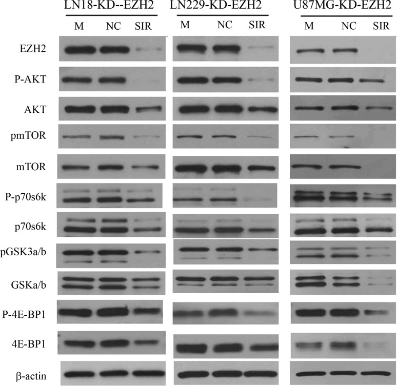
Previous studies have demonstrated that the apoptotic potential of EZH2 is highly associated with activation of the phosphatidylinositol 3-kinase (PI3K)/Akt/mTOR signaling pathway. To further investigate the regulatory relationship between EZH2 and the PI3K/Akt/mTOR pathway, we examined the expression of PI3K/Akt/mTOR pathway proteins in malignant glioma cells transfected with EZH2 siRNA. When EZH2 protein expression was significantly down-regulated by treatment with EZH2 siRNA the signaling proteins of the PI3K/Akt/mTOR pathway also showed a significant reduction in expression, indicating that PI3K/Akt/mTOR signaling proteins exert their roles downstream of EZH2 in malignant glioma growth (Figure 5).
Figure 5.
EZH2 gene silencing attenuated the expression of proteins involved in the PI3K/Akt signaling pathway. M – blank control (mock), NC – normal control (cells with scrambled siRNA), SIR – cells treated with specific siRNA. The blots are representative of 3 experiments.
PCI-24781 decreases expression of the PI3K/Akt signaling pathway
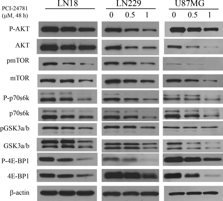
Western blotting showed that the treatment of cells with PCI-24781 for 48 h significantly reduced the expression of PI3K/Akt signaling pathway proteins compared with the control group, a finding consistent with the results for EZH2 gene silencing (Figure 6). These findings suggested that PCI-24781 reduced the expression of the PI3K/Akt signaling pathway during the growth of malignant glioma cells by inhibiting EZH2.
Figure 6.
Treatment with PCI-24781 attenuated the expression of proteins involved in the PI3K/Akt signaling pathway. The blots are representative of 3 experiments.
Discussion
Although a recent study investigated the roles of HDACI in malignant gliomas (Orzan et al., 2011), no report has focused on the cytotoxicity of PCI-24781, a novel, broad spectrum histone deacetylase inhibitor. In accordance with previous studies (Yamaguchi et al., 2010; Orzan et al., 2011), we found that PCI-24781 inhibited growth and stimulated apoptosis in the three malignant glioma cell lines, in addition to down-regulating the expression of EZH2. These effects were also seen after knockdown of EZH2.
Several studies have demonstrated that inhibition of EZH2 can promote apoptosis in glioma cells, but the mechanism involved remains poorly understood (Suvà et al., 2009; Orzan et al., 2011). In this study, we detected expression of the apoptotic markers cleaved PARP and caspase 3. The expression of these markers was increased in glioma cells with EZH2 knockdown, suggesting that EZH2 over-expression could have an anti-apoptotic function in glioma cells by regulating PARP and caspase 3.
The PI3K/Akt/mTOR pathway is an intracellular signaling pathway important intumor cell metabolism, growth, proliferation and survival (Morgensztern and McLeod, 2005; Yap et al., 2008). This pathway has been targeted for early drug development against glioblastoma (Wick et al., 2011). Previous work has shown that c-myc is a direct target of EZH2 and that the PRC-2 complex binds directly to the c-myc promoter to enhance its expression in multiform glioblastoma cancer stem cells, thereby promoting cell growth (Suvà et al., 2009). Further, abnormal c-myc-driven cell growth is sustained by induction of PI3K/Akt/mTOR survival pathways. Silencing c-myc reduced PI3K/Akt and mTOR expression and induced cell apoptosis (Ladu et al., 2008). As expected, knockdown of EZH2 or treatment with PCI-24781 reduced the expression of proteins in the PI3K/Akt/mTOR signaling pathway.
In summary, we have shown that EZH2 is highly expressed in glioma cells. Treatment with PCI-24781 attenuated cell proliferation and increased cellular apoptosis by down-regulating EZH2 expression which then promoted c-myc-driven apoptosis by suppressing PI3K/Akt/mTOR survival pathways. This effect could be simulated by EZH2 siRNA. Our results provide evidence that EZH2 depletion may be a promising intervention for the treatment of malignant gliomas. However, further studies are needed to show that the effect of PCI-24781 on PI3K signaling can be reverted by restoring EZH2 expression. The inhibitory effect of PCI-24781 on cell proliferation should be investigated in greater detail by using more physiologically relevant models, such as neurospheres of glioma stem cells or intracranial xenografts.
Acknowledgments
We would like to thank the editor and the staff for their help and support at every step of the publication process. We also greatly appreciate the comments of the reviewers. No funds were received in support of this work.
Footnotes
Senior Editor: Emmanuel Dias Neto
References
- Banuelos CA, Banáth JP, MacPhail SH, Zhao J, Reitsema T, Olive PL. Radiosensitization by the histone deacetylase inhibitor PCI-24781. Clin Cancer Res. 2007;13:6816–6826. doi: 10.1158/1078-0432.CCR-07-1126. [DOI] [PubMed] [Google Scholar]
- Bhalla K, Fiskus W, Herger B, Rao R, Ustun C, Jillella A, Atadja P. Anti-leukemia activity of histone deacetylase (HDAC) inhibitor LBH589 involves depletion of EZH2 and DNA methyltransferase (DNMT) 1 through disruption of their chaperone association with heat shock protein (hsp) 90. J Clin Oncol. 2007;25:10501. [Google Scholar]
- Bhalla S, Balasubramanian S, David K, Sirisawad M, Buggy J, Mauro L, Prachand S, Miller R, Gordon LI, Evens AM. PCI-24781 induces caspase and reactive oxygen species-dependent apoptosis through NF-κB mechanisms and is synergistic with bortezomib in lymphoma cells. Clin Cancer Res. 2009;15:3354–3365. doi: 10.1158/1078-0432.CCR-08-2365. [DOI] [PMC free article] [PubMed] [Google Scholar]
- Dolecek TA, Propp JM, Stroup NE, Kruchko C. CBTRUS statistical report: Primary brain and central nervous system tumors diagnosed in the United States in 2005–2009. Neuro-Oncology. 2012;14:1–49. doi: 10.1093/neuonc/nos218. [DOI] [PMC free article] [PubMed] [Google Scholar]
- Fiskus W, Wang Y, Sreekumar A, Buckley KM, Shi H, Jillella A, Ustun C, Rao R, Fernandez P, Chen J. Blood combined epigenetic therapy with the histone methyltransferase EZH2 inhibitor 3-deazaneplanocin A and the histone deacetylase inhibitor panobinostat against human AML cells. Blood. 2009;114:2733–2743. doi: 10.1182/blood-2009-03-213496. [DOI] [PMC free article] [PubMed] [Google Scholar]
- Hubaux R, Thu KL, Coe BP, MacAulay C, Lam S, Lam WL. EZH2 promotes E2F-driven SCLC tumorigenesis through modulation of apoptosis and cell-cycle regulation. J Thorac Oncol. 2013;8:1102–1106. doi: 10.1097/JTO.0b013e318298762f. [DOI] [PMC free article] [PubMed] [Google Scholar]
- Huse JT, Holland EC. Targeting brain cancer: Advances in the molecular pathology of malignant glioma and medulloblastoma. Nat Rev Cancer. 2010;10:319–331. doi: 10.1038/nrc2818. [DOI] [PubMed] [Google Scholar]
- Kitamura T, Connolly K, Ruffino L, Ajiki T, Lueckgen A, DiGiovanni J, Kiguchi K. The therapeutic effect of histone deacetylase inhibitor PCI-24781 on gallbladder carcinoma in BK5.erbB2 mice. J Hepatol. 2012;57:84–91. doi: 10.1016/j.jhep.2012.01.018. [DOI] [PMC free article] [PubMed] [Google Scholar]
- Ladu S, Calvisi DF, Conner EA, Farina M, Factor VM, Thorgeirsson SS. Gastroenterology E2F1 inhibits c-Myc-driven apoptosis via PIK3CA/Akt/mTOR and COX-2 in a mouse model of human liver cancer. Gastroenterology. 2008;135:1322–1332. doi: 10.1053/j.gastro.2008.07.012. [DOI] [PMC free article] [PubMed] [Google Scholar]
- Liu Y, Liu T, Bao X, He M, Li L, Yang X. Increased EZH2 expression is associated with proliferation and progression of cervical cancer and indicates a poor prognosis. Int J Gynecol Pathol. 2014;33:218–224. doi: 10.1097/PGP.0b013e31829c6574. [DOI] [PubMed] [Google Scholar]
- Lopez G, Liu J, Ren W, Wei W, Wang S, Lahat G, Zhu Q-S, Bornmann WG, McConkey DJ, Pollock RE. Combining PCI-24781, a novel histone deacetylase inhibitor, with chemotherapy for the treatment of soft tissue sarcoma. Clin Cancer Res. 2009;15:3472–3483. doi: 10.1158/1078-0432.CCR-08-2714. [DOI] [PubMed] [Google Scholar]
- Lu C, Han HD, Mangala LS, Ali-Fehmi R, Newton CS, Ozbun L, Armaiz-Pena GN, Hu W, Stone RL, Munkarah A. Regulation of tumor angiogenesis by EZH2. Cancer Cell. 2010;18:185–197. doi: 10.1016/j.ccr.2010.06.016. [DOI] [PMC free article] [PubMed] [Google Scholar]
- Morgensztern D, McLeod HL. Anti-cancer drugs PI3K/Akt/mTOR pathway as a target for cancer therapy. Anti-Cancer Drugs. 2005;16:797–803. doi: 10.1097/01.cad.0000173476.67239.3b. [DOI] [PubMed] [Google Scholar]
- Orzan F, Pellegatta S, Poliani P, Pisati F, Caldera V, Menghi F, Kapetis D, Marras C, Schiffer D, Finocchiaro G. Enhancer of Zeste 2 (EZH2) is up-regulated in malignant gliomas and in glioma stem-like cells. Neuropathol Appl Neurobiol. 2011;37:381–394. doi: 10.1111/j.1365-2990.2010.01132.x. [DOI] [PubMed] [Google Scholar]
- Ren G, Baritaki S, Marathe H, Feng J, Park S, Beach S, Bazeley PS, Beshir AB, Fenteany G, Mehra R. Polycomb protein EZH2 regulates tumor invasion via the transcriptional repression of the metastasis suppressor RKIP in breast and prostate cancer. Cancer Res. 2012;72:3091–3104. doi: 10.1158/0008-5472.CAN-11-3546. [DOI] [PubMed] [Google Scholar]
- Simon JA, Lange CA. Roles of the EZH2 histone methyltransferase in cancer epigenetics. Mutat Res - Fund Mol M. 2008;647:21–29. doi: 10.1016/j.mrfmmm.2008.07.010. [DOI] [PubMed] [Google Scholar]
- Steert K, Berg M, Mottram JC, Westrop GD, Coombs GH, Cos P, Maes L, Joossens J, Van der Veken P, Haemers A. α-Ketoheterocycles as inhibitors of Leishmania mexicana cysteine protease CPB. Chem Med Chem. 2010;5:1734–1748. doi: 10.1002/cmdc.201000265. [DOI] [PMC free article] [PubMed] [Google Scholar]
- Suvà M-L, Riggi N, Janiszewska M, Radovanovic I, Provero P, Stehle J-C, Baumer K, Le Bitoux M-A, Marino D, Cironi L. EZH2 is essential for glioblastoma cancer stem cell maintenance. Cancer Res. 2009;69:9211–9218. doi: 10.1158/0008-5472.CAN-09-1622. [DOI] [PubMed] [Google Scholar]
- Tarbell N, Barnes P, Scott RM, Goumnerova L, Pomeroy S, McL BP, Sallan S, Billett A, LaVally B, Helmus A. Advances in radiation therapy for craniopharyngiomas. Pediatr Neurosurg. 2008;21:101–107. doi: 10.1159/000120870. [DOI] [PubMed] [Google Scholar]
- Tutt A, Robson M, Garber JE, Domchek SM, Audeh MW, Weitzel JN, Friedlander M, Arun B, Loman N, Schmutzler RK. Oral poly(ADP-ribose) polymerase inhibitor olaparib in patients with BRCA1 or BRCA2 mutations and advanced breast cancer: A proof-of-concept trial. Lancet. 2010;376:235–244. doi: 10.1016/S0140-6736(10)60892-6. [DOI] [PubMed] [Google Scholar]
- van der Vlag J, Otte AP. Transcriptional repression mediated by the human polycomb-group protein EED involves histone deacetylation. Nat Genet. 1999;23:474–478. doi: 10.1038/70602. [DOI] [PubMed] [Google Scholar]
- Wagener N, Holland D, Bulkescher J, Crnkovic-Mertens I, Hoppe-Seyler K, Zentgraf H, Pritsch M, Buse S, Pfitzenmaier J, Haferkamp A, et al. The enhancer of zeste homolog 2 gene contributes to cell proliferation and apoptosis resistance in renal cell carcinoma cells. Int J Cancer. 2008;123:1545–1550. doi: 10.1002/ijc.23683. [DOI] [PubMed] [Google Scholar]
- Wick W, Weller M, Weiler M, Batchelor T, Yung AW, Platten M. Pathway inhibition: Emerging molecular targets for treating glioblastoma. Neuro-Oncol. 2011;13:566–579. doi: 10.1093/neuonc/nor039. [DOI] [PMC free article] [PubMed] [Google Scholar]
- Xia H, Zhang W, Li Y, Guo N, Yu C. EZH2 silencing with RNA interference induces G2/M arrest in human lung cancer cells in vitro. BioMed Res Int. 2014;2014:348728. doi: 10.1155/2014/348728. [DOI] [PMC free article] [PubMed] [Google Scholar]
- Yamaguchi J, Sasaki M, Sato Y, Itatsu K, Harada K, Zen Y, Ikeda H, Nimura Y, Nagino M, Nakanuma Y. Histone deacetylase inhibitor (SAHA) and repression of EZH2 synergistically inhibit proliferation of gallbladder carcinoma. Cancer Sci. 2010;101:355–362. doi: 10.1111/j.1349-7006.2009.01387.x. [DOI] [PMC free article] [PubMed] [Google Scholar]
- Yap TA, Garrett MD, Walton MI, Raynaud F, de Bono JS, Workman P. Targeting the PI3K-AKT-mTOR pathway: Progress, pitfalls, and promises. Curr Opin Pharmacol. 2008;8:393–412. doi: 10.1016/j.coph.2008.08.004. [DOI] [PubMed] [Google Scholar]
- Zhan Q, Tsai S, Lu Y, Wang C, Kwan Y, Ngai S. RuvBL2 is involved in histone deacetylase inhibitor PCI-24781-induced cell death in SK-N-DZ neuroblastoma cells. PloS One. 2013;8:e71663. doi: 10.1371/journal.pone.0071663. [DOI] [PMC free article] [PubMed] [Google Scholar]
- Zhang R, Wang R, Chang H, Wu F, Liu C, Deng D, Fan W. Downregulation of Ezh2 expression by RNA interference induces cell cycle arrest in the G0/G1 phase and apoptosis in U87 human glioma cells. Oncol Rep. 2012;28:2278–2284. doi: 10.3892/or.2012.2033. [DOI] [PubMed] [Google Scholar]


