

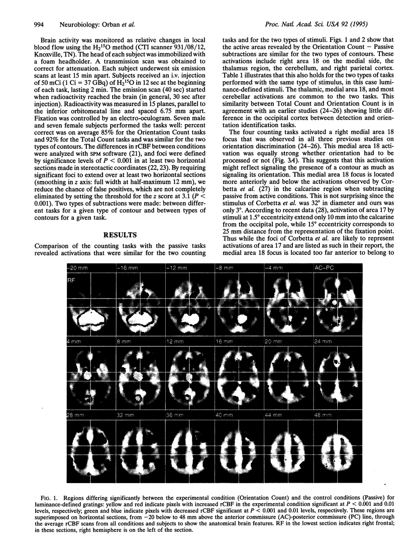
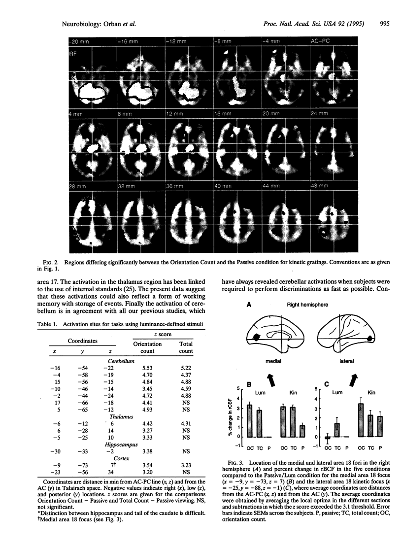
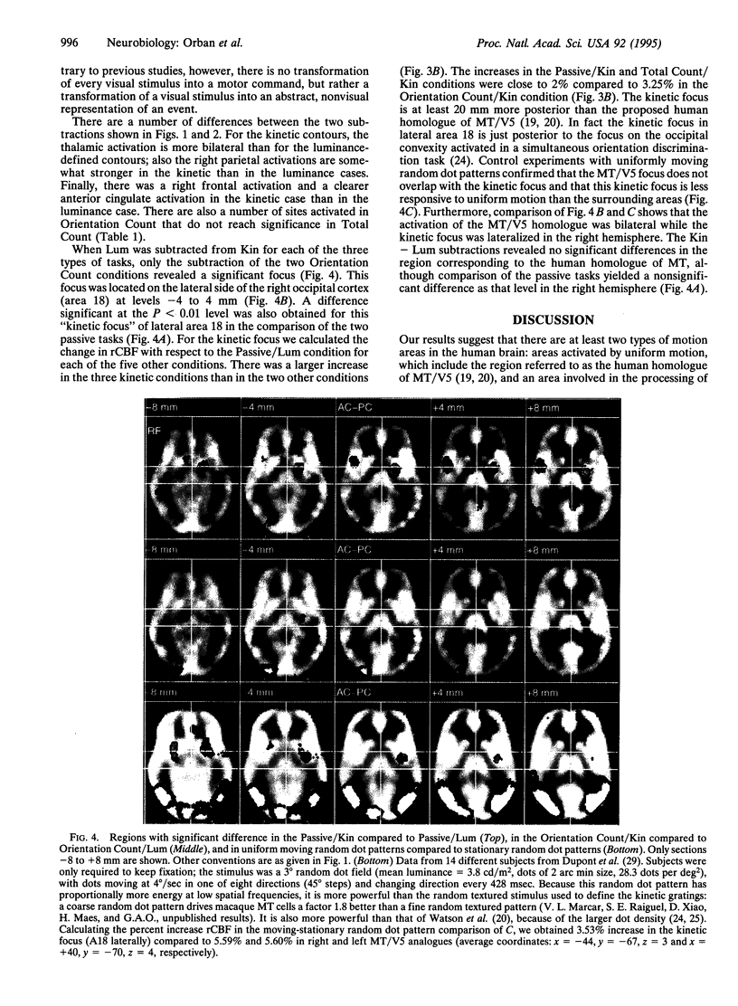
Abstract
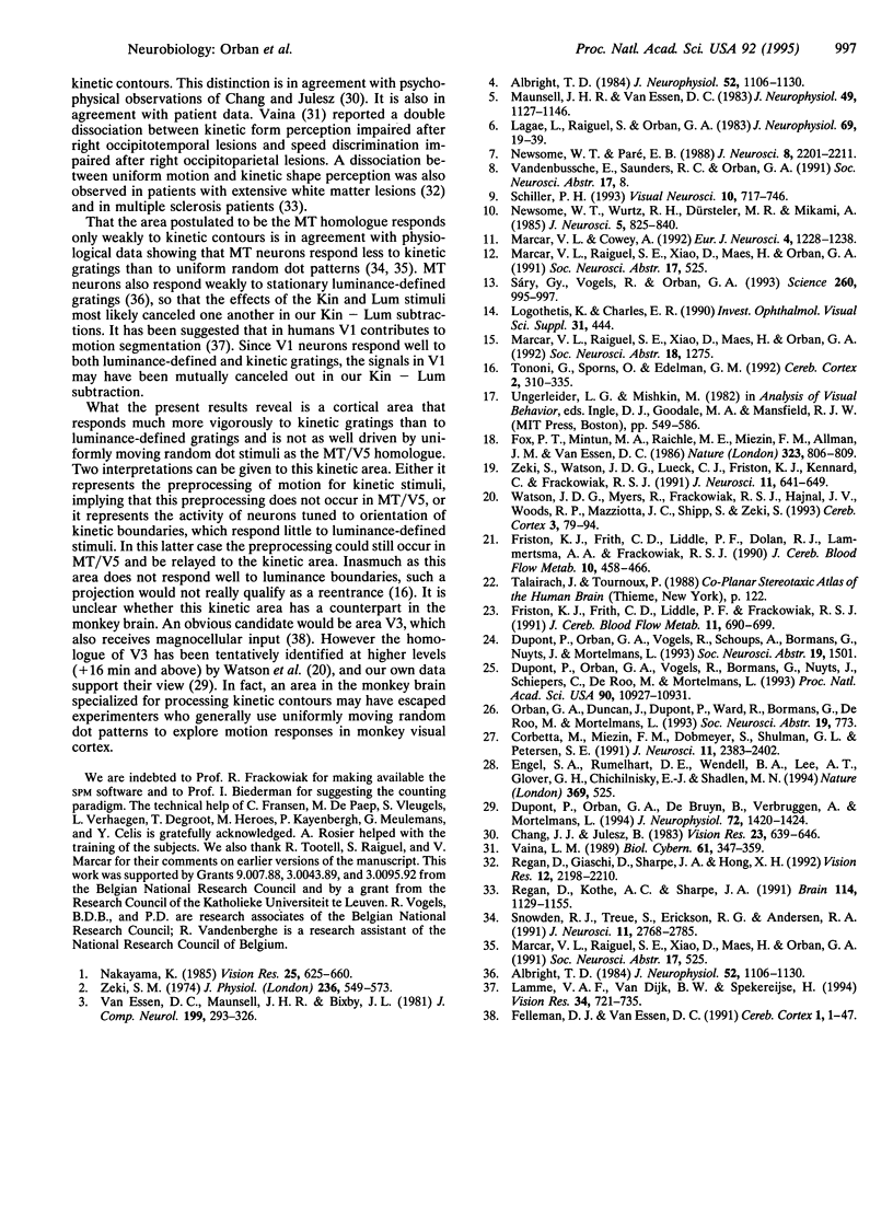
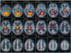
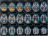
We have localized an area in the human brain involved in the processing of contours defined by motion differences (kinetic contours) by comparing with positron emission tomography the regional cerebral blood flow in tasks performed with kinetic and luminance-defined gratings. These tasks included passive viewing, counting the total number of grating stimuli, and counting the number of gratings of a given orientation. Comparison between the counting tasks and passive viewing with a given type of contour revealed a set of active areas that were similar for both luminance-defined and kinetic contours. Comparisons between these two types of contours revealed a single focus in the right hemisphere that did not overlap with the many regions activated by uniform motion. In particular this "kinetic focus" was clearly separated from the area previously defined as the human homologue of V5/middle temporal. Activity in this kinetic focus was stronger when orientation had to be processed than in the other two tasks. These results and control experiments with uniformly moving random dot patterns suggest the existence of an area in the human visual system that is activated much more by kinetic contours than by luminance contours or uniformly moving random dots. Up to now, such an area has not been described in the monkey visual system.
Full text
PDF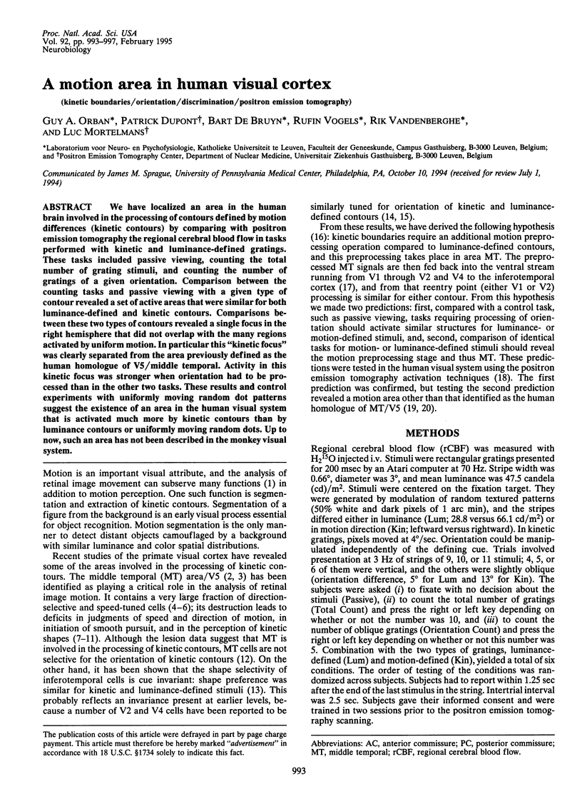
Images in this article
Selected References
These references are in PubMed. This may not be the complete list of references from this article.
- Albright T. D. Direction and orientation selectivity of neurons in visual area MT of the macaque. J Neurophysiol. 1984 Dec;52(6):1106–1130. doi: 10.1152/jn.1984.52.6.1106. [DOI] [PubMed] [Google Scholar]
- Albright T. D. Direction and orientation selectivity of neurons in visual area MT of the macaque. J Neurophysiol. 1984 Dec;52(6):1106–1130. doi: 10.1152/jn.1984.52.6.1106. [DOI] [PubMed] [Google Scholar]
- Chang J. J., Julesz B. Displacement limits, directional anisotropy and direction versus form discrimination in random-dot cinematograms. Vision Res. 1983;23(6):639–646. doi: 10.1016/0042-6989(83)90070-6. [DOI] [PubMed] [Google Scholar]
- Corbetta M., Miezin F. M., Dobmeyer S., Shulman G. L., Petersen S. E. Selective and divided attention during visual discriminations of shape, color, and speed: functional anatomy by positron emission tomography. J Neurosci. 1991 Aug;11(8):2383–2402. doi: 10.1523/JNEUROSCI.11-08-02383.1991. [DOI] [PMC free article] [PubMed] [Google Scholar]
- Dupont P., Orban G. A., De Bruyn B., Verbruggen A., Mortelmans L. Many areas in the human brain respond to visual motion. J Neurophysiol. 1994 Sep;72(3):1420–1424. doi: 10.1152/jn.1994.72.3.1420. [DOI] [PubMed] [Google Scholar]
- Dupont P., Orban G. A., Vogels R., Bormans G., Nuyts J., Schiepers C., De Roo M., Mortelmans L. Different perceptual tasks performed with the same visual stimulus attribute activate different regions of the human brain: a positron emission tomography study. Proc Natl Acad Sci U S A. 1993 Dec 1;90(23):10927–10931. doi: 10.1073/pnas.90.23.10927. [DOI] [PMC free article] [PubMed] [Google Scholar]
- Engel S. A., Rumelhart D. E., Wandell B. A., Lee A. T., Glover G. H., Chichilnisky E. J., Shadlen M. N. fMRI of human visual cortex. Nature. 1994 Jun 16;369(6481):525–525. doi: 10.1038/369525a0. [DOI] [PubMed] [Google Scholar]
- Felleman D. J., Van Essen D. C. Distributed hierarchical processing in the primate cerebral cortex. Cereb Cortex. 1991 Jan-Feb;1(1):1–47. doi: 10.1093/cercor/1.1.1-a. [DOI] [PubMed] [Google Scholar]
- Fox P. T., Mintun M. A., Raichle M. E., Miezin F. M., Allman J. M., Van Essen D. C. Mapping human visual cortex with positron emission tomography. 1986 Oct 30-Nov 5Nature. 323(6091):806–809. doi: 10.1038/323806a0. [DOI] [PubMed] [Google Scholar]
- Friston K. J., Frith C. D., Liddle P. F., Dolan R. J., Lammertsma A. A., Frackowiak R. S. The relationship between global and local changes in PET scans. J Cereb Blood Flow Metab. 1990 Jul;10(4):458–466. doi: 10.1038/jcbfm.1990.88. [DOI] [PubMed] [Google Scholar]
- Friston K. J., Frith C. D., Liddle P. F., Frackowiak R. S. Comparing functional (PET) images: the assessment of significant change. J Cereb Blood Flow Metab. 1991 Jul;11(4):690–699. doi: 10.1038/jcbfm.1991.122. [DOI] [PubMed] [Google Scholar]
- Lagae L., Raiguel S., Orban G. A. Speed and direction selectivity of macaque middle temporal neurons. J Neurophysiol. 1993 Jan;69(1):19–39. doi: 10.1152/jn.1993.69.1.19. [DOI] [PubMed] [Google Scholar]
- Lamme V. A., Van Dijk B. W., Spekreijse H. Organization of contour from motion processing in primate visual cortex. Vision Res. 1994 Mar;34(6):721–735. doi: 10.1016/0042-6989(94)90211-9. [DOI] [PubMed] [Google Scholar]
- Marcar Valentine L., Cowey Alan. The Effect of Removing Superior Temporal Cortical Motion Areas in the Macaque Monkey: II. Motion Discrimination Using Random Dot Displays. Eur J Neurosci. 1992;4(12):1228–1238. doi: 10.1111/j.1460-9568.1992.tb00148.x. [DOI] [PubMed] [Google Scholar]
- Maunsell J. H., Van Essen D. C. Functional properties of neurons in middle temporal visual area of the macaque monkey. I. Selectivity for stimulus direction, speed, and orientation. J Neurophysiol. 1983 May;49(5):1127–1147. doi: 10.1152/jn.1983.49.5.1127. [DOI] [PubMed] [Google Scholar]
- Nakayama K. Biological image motion processing: a review. Vision Res. 1985;25(5):625–660. doi: 10.1016/0042-6989(85)90171-3. [DOI] [PubMed] [Google Scholar]
- Newsome W. T., Paré E. B. A selective impairment of motion perception following lesions of the middle temporal visual area (MT). J Neurosci. 1988 Jun;8(6):2201–2211. doi: 10.1523/JNEUROSCI.08-06-02201.1988. [DOI] [PMC free article] [PubMed] [Google Scholar]
- Newsome W. T., Wurtz R. H., Dürsteler M. R., Mikami A. Deficits in visual motion processing following ibotenic acid lesions of the middle temporal visual area of the macaque monkey. J Neurosci. 1985 Mar;5(3):825–840. doi: 10.1523/JNEUROSCI.05-03-00825.1985. [DOI] [PMC free article] [PubMed] [Google Scholar]
- Regan D., Giaschi D., Sharpe J. A., Hong X. H. Visual processing of motion-defined form: selective failure in patients with parietotemporal lesions. J Neurosci. 1992 Jun;12(6):2198–2210. doi: 10.1523/JNEUROSCI.12-06-02198.1992. [DOI] [PMC free article] [PubMed] [Google Scholar]
- Regan D., Kothe A. C., Sharpe J. A. Recognition of motion-defined shapes in patients with multiple sclerosis and optic neuritis. Brain. 1991 Jun;114(Pt 3):1129–1155. doi: 10.1093/brain/114.3.1129. [DOI] [PubMed] [Google Scholar]
- Schiller P. H. The effects of V4 and middle temporal (MT) area lesions on visual performance in the rhesus monkey. Vis Neurosci. 1993 Jul-Aug;10(4):717–746. doi: 10.1017/s0952523800005423. [DOI] [PubMed] [Google Scholar]
- Snowden R. J., Treue S., Erickson R. G., Andersen R. A. The response of area MT and V1 neurons to transparent motion. J Neurosci. 1991 Sep;11(9):2768–2785. doi: 10.1523/JNEUROSCI.11-09-02768.1991. [DOI] [PMC free article] [PubMed] [Google Scholar]
- Sáry G., Vogels R., Orban G. A. Cue-invariant shape selectivity of macaque inferior temporal neurons. Science. 1993 May 14;260(5110):995–997. doi: 10.1126/science.8493538. [DOI] [PubMed] [Google Scholar]
- Tononi G., Sporns O., Edelman G. M. Reentry and the problem of integrating multiple cortical areas: simulation of dynamic integration in the visual system. Cereb Cortex. 1992 Jul-Aug;2(4):310–335. doi: 10.1093/cercor/2.4.310. [DOI] [PubMed] [Google Scholar]
- Vaina L. M. Selective impairment of visual motion interpretation following lesions of the right occipito-parietal area in humans. Biol Cybern. 1989;61(5):347–359. doi: 10.1007/BF00200800. [DOI] [PubMed] [Google Scholar]
- Van Essen D. C., Maunsell J. H., Bixby J. L. The middle temporal visual area in the macaque: myeloarchitecture, connections, functional properties and topographic organization. J Comp Neurol. 1981 Jul 1;199(3):293–326. doi: 10.1002/cne.901990302. [DOI] [PubMed] [Google Scholar]
- Watson J. D., Myers R., Frackowiak R. S., Hajnal J. V., Woods R. P., Mazziotta J. C., Shipp S., Zeki S. Area V5 of the human brain: evidence from a combined study using positron emission tomography and magnetic resonance imaging. Cereb Cortex. 1993 Mar-Apr;3(2):79–94. doi: 10.1093/cercor/3.2.79. [DOI] [PubMed] [Google Scholar]
- Zeki S. M. Functional organization of a visual area in the posterior bank of the superior temporal sulcus of the rhesus monkey. J Physiol. 1974 Feb;236(3):549–573. doi: 10.1113/jphysiol.1974.sp010452. [DOI] [PMC free article] [PubMed] [Google Scholar]
- Zeki S., Watson J. D., Lueck C. J., Friston K. J., Kennard C., Frackowiak R. S. A direct demonstration of functional specialization in human visual cortex. J Neurosci. 1991 Mar;11(3):641–649. doi: 10.1523/JNEUROSCI.11-03-00641.1991. [DOI] [PMC free article] [PubMed] [Google Scholar]