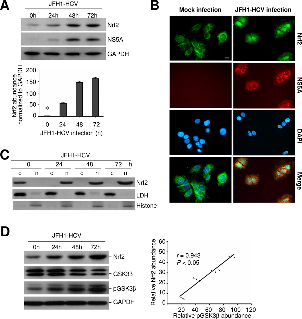
Figure 1. HCV infection induces Nrf2 antioxidant response, characterized by nuclear accumulation of Nrf2 in human hepatic cells, associated with inhibitory phosphorylation of GSK3β.
Huh7.5.1 cells were mock infected or infected with JFH-1 at a multiplicity of infection (m.o.i.) of 0.5. (A) At different time points, cellular lysates were prepared and analyzed by western immunoblot (WB) for Nrf2, NS5A and GAPDH; Relative abundance of Nrf2 normalized by GAPDH was determined by densitometric analysis of immunoblots; *P<0.05 versus all other groups, (n=3); (B) Fluorescent immunocytochemistry staining for Nrf2 (green signals) and NS5A (red signals) in mock infected or JFH-1 infected Huh7.5.1 cells, which were counterstained with DAPI (blue signals); Bar = 10 μm. (C) Cytoplasmic(c) and nuclear(n) protein fractions were prepared from JFH-1 infected cells and subjected to immunoblot analysis for Nrf2 or for nuclear protein histone and cytoplasmic protein lactate dehydrogenase (LDH), which served as quality control respectively for cytoplasmic and nuclear protein extraction. (D) Cell lysates were analyzed by immunoblot analysis for Nrf2, GSK3β, p-GSK3β (Ser9) and GAPDH; Relative abundance of Nrf2 and pGSK3β normalized by GAPDH and GSK3β respectively was determined by densitometric analysis and linear regression analysis indicates that inhibitory phosphorylation of GSK3β positively correlates with the expression of Nrf2 (P < 0.05, n=3).

