Abstract
Introduction
Studies employing serological, DTH or conventional PCR techniques suggest a vast proportion of Leishmania infected individuals living in regions endemic for Visceral Leishmaniasis (VL) remain asymptomatic. This study was designed to assess whether quantitative PCR (qPCR) can be used for detection of asymptomatic or early Leishmania donovani infection and as a predictor of progression to symptomatic disease.
Methods
The study included 1469 healthy individuals living in endemic region (EHC) including both serology-positive and -negative subjects. TaqMan based qPCR assay was done on peripheral blood of each subject using kDNA specific primers and probes.
Results
A large proportion of EHC 511/1469 (34.78%) showed qPCR positivity and 56 (3.81% of 1469 subjects) had more than 1 calculated parasite genome/ml of blood. However, the number of individuals with parasite load above 5 genomes/ml was only 20 (1.36% of 1469). There was poor agreement between serological testing and qPCR (k = 0.1303), and 42.89% and 31.83% EHC were qPCR positive in seropositive and seronegative groups, respectively. Ten subjects had developed to symptomatic VL after 12 month of their follow up examination, of which eight were initially positive according to qPCR and among these, five had high parasite load.
Discussion
Thus, qPCR can help us to detect significant early parasitaemia, thereby assisting us in recognition of potential progressors to clinical disease. This test could facilitate early intervention, decreased morbidity and mortality, and possibly interruption of disease transmission.
Author Summary
Anthroponotic VL caused by Leishmania donovani in the Indian subcontinent accounts for 70% of the world burden of VL. Among the estimated 100,000 cases of VL acquired annually in India, 90% occur in the state of Bihar. Leishmania infection can result in either symptomatic or asymptomatic infection. L. donovani infection can also manifest as post-kala azar dermal leishmaniasis, a chronic cutaneous form thought to provide the reservoir for anthroponotic transmission of VL in regions endemic for this parasite species. We hypothesized that, in areas endemic for L. donovani, asymptomatic infections might also play a crucial role in disease transmission. This study describes use of quantitative PCR (qPCR) to determine the infection status in individuals living in an endemic region of India. We hypothesized that parasite load estimation by qPCR of peripheral blood cells among healthy individuals living in the endemic region might reveal the true frequency of infections through direct evidence of parasitemia. We reasoned this test would detect both asymptomatic non-progressors as well as asymptomatic individuals who will progress to fully symptomatic VL. Serologic testing by ELISA or DAT showed poor agreement with molecular detection of parasite DNA by qPCR, suggesting the tests differentiate between infection and immune response. Amongst ten healthy individuals who progressed to VL, only six were serologically positive whereas eight were initially qPCR positive, among whom five had high parasite loads in their blood. Thus, deployment of qPCR technique to estimate the presence and level of parasitemia in healthy individuals from Leishmania endemic regions may contribute to early case detection, thereby reducing morbidity and mortality. Consistent with the goals of the VL control and elimination program, this early intervention approach could help interrupt disease transmission.
Introduction
The Leishmania spp. parasites of humans are endemic in 98 countries, and more than 350 million people are at risk of infection [1]. Leishmaniasis is a neglected tropical disease, and the most severe form visceral leishmaniasis (VL, also known as kala-azar) is fatal if untreated. VL is primarily an anthroponotic infection caused by Leishmania donovani in India, transmitted by the sand fly vector Phelobotomus argentipes [2], [3].The state of Bihar in India accounts for 90% of cases in the country [4]. A majority of infected individuals do not develop clinical illness [5], [6], [7]. According to a serology-based epidemiological survey, the prevalence of asymptomatic Leishmania donovani infection in Bihar is 110 per 1,000 persons, and the rate of progression to symptomatic VL is 17.85 per 1,000 persons [8]. The kinetics of parasite amplification during the progression from infection to disease is as yet uncharacterized. We have recently shown that a highly quantitative qPCR test of blood can track the decrease in parasite load during successful treatment of infection [9]. The current study was based on the hypothesis that the number or the kinetics of circulating parasites in asymptomatically infected individuals, as measured by qPCR, might provide the most sensitive early indicator of infected subjects apt to progress to full blown disease.
Alternate techniques to detect parasites in persons with VL include direct histological examination and/or culture of bone marrow and splenic aspirates. However these methods are not feasible for screening methods or epidemiological research due to their invasive nature. Serological methods are simple, non-invasive means of detecting specific antibodies, but it is already shown that there is a lack of correlation between serology and nucleic acid methods for parasite detection [10], [11], [12]. This could reflect the inability of serology to distinguish past from ongoing infection, and therefore might result in overestimation of the number of infected asymptomatic individuals.
A large proportion of infected individuals are reportedly asymptomatic according to both serology and PCR surveys in India and nearby endemic countries [13]. Recent epidemiological reports from Brazil, Spain, and France have shown that detectable parasite DNA is present in the blood of asymptomatic infected individuals [14], [15], [16]. qPCR based epidemiological studies in the Mediterranean region have described a threshold and reference value for asymptomatic infection [17]. A similar study from our population in Bihar suggested the equivalent of 5 L. donovani parasite genomes detected/ml of blood is the threshold for clinical symptoms of VL to occur [18]. Data from our prior work in India suggest that serologic status is not a good predictor of conversion to symptomatic VL. Indeed, only 3.48% of seropositive individuals converted to active VL, whereas the conversion rate was 2.57% among seronegative individuals from the same endemic region [19]. We therefore investigated the potential for molecular quantification of parasite genome equivalents in blood as a more sensitive measure of asymptomatic infection likely to progress to disease.
Early case detection and treatment are the most important control measures for Leishmaniasis. Thus, the inability to identify individuals with asymptomatic infection, and among these to discern the individuals that are likely to progress to disease, presents a problem for clinical management. In response to this need, the current study constitutes a comparison of qPCR, serological testing with direct agglutination test (DAT), and the rK39 ELISA as predictors of progression from asymptomatic infection to fully symptomatic VL. We performed this study in a population of individuals living in the highly endemic Muzaffarpur region of the state of Bihar, India.
Materials and Methods
Study site and sample collection
The work was carried out in the Department of Medicine, Banaras Hindu University, Varanasi and at its field site Kala-Azar Medical Research Centre, Muzaffurpur, Bihar and villages of Muzaffarpur district. The study was approved by the Ethics Committee of the Institute of Medical Sciences, Banaras Hindu University, the University of Iowa and the National Institutes of Health. The IRB at Banaras Hindu University is registered with the US NIH. Written informed consent was obtained from each participating individual.
Sample collection
The study was carried out in villages of Muzaffarpur district, which is endemic for VL. To identify individuals who had recently seroconverted, an epidemiological sero-survey was performed for two consecutive years (2009 to 2012). Villages from which large numbers of VL cases originated were identified from hospital records at Kala Azar Medical Research Centre. The research team enrolled all consenting adults age 18 and above in these villages. In the first survey, serology was done using DAT and rK39 ELISA from figure prick blood collected on filter paper. All individuals who were seronegative on the first survey were selected for testing for seroconversion by DAT and rK39 during the second serosurvey conducted 12 months later. To extract blood leukocyte DNA, two ml of blood were collected in the citrate-containing tubes from 401 recent seroconverters (seropositive) as well as 1068 randomly selected seronegative individuals within 15 days of serologic test. Buffy coat cells were isolated, and were transported from Muzaffarpur on ice to the central laboratory in Varanasi and stored at −20°C until use. 36 nonendemic healthy person's blood were also taken for qPCR assay.
Serological test (DAT/ELISA) and molecular test (qPCR)
Sera were eluted from filter papers containing finger prick blood and used to perform serology by DAT and rK39 ELISA as described previously [20], [21]. Individuals who are either DAT or rK39 ELISA positive were considered seropositive.
DNA was extracted using the QIAamp DNA mini kit (Qiagen, Hilden Germany) as per the manufacturer's instructions Only those DNA samples that had an optical density (OD) 260/280 ratio of 1.8–2.0 and an OD 260/230 ratio >1.5 by spectrophotometer measurements (ND-2000 spectrophotometer; Thermo Scientific, Waltham, MA, USA) were taken for qPCR experiments. The TaqMan based qPCR assay was performed in a final volume of 10 µL containing 5 µl TaqMan master mixture (2×) [Applied Biosystems (ABI), Carlsbad, CA, USA], 4 µl of DNA template and 0.25 µl (5 µM) of forward and reverse primer and 0.375 µl of probe (Integrated DNA Technologies, Coralville, IA, USA) (Table 1.). Primer-probe sequences are listed in Table 1. Amplification was conducted in a 7500 Real-Time PCR system [Applied Biosystems (ABI), Carlsbad, CA, USA]. The standard curve method for absolute quantification of parasite numbers was used as described previously [9]. All assays included no-DNA template controls, as well DNA from a negative control unexposed healthy subject. Cutoff values to consider a test positive were Cyclic threshold (Ct) value of 39. According to the standard curve, 0.001 parasite genome equivalents in the well corresponded to a CT value of 39.
Table 1. Taqman primers and probe for detection of Leishmania DNA in endemic healthy controls.
| Primers | Probe | |
| kDNA4 forward | GGGTGCAGAAATCCCGTTCA | ACCCCCAGTTTCCCGCCCCG |
| kDNA4 reverse | CCCGGCCCTATTTTACACCA |
Sequences correspond to the kDNA4 minicircle DNA primers and probe [21].
Statistical analysis
Data analysis was done by non parametric Mann-Whitney test using SPSS 16 (IBM, Somers, NY, USA) and Prism (Graph pad software).
Results
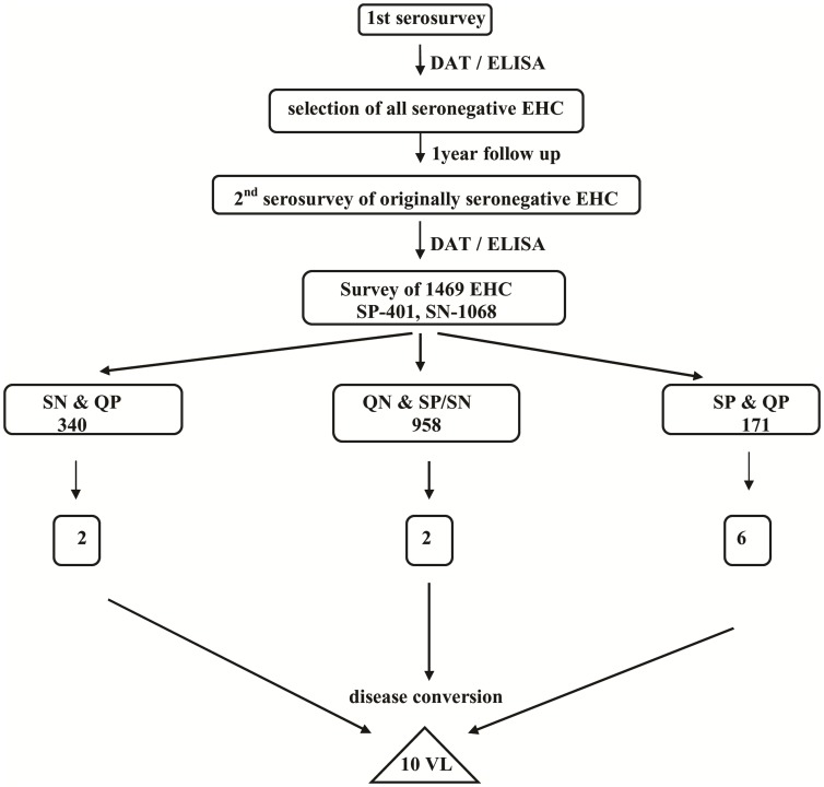
A flow chart of the progression of results is shown in Fig. 1. Serological results were interpreted in light of our original description of the first serosurvey. Cutoff values for positive serology were chosen considering results from study of negative control unexposed Indian subjects, positive control subjects with acute or successfully treated VL, and recommendations from the serological test manufacturers [20]. Considering conversion of either the DAT or the rK39 ELISA as a seroloconversion, 401 subjects converted from seronegative to seropositive between the first and the second serosurvey, whereas the remaining 1068 individuals remained seronegative.
Figure 1. Flow chart of study of serology in endemic healthy control individuals (EHC) from baseline serosurvey (year 1) to identification of progressors.
SP – seropositive; SN – seronegative; QP – qPCR positive (CT cutoff 39); QN – qPCR negative; VL – progressors to symptomatic visceral leishmaniasis.
Quantitative PCR of DNA extracted from circulating blood cells was used to assess the proportions of individuals from the endemic region with evidence of asymptomatic parasitemia, and the correlation with serological conversion. The kDNA4 probe set and taqman assay was chosen from our previously published diagnostic criteria, because of the efficient amplification of L. donovani sequences and the lack of primer-dimers complicating quantification of low numbers of parasites [22]. Data were carefully controlled, and results of individual qPCR runs were only accepted when there was a lack of kDNA amplification in no-DNA and negative controls. A standard curve was run with each assay, using the same stock of promastigote DNA extracted from an Indian isolate, to ensure consistency between assays. Data were expressed as “genome equivalents” compared to this uniform DNA standard. Notably, more than four “genome” per ml were present in individuals with symptomatic VL according to our prior publication [9].
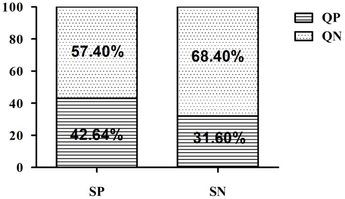
Among a total of 1469 healthy individuals living in the endemic villages, 511 (34.78%) were positive by qPCR for amplification of any parasite DNA (CT less than 39). 171/401 (42.8%) were from seropositive group and the remaining 340/1068 (31.6%) were seronegative (Fig. 2, Table 2). The median value of parasite genomes/ml of blood was less than one and found to be 0.11 and 0.15 in seropositive and seronegative group respectively. Ten individuals who were initially belong to both seropositive and seronegative category progressed to symptomatic VL by the time of the follow up. Six (60%) were both qPCR and serology positive, two (20%) were qPCR positive but seronegative, whereas two (20%) were both qPCR and serologically negative (Table 3). Among these qPCR positive progressor five of the progessors had parasitemia levels equal to or more than the threshold value for ocuurence of symptomatic VL due to L.donovani.
Figure 2. Bar graph to show Leishmania detected by qPCR in different group of EHC on the basis of their serological status (QP-qPCR positive, QN-qPCR negative, SP-sero positive, SN-sero negative).

Table 2. Parasitemia range in blood buffy coat samples from healthy controls from endemic or nonendemic regions.
| Range of parasite genome equivalents in blood cells from individuals with non-zero results of parasite qPCR test | ||||||||
| positive | negative | >0–<1 | 1–5 | 5–10 | 11–100 | 101–1000 | >1000 | |
| Seropositive EHC (n = 401) | 171 (42.6%) | 230 | 149 (1 *) | 16 (1 *) | 2 (1*) | 1 (1 *) | 3 (2 *) | 0 |
| SeronegativeEHC (n = 1068) | 340 (31.8%) | 728 (2*) | 306 (1 *) | 24 | 5 (1*) | 4 | 1 | 0 |
| Seronegative NEHC (n = 36) | zero | all | - | - | - | - | - | - |
(*converted into VL).
Table 3. Pogressors (disease conversion, VL cases) details: Parasite load and serostatus of EHC who were healthy at the first survey, but developed symptomatic VL at the time of the 12-month follow up.
| S.No. | disease conversion time (after blood collection) | qPCR (genome equivalents/mlof blood) | Sero statuspos/neg |
| 1 | after one month | 10.5 | Pos |
| 2 | with in one month | 6.6 | Pos |
| 3 | after five months | 97 | Pos |
| 4 | after six months | 0.38 | Pos |
| 5 | with in one month | 754.73 | Pos |
| 6 | with in one month | 146.32 | Pos |
| 7 | after three months | 5.09 | Neg |
| 8 | after three months | 0.613 | Neg |
| 9 | after five months | 0 | Neg |
| 10 | after two months | 0 | Neg |
(pos - positive on either serologic test; neg – negative on both serologic tests).
The non-randomness of qPCR results is illustrated by the fact that all noendemic healthy were negative for qPCR.
Discussion
In this study Leishmania DNA was detected in large proportions of both seropositive and seronegative endemic healthy groups (Fig. 1). In contrast, to nonendemic healthy who were negative for the test. Similar findings were reported in a study of L. infantum infection, a cause of VL in the Mediterranean and in Latin America [17]. In one of our earlier study we showed that the parasite load in individuals with acute symptomatic VL due to L. donovani was at least 20, and at day 30 of treatment was >1.12 genome equivalents/ml [9]. In other study we found 5 parasite genome/ml of blood as the threshold value to differentiate asymptomatic from symptomatic [18]. Mary et al. cited a persistent level of more than 1 parasite/ml as a risk for relapse of L. infantum disease [17]. Our ability to quantify the parasite load in asymptomatic individuals led us to examine a potential threshold for progression to active infection in previously uninfected individuals.
A positive serologic test for L. donovani in individuals living in endemic regions who have no symptoms of VL could indicate prior exposure without substantial active infection, ongoing asymptomatic infection which will not lead to disease, or early infection that will progress. In this situation it would be extremely valuable to perform additional diagnostic testing that could be used as a marker of infection, and also be capable of differentiating those likely to progress from those at low risk for progression to disease. Given our results suggesting the magnitude of parasitemia is related to the risk of disease, a quantitative test such as the qPCR reported herein represents a candidate test for this distinction.
Both our study and the reported study of L. infantum parasitemia cite very low numbers of parasite genome equivalents in the blood as indicative of infection. It is important to appreciate the distinction between the calculated number of parasite genomes is not equivalent to the actual number of parasites in a ml of drawn blood. We previously reported that the number of kDNA copies varies between amastigotes and promastigotes, and that copy number is highly variable between strains of the same parasite species [22]. Given this variability as well as the fact that there is an anticipated loss of DNA in the extraction process itself, one can assume that the numbers of genomes quantified on a standard curve will be relatively quantitative compared to comparison samples treated in the same manner. However one cannot draw conclusions about the absolute numbers of parasites present in the subject based on these relative numbers. It is nonetheless important to use a standard DNA so as to obtain as equivalent quantitative measures between assays as possible.
A study of asymptomatic L. donovani infection in Nepal cited poor agreement between serological and molecular tests, i.e. DAT and routine PCR [5]. Our study similarly showed a lack of agreement between serology and qPCR (k = 0.1303). Herein 42.8% or 31.6% of healthy subjects from the endemic neighborhood who were seropositive or seronegative, respectively, contained Leishmania specific DNA in their blood (Fig. 2.). Potential reasons that seropositive individuals might become qPCR negative could include degradation and clearance of Leishmania DNA after infection, corresponding with development of protective immunity. A positive qPCR test in seronegative individuals could occur if the individual was bitten by a Leishmania infected sand fly, but either immunity has not yet developed or antibody levels are too low to be detectable by the methods employed. Analogous to infection with hepatitis B, it is possible that parasite DNA, detected by PCR of peripheral blood, could be the first marker of the infection prior to antibody seroconversion. Consistent with this hypothesis, during canine VL, kDNA-PCR is significantly more sensitive than the other parasitological and serological methods, allowing the identification of infected dogs even before the appearance of antibodies [23].
Quantification on the standard curve revealed that among qPCR positives, 56 subjects (10.95% of total qPCR positive) had more than one parasite genome/ml of blood, and among them 20 (3.91%) had five or more parasites (Table 2.). Although progression to disease occurred both in seropositive and seronegative groups, 8/10 (80%) of those converting to clinical VL were qPCR positive and 5/10 (50%) had relatively high parasite loads. This suggests that asymptomatic individuals who have high parasite load may be more likely to progress to disease than individuals whose parasite loads are low (Table 3.). Other reports of asymptomatic infection suggest that parasite DNA does not often persist for more than one year, but that rarely detectable asymptomatic infection may last for decades [24]. Further their follow up is necessary to know their conversion into symptomatic cases or they remain asymptomatic.
Our recent serological study from same population area shows there is an increased risk of progressing to disease among individuals with high titers of DAT or rk39 serology [21]. Although our study suggested that DAT/ELISA titers are less sensitive and specific than qPCR with high parasite load for detection of progressors, neither approach was perfect. It may be that a combination of qPCR to detect the presence and quantity of parasite nucleic acid, coupled with serology to identify individuals with very high titers, may be a practical and sensitive means of detecting infection, for use in early case detection. The qPCR measure serves as well as an effective tool to monitor clinical management. Early case detection and treatment are the most important control measures for leishmaniasis. In anthroponotic leishmaniasis in which humans are the only reservoir, early detection by qPCR should also be explored as a means of identifying individuals who might also pose a reservoir for disease transmission.
Limitations of qPCR include high initial investment, relatively higher cost per test compared to serology. Requirement of skilled personnel can be another limiting factor, however, if completely equipped and manned central laboratories are established at strategic locations to cater to one or several districts, a reliable diagnosis can be provided to population living in endemic regions for VL which will give more possibility of identification of symptomatic condition of VL disease in infected persons.
Supporting Information
STROBE Checklist.
(DOC)
Data Availability
The authors confirm that all data underlying the findings are fully available without restriction. All relevant data are within the paper and its Supporting Information files.
Funding Statement
The study was funded in part by National Institute of Allergy and Infectious disease (NIAID), DMID funding mechanism: Tropical Medicine Research Center Grant number: P50AI074321. Partial funding was also derived from NIH grant R01 AI076233 (MEW). Authors Medhavi Sudarshan and Bhawana Singh got financial support from Council of Scientific and Industrial Research (CSIR), New Delhi, India. Toolika Singh and Abhishek Singh got financial support from Indian Council of Medical Research(ICMR), New Delhi, India. The funders had no role in study design, data collection and analysis, decision to publish, or preparation of the manuscript.
References
- 1. Alvar J, Velez ID, Bern C, Herrero M, Desjeux P, et al. (2012) Leishmaniasis worldwide and global estimates of its incidence. PLoS One 7: e35671. [DOI] [PMC free article] [PubMed] [Google Scholar]
- 2. Swaminath CS, Shortt HE, Anderson LA (2006) Transmission of Indian kala-azar to man by the bites of Phlebotomus argentipes, ann and brun. 1942. Indian J Med Res 123: 473–477. [PubMed] [Google Scholar]
- 3. Dinesh DS, Kar SK, Kishore K, Palit A, Verma N, et al. (2000) Screening sandflies for natural infection with Leishmania donovani, using a non-radioactive probe based on the total DNA of the parasite. Ann Trop Med Parasitol 94: 447–451. [DOI] [PubMed] [Google Scholar]
- 4. Sundar S, Agrawal G, Rai M, Makharia MK, Murray HW (2001) Treatment of Indian visceral leishmaniasis with single or daily infusions of low dose liposomal amphotericin B: randomised trial. BMJ 323: 419–422. [DOI] [PMC free article] [PubMed] [Google Scholar]
- 5. Ostyn B, Gidwani K, Khanal B, Picado A, Chappuis F, et al. (2011) Incidence of symptomatic and asymptomatic Leishmania donovani infections in high-endemic foci in India and Nepal: a prospective study. PLoS Negl Trop Dis 5: e1284. [DOI] [PMC free article] [PubMed] [Google Scholar]
- 6. Das VN, Siddiqui NA, Verma RB, Topno RK, Singh D, et al. (2011) Asymptomatic infection of visceral leishmaniasis in hyperendemic areas of Vaishali district, Bihar, India: a challenge to kala-azar elimination programmes. Trans R Soc Trop Med Hyg 105: 661–666. [DOI] [PubMed] [Google Scholar]
- 7. Stauch A, Sarkar RR, Picado A, Ostyn B, Sundar S, et al. (2011) Visceral leishmaniasis in the Indian subcontinent: modelling epidemiology and control. PLoS Negl Trop Dis 5: e1405. [DOI] [PMC free article] [PubMed] [Google Scholar]
- 8. Topno RK, Das VN, Ranjan A, Pandey K, Singh D, et al. (2010) Asymptomatic infection with visceral leishmaniasis in a disease-endemic area in bihar, India. Am J Trop Med Hyg 83: 502–506. [DOI] [PMC free article] [PubMed] [Google Scholar]
- 9. Sudarshan M, Weirather JL, Wilson ME, Sundar S (2011) Study of parasite kinetics with antileishmanial drugs using real-time quantitative PCR in Indian visceral leishmaniasis. J Antimicrob Chemother 66: 1751–1755. [DOI] [PMC free article] [PubMed] [Google Scholar]
- 10. Badaro R, Reed SG, Carvalho EM (1983) Immunofluorescent antibody test in American visceral leishmaniasis: sensitivity and specificity of different morphological forms of two Leishmania species. Am J Trop Med Hyg 32: 480–484. [DOI] [PubMed] [Google Scholar]
- 11. Mary C, Lamouroux D, Dunan S, Quilici M (1992) Western blot analysis of antibodies to Leishmania infantum antigens: potential of the 14-kD and 16-kD antigens for diagnosis and epidemiologic purposes. Am J Trop Med Hyg 47: 764–771. [DOI] [PubMed] [Google Scholar]
- 12. Bhattarai NR, Van der Auwera G, Khanal B, De Doncker S, Rijal S, et al. (2009) PCR and direct agglutination as Leishmania infection markers among healthy Nepalese subjects living in areas endemic for Kala-Azar. Trop Med Int Health 14: 404–411. [DOI] [PubMed] [Google Scholar]
- 13. Srivastava P, Gidwani K, Picado A, Van der Auwera G, Tiwary P, et al. (2013) Molecular and serological markers of Leishmania donovani infection in healthy individuals from endemic areas of Bihar, India. Trop Med Int Health 18: 548–554. [DOI] [PubMed] [Google Scholar]
- 14. Badaro R, Jones TC, Carvalho EM, Sampaio D, Reed SG, et al. (1986) New perspectives on a subclinical form of visceral leishmaniasis. J Infect Dis 154: 1003–1011. [DOI] [PubMed] [Google Scholar]
- 15. Moral L, Rubio EM, Moya M (2002) A leishmanin skin test survey in the human population of l'Alacanti region (Spain): implications for the epidemiology of Leishmania infantum infection in southern Europe. Trans R Soc Trop Med Hyg 96: 129–132. [DOI] [PubMed] [Google Scholar]
- 16. Biglino A, Bolla C, Concialdi E, Trisciuoglio A, Romano A, et al. (2010) Asymptomatic Leishmania infantum infection in an area of northwestern Italy (Piedmont region) where such infections are traditionally nonendemic. J Clin Microbiol 48: 131–136. [DOI] [PMC free article] [PubMed] [Google Scholar]
- 17. Mary C, Faraut F, Drogoul MP, Xeridat B, Schleinitz N, et al. (2006) Reference values for Leishmania infantum parasitemia in different clinical presentations: quantitative polymerase chain reaction for therapeutic monitoring and patient follow-up. Am J Trop Med Hyg 75: 858–863. [PubMed] [Google Scholar]
- 18. Sudarshan M, Sundar S (2014) Parasite load estimation by qPCR differentiates between asymptomatic and symptomatic infection in Indian Visceral Leishmaniasis. Diagn Microbiol Infect Dis. 80: 40–2. [DOI] [PMC free article] [PubMed] [Google Scholar]
- 19. Gidwani K, Kumar R, Rai M, Sundar S (2009) Longitudinal seroepidemiologic study of visceral leishmaniasis in hyperendemic regions of Bihar, India. Am J Trop Med Hyg 80: 345–346. [PubMed] [Google Scholar]
- 20. Hasker E, Kansal S, Malaviya P, Gidwani K, Picado A, et al. (2013) Latent infection with Leishmania donovani in highly endemic villages in Bihar, India. PLoS Negl Trop Dis 7: e2053. [DOI] [PMC free article] [PubMed] [Google Scholar]
- 21. Hasker E, Malaviya P, Gidwani K, Picado A, Ostyn B, et al. (2014) Strong Association between Serological Status and Probability of Progression to Clinical Visceral Leishmaniasis in Prospective Cohort Studies in India and Nepal. PLoS Negl Trop Dis 8: e2657. [DOI] [PMC free article] [PubMed] [Google Scholar]
- 22. Weirather JL, Jeronimo SM, Gautam S, Sundar S, Kang M, et al. (2011) Serial quantitative PCR assay for detection, species discrimination, and quantification of Leishmania spp. in human samples. J Clin Microbiol 49: 3892–3904. [DOI] [PMC free article] [PubMed] [Google Scholar]
- 23. Fallah E, Khanmohammadi M, Rahbari S, Farshchian M, Farajnia S, et al. (2011) Serological survey and comparison of two polymerase chain reaction (PCR) assays with enzyme-linked immunosorbent assay (ELISA) for the diagnosis of canine visceral leishmaniasis in dogs. African Journal of Biotechnology 10: 648–656. [Google Scholar]
- 24. Guevara P, Ramirez JL, Rojas E, Scorza JV, Gonzalez N, et al. (1993) Leishmania braziliensis in blood 30 years after cure. Lancet 341: 1341. [DOI] [PubMed] [Google Scholar]
Associated Data
This section collects any data citations, data availability statements, or supplementary materials included in this article.
Supplementary Materials
STROBE Checklist.
(DOC)
Data Availability Statement
The authors confirm that all data underlying the findings are fully available without restriction. All relevant data are within the paper and its Supporting Information files.


