

Abstract
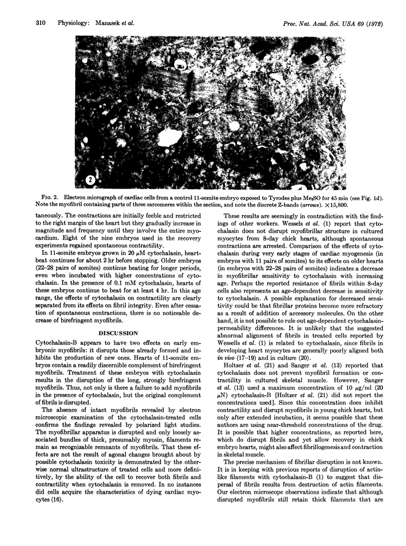
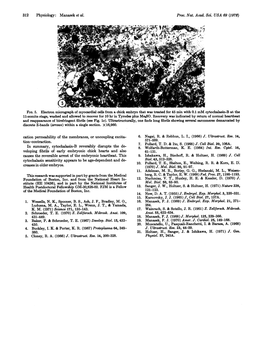
Developing cardiac muscle cells of 11- to 13-somite chick embryos are sensitive to cytochalasin-B. In cultured chick embryos, ranging in development from 11 to 13 somites, hearts stop beating in the presence of this agent. Both polarized light and electron microscopic examination show that cytochalasin-B disrupts existing myofibrils and inhibits the formation of new ones. Discrete Z-bands are not present in treated heart cells and thick, presumably myosin, filaments are found in disarray. These effects are reversible; after cytochalasin-B is removed from the medium, heartbeat recovers and myofibrils with discrete Z-bands reappear. Fibrillar sensitivity appears to be a function of age since fibrils in hearts of embryos having from 22 to 28 pairs of somites are more resistant.
Keywords: electron microscopy, polarized light, Z-bands, heartbeat
Full text
PDF


Images in this article


Selected References
These references are in PubMed. This may not be the complete list of references from this article.
- Adelman M. R., Borisy G. G., Shelanski M. L., Weisenberg R. C., Taylor E. W. Cytoplasmic filaments and tubules. Fed Proc. 1968 Sep-Oct;27(5):1186–1193. [PubMed] [Google Scholar]
- Baker P. C., Schroeder T. E. Cytoplasmic filaments and morphogenetic movement in the amphibian neural tube. Dev Biol. 1967 May;15(5):432–450. doi: 10.1016/0012-1606(67)90036-x. [DOI] [PubMed] [Google Scholar]
- Buckley I. K., Porter K. R. Cytoplasmic fibrils in living cultured cells. A light and electron microscope study. Protoplasma. 1967;64(4):349–380. doi: 10.1007/BF01666538. [DOI] [PubMed] [Google Scholar]
- Cloney R. A. Cytoplasmic filaments and cell movements: epidermal cells during ascidian metamorphosis. J Ultrastruct Res. 1966 Feb;14(3):300–328. doi: 10.1016/s0022-5320(66)80051-5. [DOI] [PubMed] [Google Scholar]
- Ishikawa H., Bischoff R., Holtzer H. Formation of arrowhead complexes with heavy meromyosin in a variety of cell types. J Cell Biol. 1969 Nov;43(2):312–328. [PMC free article] [PubMed] [Google Scholar]
- Manasek F. J. Embryonic development of the heart. I. A light and electron microscopic study of myocardial development in the early chick embryo. J Morphol. 1968 Jul;125(3):329–365. doi: 10.1002/jmor.1051250306. [DOI] [PubMed] [Google Scholar]
- Manasek F. J. Histogenesis of the embryonic myocardium. Am J Cardiol. 1970 Feb;25(2):149–168. doi: 10.1016/0002-9149(70)90576-x. [DOI] [PubMed] [Google Scholar]
- Manasek F. J. Myocardial cell death in the embryonic chick ventricle. J Embryol Exp Morphol. 1969 Apr;21(2):271–284. [PubMed] [Google Scholar]
- Muscatello U., Pasquali-Ronchetti I., Barasa A. An electron microscope study of myoblasts from chick embryo heart cultured in vitro. J Ultrastruct Res. 1968 Apr;23(1):44–59. doi: 10.1016/s0022-5320(68)80030-9. [DOI] [PubMed] [Google Scholar]
- Nachmias V. T., Huxley H. E. Electron microscope observations on actomyosin and actin preparations from Physarum polycephalum, and on their interaction with heavy meromyosin subfragment I from muscle myosin. J Mol Biol. 1970 May 28;50(1):83–90. doi: 10.1016/0022-2836(70)90105-1. [DOI] [PubMed] [Google Scholar]
- Nagai R., Rebhun L. I. Cytoplasmic microfilaments in streaming Nitella cells. J Ultrastruct Res. 1966 Mar;14(5):571–589. doi: 10.1016/s0022-5320(66)80083-7. [DOI] [PubMed] [Google Scholar]
- Pollard T. D., Shelton E., Weihing R. R., Korn E. D. Ultrastructural characterization of F-actin isolated from Acanthamoeba castellanii and identification of cytoplasmic filaments as F-actin by reaction with rabbit heavy meromyosin. J Mol Biol. 1970 May 28;50(1):91–97. doi: 10.1016/0022-2836(70)90106-3. [DOI] [PubMed] [Google Scholar]
- Sanger J. W., Holtzer S., Holtzer H. Effects of cytochalasin B on muscle cells in tissue culture. Nat New Biol. 1971 Jan 27;229(4):121–123. doi: 10.1038/newbio229121a0. [DOI] [PubMed] [Google Scholar]
- Schroeder T. E. The contractile ring. I. Fine structure of dividing mammalian (HeLa) cells and the effects of cytochalasin B. Z Zellforsch Mikrosk Anat. 1970;109(4):431–449. [PubMed] [Google Scholar]
- WAINRACH S., SOTELO J. R. Electron microscope study of the developing chick embryo heart. Z Zellforsch Mikrosk Anat. 1961;55:622–634. doi: 10.1007/BF00384502. [DOI] [PubMed] [Google Scholar]
- Wessells N. K., Spooner B. S., Ash J. F., Bradley M. O., Luduena M. A., Taylor E. L., Wrenn J. T., Yamada K. Microfilaments in cellular and developmental processes. Science. 1971 Jan 15;171(3967):135–143. doi: 10.1126/science.171.3967.135. [DOI] [PubMed] [Google Scholar]
- Wohlfarth-Bottermann K. E. Cell structures and their significance for ameboid movement. Int Rev Cytol. 1964;16:61–131. doi: 10.1016/s0074-7696(08)60294-6. [DOI] [PubMed] [Google Scholar]