

Abstract
Female breast tissue is composed of variable proportions of fat and fibroglandular tissue, and in general, an increased ratio of fibroglandular tissue to fat corresponds to increased mammographic density. Studies suggest that mammographic density is an independent risk factor for breast cancer, and the sensitivity of mammography can be lower with heterogeneously dense or extremely dense breasts. Nineteen states have legal statutes requiring that patients be notified if they have dense breasts, including the state of Texas. Henda's law, mandated on January 1, 2012 in Texas, suggests that patients with dense breasts could benefit from additional screening tests such as breast magnetic resonance imaging (MRI). Our study examined the impact of Henda's law by comparing the number of screening breast MRIs performed for dense breasts before and after the law's implementation. Results showed a 23-fold increase in the number of dense breast MRIs in the 2 years that this new legislation was in effect. This increase could have substantial implications for the health care economy, and further studies are needed to determine the cost-effectiveness of this additional screening tool.
Female breast tissue is composed of varying amounts of fat and fibroglandular tissue. The fibroglandular tissue appears white or “dense” on a mammogram, while fat appears dark (1). The proportions of white to dark are described as “mammographic density” (2). Although there can be some interobserver subjectivity, overall breast composition is categorized as either fatty replaced (0–25% fibroglandular tissue), scattered fibroglandular densities (25%–50% fibroglandular tissue), heterogeneously dense (50%–75% fibroglandular tissue), or extremely dense (>75% fibroglandular tissue). Studies suggest that the extent and percentage of mammographic density are independent risk factors for breast cancer (2–4), and women with extremely dense breasts have a cancer risk that is 1.8 to 6 times higher than that of women with the least amount of fibroglandular tissue (5). The sensitivity of mammography can be lower as mammographic density increases, as dense breasts could potentially obscure underlying abnormalities. To date, 19 states have passed legal statutes requiring that patients be notified if they have dense breasts, and efforts are underway to introduce legislation at the federal level. Texas' legislation, known as Henda's law, requires mammography facilities as of January 1, 2012, to notify a patient and her health care provider that the patient demonstrates dense breast tissue and might benefit from supplemental screening tests such as breast magnetic resonance imaging (MRI). Our study evaluated the trend in the number of screening MRIs performed for dense breasts at Baylor University Medical Center at Dallas in 2011 (before Henda's law) and in 2012 and 2013 (after the law's implementation).
METHODS
To determine the overall trend in the number of MRIs performed for the purpose of evaluating dense breast tissue, all breast MRIs between 2011 and 2013 were examined at Baylor University Medical Center at Dallas and were divided into three categories: 1) total number of breast MRIs; 2) total number of screening breast MRIs (defined as studies of asymptomatic patients with no personal history of breast cancer); and 3) total number of screening breast MRIs ordered for the evaluation of dense breasts (excluding high-risk patients with a strong family history of breast cancer or who were known to be BRCA positive).
RESULTS
In 2011 (pre-Henda's law), a total of 255 breast MRIs were performed, 32 of which were considered to be screening breast MRIs. Only 2 of the patients with screening breast MRIs received MRIs for evaluation of dense breasts (Table). After the implementation of Henda's law in 2012, not only was there an increase in the total number of breast MRIs, but there was also an increase in the number of screening MRIs performed for evaluation of dense breasts (Table). There was a continued increase in the number of screening breast MRIs performed in 2013, with 46 MRIs performed for the evaluation of dense breasts out of 78 total screening breast MRIs (Table). This represents a 23-fold increase in the number of MRIs ordered for dense breasts when compared to the pre-Henda's law data from 2011 (Figure).
Table.
Breast MRI data at Baylor University Medical Center at Dallas before and after implementation of Henda's law
| Month | Total breast MRIs | Screening breast MRIs | Dense breast MRIs |
|---|---|---|---|
| 2011: Before implementation | |||
| Jan | 19 | 2 | 0 |
| Feb | 12 | 1 | 0 |
| Mar | 25 | 0 | 0 |
| Apr | 13 | 3 | 0 |
| May | 24 | 4 | 0 |
| Jun | 20 | 1 | 0 |
| Jul | 13 | 3 | 1 |
| Aug | 31 | 4 | 0 |
| Sep | 23 | 3 | 0 |
| Oct | 26 | 2 | 0 |
| Nov | 31 | 3 | 0 |
| Dec | 18 | 6 | 1 |
| Total | 255 | 32 | 2 |
| 2012: After implementation | |||
| Jan | 19 | 4 | 1 |
| Feb | 25 | 5 | 0 |
| Mar | 27 | 2 | 0 |
| Apr | 37 | 9 | 0 |
| May | 35 | 10 | 3 |
| Jun | 39 | 4 | 0 |
| Jul | 34 | 6 | 2 |
| Aug | 30 | 2 | 1 |
| Sep | 18 | 4 | 0 |
| Oct | 31 | 6 | 1 |
| Nov | 31 | 9 | 1 |
| Dec | 34 | 4 | 0 |
| Total | 360 | 65 | 9 |
| 2013: After implementation | |||
| Jan | 30 | 6 | 2 |
| Feb | 27 | 2 | 0 |
| Mar | 26 | 7 | 3 |
| Apr | 32 | 5 | 1 |
| May | 30 | 1 | 0 |
| Jun | 28 | 2 | 0 |
| Jul | 38 | 12 | 4 |
| Aug | 23 | 6 | 6 |
| Sep | 34 | 7 | 5 |
| Oct | 39 | 9 | 5 |
| Nov | 39 | 10 | 10 |
| Dec | 41 | 11 | 10 |
| Total | 387 | 78 | 46 |
Total breast MRIs indicates the total number of both screening and diagnostic magnetic resonance imaging (MRI) scans divided by month; screening breast MRIs, the total number of screening breast MRIs divided by month; dense breast MRIs, the total number of dense breast screening MRIs divided by month.
Figure.
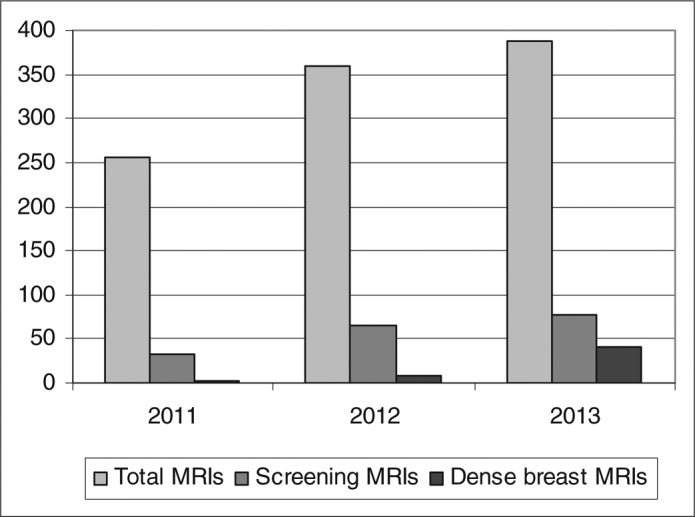
Bar graph representing breast magnetic resonance imaging (MRI) performed at Baylor University Medical Center at Dallas both prior to (2011) and after (2012 and 2013) the implementation of Henda's law. Total MRIs indicates the total yearly number of both screening and diagnostic breast MRIs; screening MRIs, the total yearly number of screening breast MRIs; dense breast MRIs, the total yearly number of dense breast screening MRIs.
DISCUSSION
Increased breast tissue density is a frequent mammographic finding, with 26% to 32% of women in the general population having a breast tissue density ≥50% (2). The sensitivity of mammography can decrease as the density of breast tissue increases, with one study showing sensitivity values ranging from 87% in breast tissue composed almost entirely of fat to 62.9% in extremely dense breast tissue (6). Henda's law took effect in the state of Texas on January 1, 2012, to ensure that affected patients and their primary care physicians were informed of the possible limitations of mammography and the potential benefit of supplemental screening examinations such as breast MRI. Contrast-enhanced MRI has been shown to be more sensitive than mammography in the detection of early breast tumors due to the different contrast enhancement pattern of an underlying breast cancer in comparison to normal breast parenchyma (1, 7, 8). Our analysis of the number of breast MRIs performed for the evaluation of dense breasts during the year 2011 (pre-Henda's law) as well as during the 2 years after the implementation of Henda's Law (2012 and 2013) showed a 23-fold increase after the law was passed, demonstrating the effect of Henda's law on the overall course of preventive breast care.
The implications of this increase could be potentially staggering to the health care economy. To date, only one state, Connecticut, has mandated that insurance cover adjunctive screening in these higher-risk patients. Among women in their 40s, 74% have dense breasts, and the percentage decreases to 57% for women in their 50s (9). Simply put, most women over 40 are affected by this need for additional screening. Without clear guidelines and definitive measures to control costs, we could no longer engage in screening as recommended by the World Health Organization, which states that any screening examination should be not only safe and effective but also relatively inexpensive (10).
References
- 1.Pike MC, Pearce CL. Mammographic density, MRI background parenchymal enhancement and breast cancer risk. Ann Oncol. 2013;24(Suppl 8):viii37–viii41. doi: 10.1093/annonc/mdt310. [DOI] [PMC free article] [PubMed] [Google Scholar]
- 2.Vachon CM, van Gils CH, Sellers TA, Ghosh K, Pruthi S, Brandt KR, Pankratz VS. Mammographic density, breast cancer risk and risk prediction. Breast Cancer Res. 2007;9(6):217–225. doi: 10.1186/bcr1829. [DOI] [PMC free article] [PubMed] [Google Scholar]
- 3.Boyd NF, Lockwood GA, Byng JW, Tritchler DL, Yaffe MJ. Mammographic densities and breast cancer risk. Cancer Epidemiol Biomarkers Prev. 1998;7(12):1133–1144. [PubMed] [Google Scholar]
- 4.Wolfe JN. Breast patterns as an index of risk for developing breast cancer. AJR Am J Roentgenol. 1976;126(6):1130–1137. doi: 10.2214/ajr.126.6.1130. [DOI] [PubMed] [Google Scholar]
- 5.Boyd NF, Dite GS, Stone J, Gunasekara A, English DR, McCredie MR, Giles GG, Tritchler D, Chiarelli A, Yaffe MJ, Hopper JL. Heritability of mammographic density, a risk factor for breast cancer. N Engl J Med. 2002;347(12):886–894. doi: 10.1056/NEJMoa013390. [DOI] [PubMed] [Google Scholar]
- 6.Carney PA, Miglioretti DL, Yankaskas BC, Kerlikowske K, Rosenberg R, Rutter CM, Geller BM, Abraham LA, Taplin SH, Dignan M, Cutter G, Ballard-Barbash R. Individual and combined effects of age, breast density, and hormone replacement therapy use on the accuracy of screening mammography. Ann Intern Med. 2003;138(3):168–175. doi: 10.7326/0003-4819-138-3-200302040-00008. [DOI] [PubMed] [Google Scholar]
- 7.DeMartini W, Lehman C. A review of current evidence-based clinical applications for breast magnetic resonance imaging. Top Magn Reson Imaging. 2008;19(3):143–150. doi: 10.1097/RMR.0b013e31818a40a5. [DOI] [PubMed] [Google Scholar]
- 8.Kang SS, Ko EY, Han BK, Shin JH, Hahn SY, Ko ES. Background parenchymal enhancement on breast MRI: influence of menstrual cycle and breast composition. J Magn Reson Imaging. 2014;39(3):526–534. doi: 10.1002/jmri.24185. [DOI] [PubMed] [Google Scholar]
- 9.Checka CM, Chun JE, Schnabel FR, Lee J, Toth H. The relationship of mammographic density and age: implications for breast cancer screening. AJR Am J Roentgenol. 2012;198(3):W292–W295. doi: 10.2214/AJR.10.6049. [DOI] [PubMed] [Google Scholar]
- 10.World Health Organization. Screening for various cancers. Available at http://www.who.int/cancer/detection/variouscancer/en/