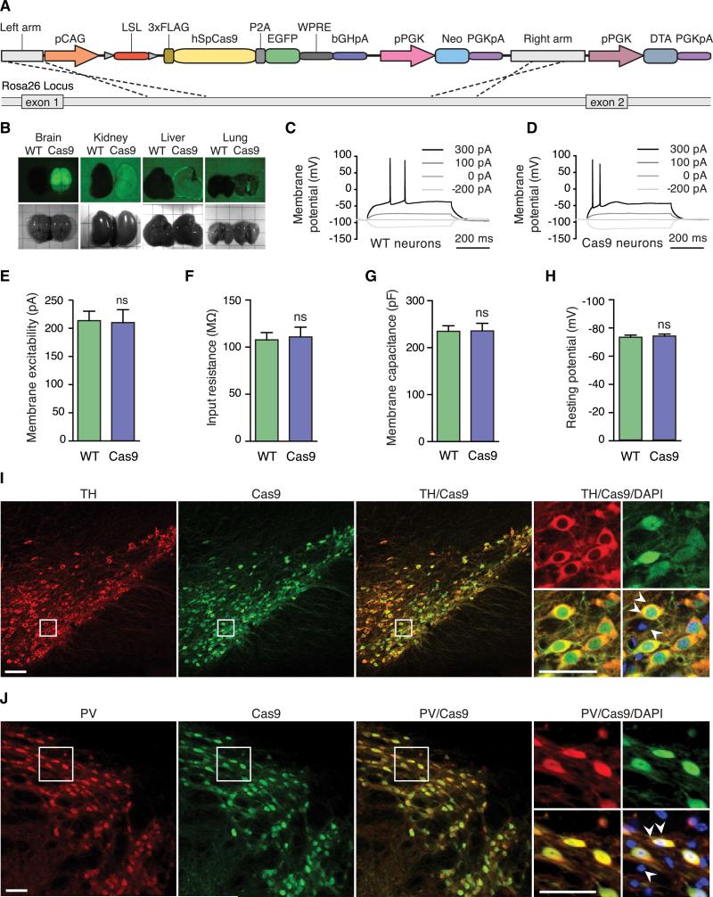
Figure 1. Generation and Characterization of Cre-Dependent and Constitutive Cas9-Expressing Mice.
(A) Schematic of the Cre-dependent Cas9 Rosa26 targeting vector.
(B) Bright-field and fluorescence stereomicroscope images of tissues dissected from constitutive Cas9-expressing (left) and wild-type (right) mice, showing Cas9-P2A-EGFP expression only in Cas9 mice.
(C and D) Representative current-clamp recordings and evoked action potentials from wild-type (C) and constitutive Cas9-expressing (D) neurons, showing no difference.
(E–H) Electrophysiological characterization of hippocampal neurons in acute slices from constitutive Cas9-expressing and wild-type neurons, showing no significant difference in: membrane excitability (E), input resistance (F), membrane capacitance (G), and resting potential (H). Data are plotted as mean ± SEM; n = 12 neurons from two wild-type mice and n = 15 neurons from two constitutive Cas9-expressing mice. n.s., not significant. See also Table S1.
(I) Representative immunofluorescence images of the substantia nigra in progenies from a Cre-dependent Cas9 mouse crossed with a TH-IRES-Cre driver mouse, showing Cas9 expression is restricted to TH-positive cells. Double arrowheads indicate a cell coexpressing TH and Cas9-P2A-EGFP. Single arrowhead indicates a cell expressing neither TH nor Cas9-P2A-EGFP. Scale bar, 100 μm.
(J) Representative immunofluorescence images of the reticular thalamus in progenies from a Cre-dependent Cas9 mouse crossed with a PV-Cre driver mouse, showing that Cas9 expression is restricted to PV-positive cells. Double arrowheads indicate a cell expressing PV and Cas9-P2A-EGFP. Single arrowhead indicates a cell expressing neither PV nor Cas9-P2A-EGFP. Scale bar, 50 μm.

