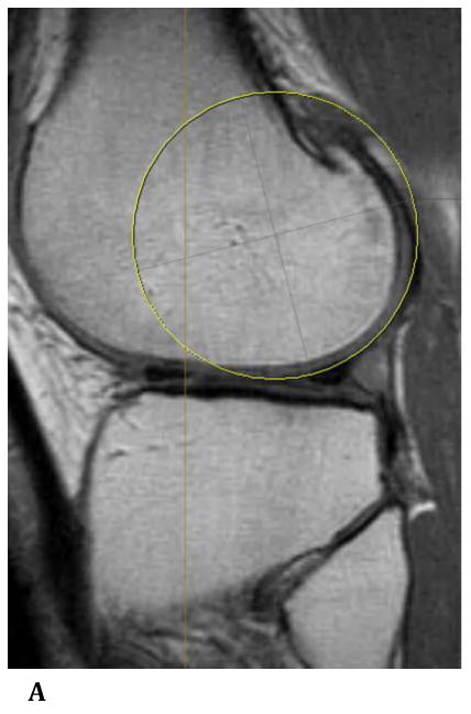
Figure 2.


Sagittal plane, T1 weighted MRI images demonstrating a best-fit circle around the cartilaginous surface of the nonaffected lateral femoral condyle (a), and propagation of this circle to the affected medial femoral condyle (b). Cartilage wear is appreciated at the posterior aspect of the medial femoral condyle.
