Abstract
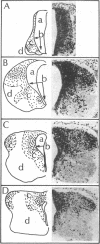
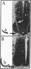
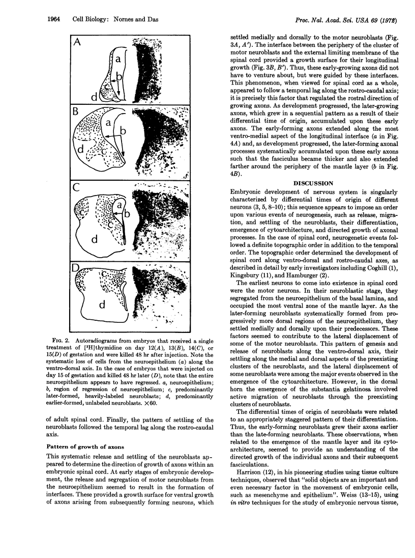
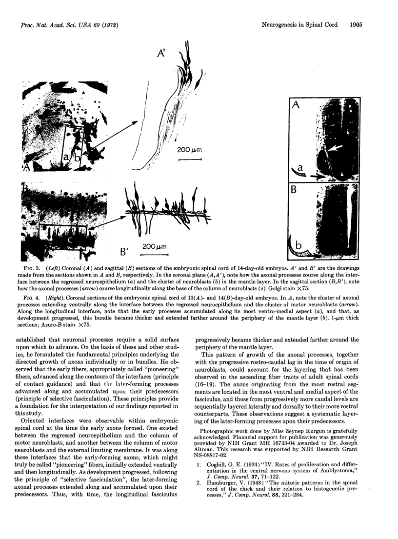
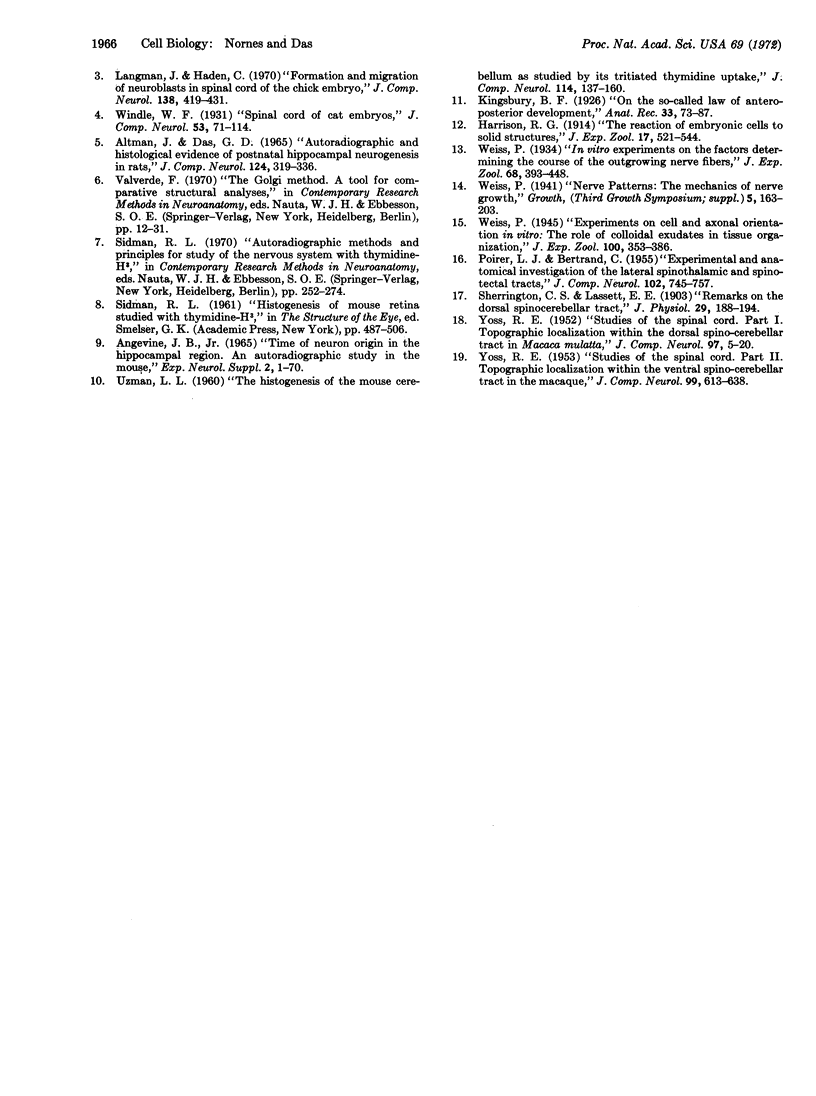
Neurogenesis was studied in rat spinal cord by correlation of the observations from [3H]thymidine autoradiography and Golgi techniques. The temporal pattern of the origin of neurons systematically lagged along the ventro-dorsal and rostro-caudal axes. The temporal pattern was correlated with the topographic pattern of the release and segregation of the newly formed neuroblasts from the neuroepithelium. These temporal and topographic patterns of neuroblast genesis imparted an order to the settling of the neuroblasts. Further, the emerging cytoarchitecture of the differentiating neuroblasts resulted in the formation of oriented interfaces along which the axons grew. The “pioneering” fibers grew along the interfaces formed by the regressed neuroepithelium and the assembled neuroblasts. As development progressed, the axons formed later “selectively fasciculated” along the “pioneering” fibers in a staggered pattern as a result of their differential times of origin. These temporal and topographic patterns occurred in a manner such that the preexisting structure served as an “organizing structure” or foundation upon which the axonal processes formed later in the developmental sequence were oriented and assembled.
Keywords: rat, neuroepithelium, autoradiography, selective fasciculation
Full text
PDF
Images in this article
Selected References
These references are in PubMed. This may not be the complete list of references from this article.
- Altman J., Das G. D. Autoradiographic and histological evidence of postnatal hippocampal neurogenesis in rats. J Comp Neurol. 1965 Jun;124(3):319–335. doi: 10.1002/cne.901240303. [DOI] [PubMed] [Google Scholar]
- Angevine J. B., Jr Time of neuron origin in the hippocampal region. An autoradiographic study in the mouse. Exp Neurol Suppl. 1965 Oct;(Suppl):1–70. [PubMed] [Google Scholar]
- Langman J., Haden C. C. Formation and migration of neuroblasts in the spinal cord of the chick embryo. J Comp Neurol. 1970 Apr;138(4):419–425. doi: 10.1002/cne.901380403. [DOI] [PubMed] [Google Scholar]
- POIRIER L. J., BERTRAND C. Experimental and anatomical investigation of the lateral spino-thalamic and spino-tectal tracts. J Comp Neurol. 1955 Jun;102(3):745–757. doi: 10.1002/cne.901020308. [DOI] [PubMed] [Google Scholar]
- Sherrington C. S., Laslett E. E. Remarks on the dorsal spino-cerebellar tract. J Physiol. 1903 Mar 16;29(2):188–194. doi: 10.1113/jphysiol.1903.sp000950. [DOI] [PMC free article] [PubMed] [Google Scholar]
- YOSS R. E. Studies of the spinal cord. I. Topographic localization within the dorsal spino-cerebellar tract in Macaca mulatta. J Comp Neurol. 1952 May;97(1):5–20. doi: 10.1002/cne.900970103. [DOI] [PubMed] [Google Scholar]
- YOSS R. E. Studies of the spinal cord. II. Topographic localization within the ventral spino-cerebellar tract in the macaque. J Comp Neurol. 1953 Dec;99(3):613–638. doi: 10.1002/cne.900990308. [DOI] [PubMed] [Google Scholar]


